Next Article in Journal
Iain Hamilton McVicar (12/08/1958–11/06/2022)
Previous Article in Journal
An Alternative Approach Using Circummandibular Wiring for Treatment of Dentoalveolar Fractures in Children: A Case Report
 
 
Craniomaxillofacial Trauma & Reconstruction is published by MDPI from Volume 18 Issue 1 (2025). Previous articles were published by another publisher in Open Access under a CC-BY (or CC-BY-NC-ND) licence, and they are hosted by MDPI on mdpi.com as a courtesy and upon agreement with Sage.
Article

Endoscopically Assisted Marginal Mandibulectomy Using an Intraoral Approach Alone for Squamous Cell Carcinoma of the Posterior Mandibular Gingiva: A Technical Note

by
Atsushi Shudo
1,2,3
1
Department of Oral and Maxillofacial Surgery, Kishiwada Tokushukai Hospital, 4-27-1 Kamori-cho, Kishiwada, Osaka 596-0042, Japan
2
Department of Oral and Maxillofacial Surgery, Hyogo College of Medicine, Hyogo, Japan
3
Dental Association of Kishiwada City, Osaka, Japan
Craniomaxillofac. Trauma Reconstr. 2022, 15(2), 175-183; https://doi.org/10.1177/19433875211015045
Submission received: 1 November 2020 / Revised: 1 December 2020 / Accepted: 1 January 2021 / Published: 2 May 2021

Abstract

:
Study Design: Technical note. Objective: Marginal mandibulectomy is a surgical procedure for treatment of mandibular gingival tumors. The intraoral approach to the posterior region of the mandible for marginal mandibulectomy is difficult due to limited access and operating field visibility; the conventional surgical procedure may require some skin incisions. This report discusses the effectiveness of endoscopic assistance in marginal mandibulectomy. Methods: This article describes endoscopically assisted marginal mandibulectomy using an intraoral approach alone for squamous cell carcinoma of the posterior mandibular gingiva. Results: The advantages of this surgical method are twofold: (1) superior visibility to the lower edge of the mandible without any skin incision; and (2) safe surgical confirmation of important anatomy on the buccolingual side (e.g., mental foramen, lingual nerve, mandibular foramen, and neurovascular bundle). This minimally invasive approach without any skin incision, as well as the superior visibility of the operating field, are important advantages of endoscopically assisted marginal mandibulectomy that cannot be obtained by other surgical methods. Conclusions: Endoscopically assisted marginal mandibulectomy using an intraoral approach alone may be useful because it avoids damage to facial skin and improves safety by employing an enlarged bright field.

Introduction

Endoscopically assisted surgery is a minimally invasive surgical method utilized in various surgical fields, including the oral and maxillofacial surgery field where aesthetic aspects are essential considerations during treatment. Oral and maxillofacial endoscopic techniques are used for tumor resection, trauma, orthognathic surgery, sialoendoscopy, temporomandibular joint surgery, foreign body removal, and dentoalveolar surgery; all of these applications require sufficient visualization and a minimally invasive approach.[1,2,3,4,5] Technological advances have allowed wide adaptation, but endoscopic techniques are not commonly applied to marginal mandibulectomy (MM). This article describes an endoscopically assisted MM using an intraoral approach alone, then discusses the advantages of this approach. This surgical method may be useful because it avoids damage to facial skin and improves safety by employing an enlarged bright field.

Material and Methods

This surgical method can be effectively used for treatment of patients in whom an extended MM is required to reach the lower portion of the mandible. This article demonstrates a typical rim mandibulectomy procedure performed with endoscopic assistance. The endoscopic instruments used were 4.0-mm-diameter and 18-cm-length rigid endoscopes, straight (Karl Storz, Straight Forward Telescope 0°), 45-degree-angled (Karl Storz, Forward-Oblique Telescope 45°), and 70-degree-angled (Karl Storz, Lateral Telescope 70°) (Figure 1).

Case Presentation

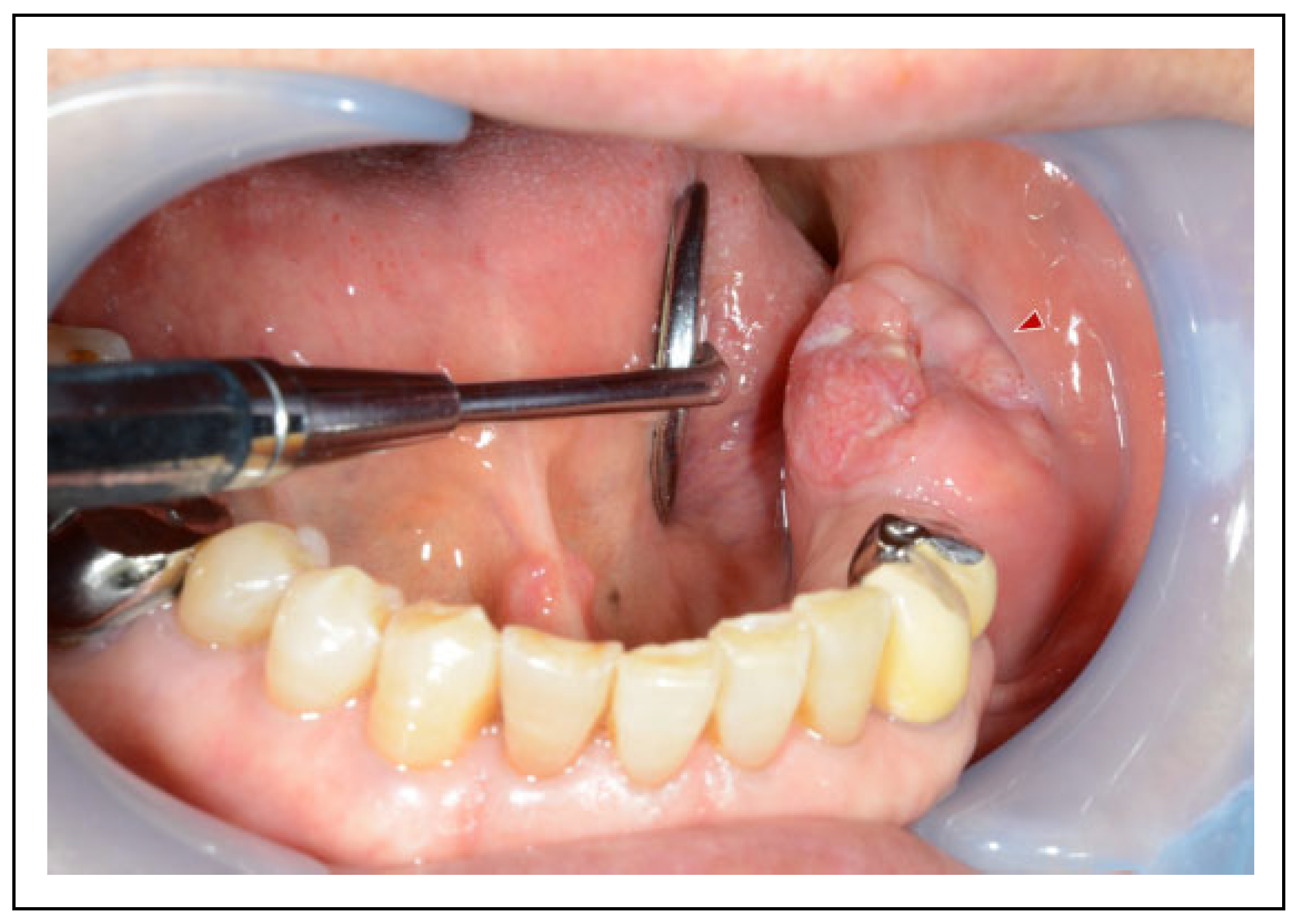
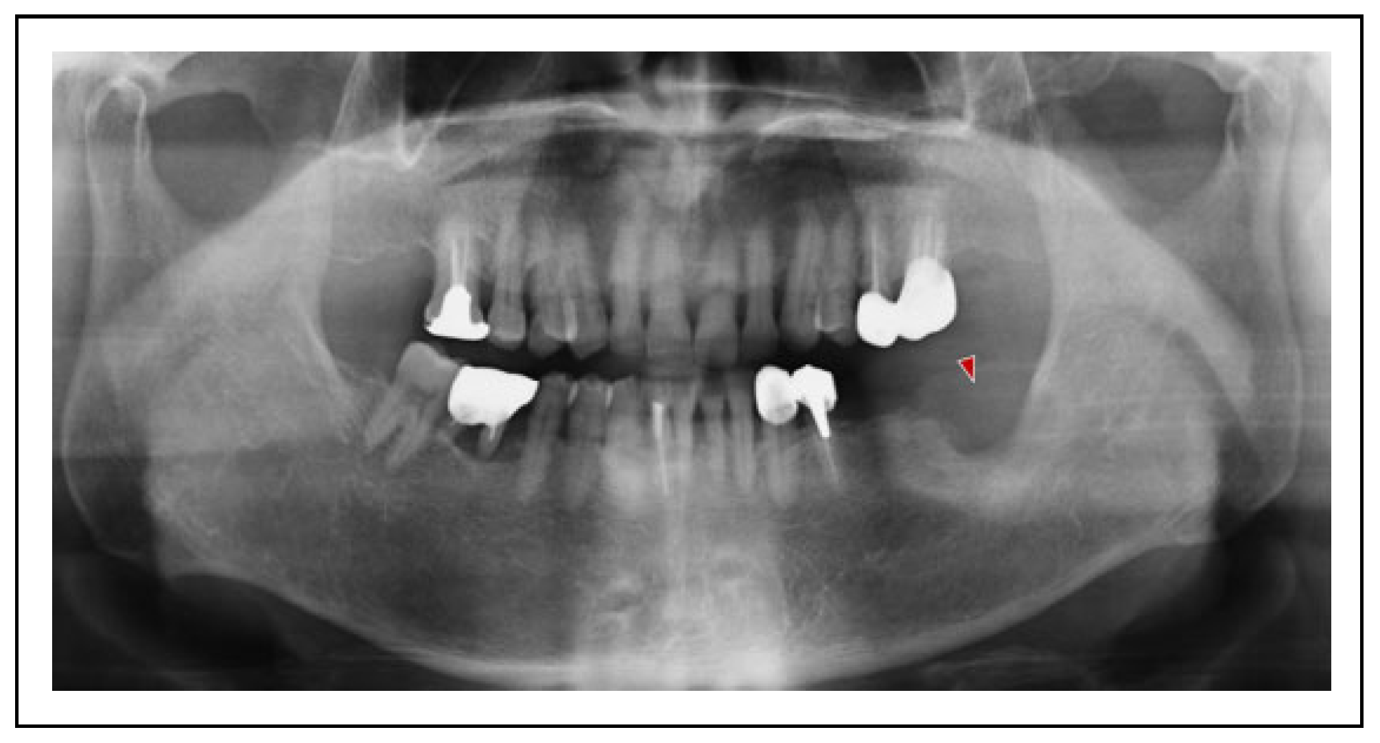
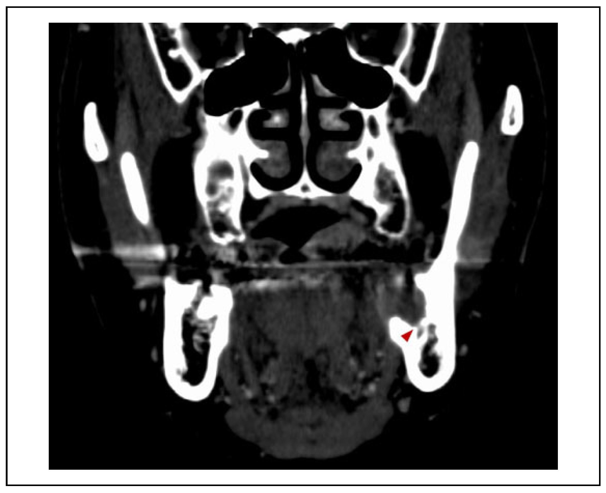
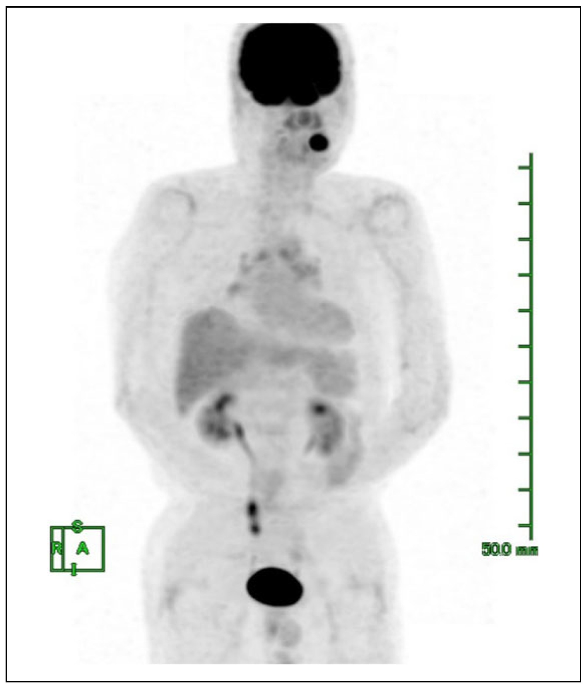
A 70-year-old man was referred to our outpatient office for further evaluation of incomplete healing at more than 4 weeks after tooth extraction. Initial clinical examination revealed an indurated mass measuring 25 × 15 mm on the left mandibular gingiva (Figure 2). There was no neuro-sensory disturbance of the left inferior alveolar nerve. Significantly swollen or indurated cervical lymph nodes were not identified. Histopathological analysis of a biopsy sample revealed findings that supported a diagnosis of squamous cell carcinoma. Imaging examinations were performed including orthopantomography, computed tomography (CT), magnetic resonance imaging, and 18F-fluorodeoxyglucose positron emission tomography/ CT (FDG-PET/CT). Orthopantomography and plain CT showed erosive bone resorption of the left mandibular bone and revealed that the primary lesion was near the mandibular canal (Figure 3 and Figure 4); however, contrastenhanced CT confirmed that the enhanced rim of the tumor was located above the mandibular canal (Figure 5). The minimum length from the site of bone resorption to the lower edge of the mandible was approximately 24 mm. FDG-PET/CT did not show obvious findings suggestive of cervical lymph node metastasis or distant metastases (Figure 6).
The clinical diagnosis was gingival squamous cell carcinoma stage IVA (cT4aN0M0), in accordance with the 8th edition of the American Joint Committee on Cancer/Union for International Cancer Control (AJCC/UICC) TNM staging system.[6] At this cancer stage, extraoral segmental mandibulectomy might be routinely recommended. However, previous studies suggested that the proportion of T4a tumors was high for gingival squamous cell carcinoma and the current AJCC/UICC T staging system thus appeared to have restricted prognostic utility; when the bone invasion criteria were excluded from the definition within the AJCC/UICC system, tumors without mandibular canal invasion would be downstaged from T4a to T2.[7,8] In our patient, clinical findings and imaging examinations did not suggest mandibular nerve invasion, although bone resorption was located near the mandibular canal. In addition, the patient did not approve the skin incision because of the associated complications; he requested the avoidance of skin damage, as much as possible.
Therefore, we proposed the following treatment plan: endoscopically assisted MM including resection of part of the mandibular canal using an intraoral approach alone, based on previous reports that MM could be applied even in patients with cT4a involving erosive cancerous bone invasion.[9,10] We noted that internal plate fixation using a fracture plate with a load-bearing approach might be performed if a mandibular fracture occurred due to thinning of the mandibular marginal bone; this would involve a transbuccal approach using a trocar. Moreover, if intraoperative rapid pathological assessment showed tumor cells in the resected margin of the mandibular neurovascular bundle, the surgical procedure might be converted to extraoral segmental mandibulectomy and reconstruction with a 2.4-mm titanium plate. Prophylactic neck dissection would not be performed; however, if postoperative histopathological examination supported a diagnosis of pT4, chemoradiotherapy would be administered as postoperative adjuvant therapy. Active surveillance would be initiated; if cervical lymph node metastasis were suspected, additional neck dissection would be performed. The patient consented to the proposed treatment plan.

Surgical Procedure

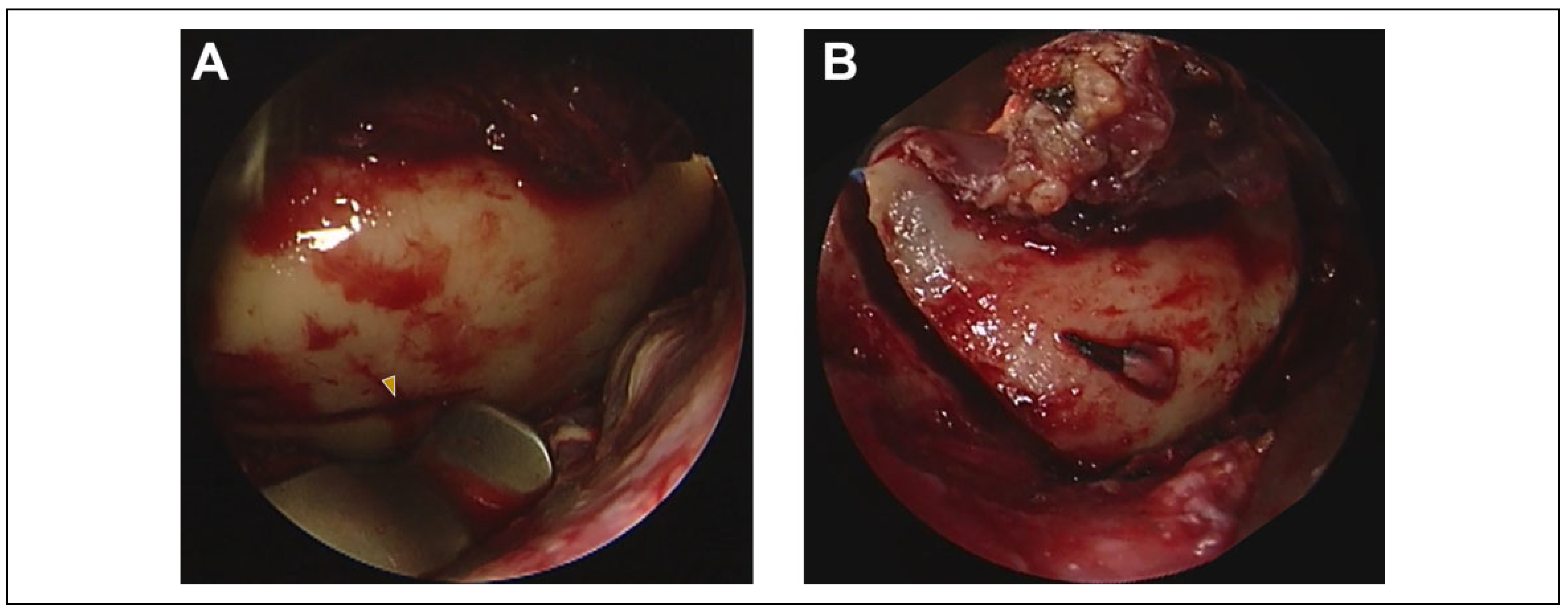
The surgery was performed by 1 surgeon and 2 assistants. One assistant performed retraction and suction, while the other assistant maneuvered the endoscope. The anesthesia machine was placed near the patient’s feet, thus allowing the surgeon or assistant to stand near the patient’s head. General anesthesia was induced with nasal intubation, combined with routine local anesthesia; the mucosal surface was then incised with a safety margin of 10 mm. The mucoperiosteal flap was separated subperiosteally from the mandibular surface on the buccolingual side. The surrounding bone surface outside of the tumor was sufficiently exposed, enabling advancement to the osteotomy procedure. Until this point, the procedure could be performed under direct vision, in accordance with a standard oral surgery approach.
To prevent bleeding from the inferior alveolar artery and vein during mandibular bone resection, the mandibular neurovascular bundle was ligated and incised at the position of the mandibular foramen, with confirmation via straight endoscopy (Figure 7). When resection advanced toward the lower edge of the mandible, a 45-degree-angled or 70-degree-angled endoscope was used. The bone resection line was marked using a side cutting bur and an oscillating saw in the endoscopically enlarged bright field to ensure a safety margin of ≥10 mm on the bone surface (Figure 8). Dedicated dissectors with a distal spatula may constitute a useful endoscope attachment; however, if these instruments are not deployed, a progeny retractor fixed to the endoscope with skin closure strips may offer a suitable alternative (Figure 9). The visibility differs depending on the surgical position (i.e., 45-degree-angled or 70-degree-angled). In this method, a sufficient bright field could be obtained with soft tissue retraction and light from a light source. After partial resection using both oscillating and reciprocating saws resulted in residual lingual cortical bone, complete bone resection was achieved with a bone chisel and a mallet (Figure 10). Intact bone marrow was evident on the resected surface; thus, tumor invasion was not suspected (Figure 11). Intraoperative rapid pathological assessment revealed no tumor cells in the resected margin of the mandibular neurovascular bundle and surrounding bone marrow. No intraoperative mandibular fractures occurred, and the surgery was completed without internal plate fixation.

Postoperative Course

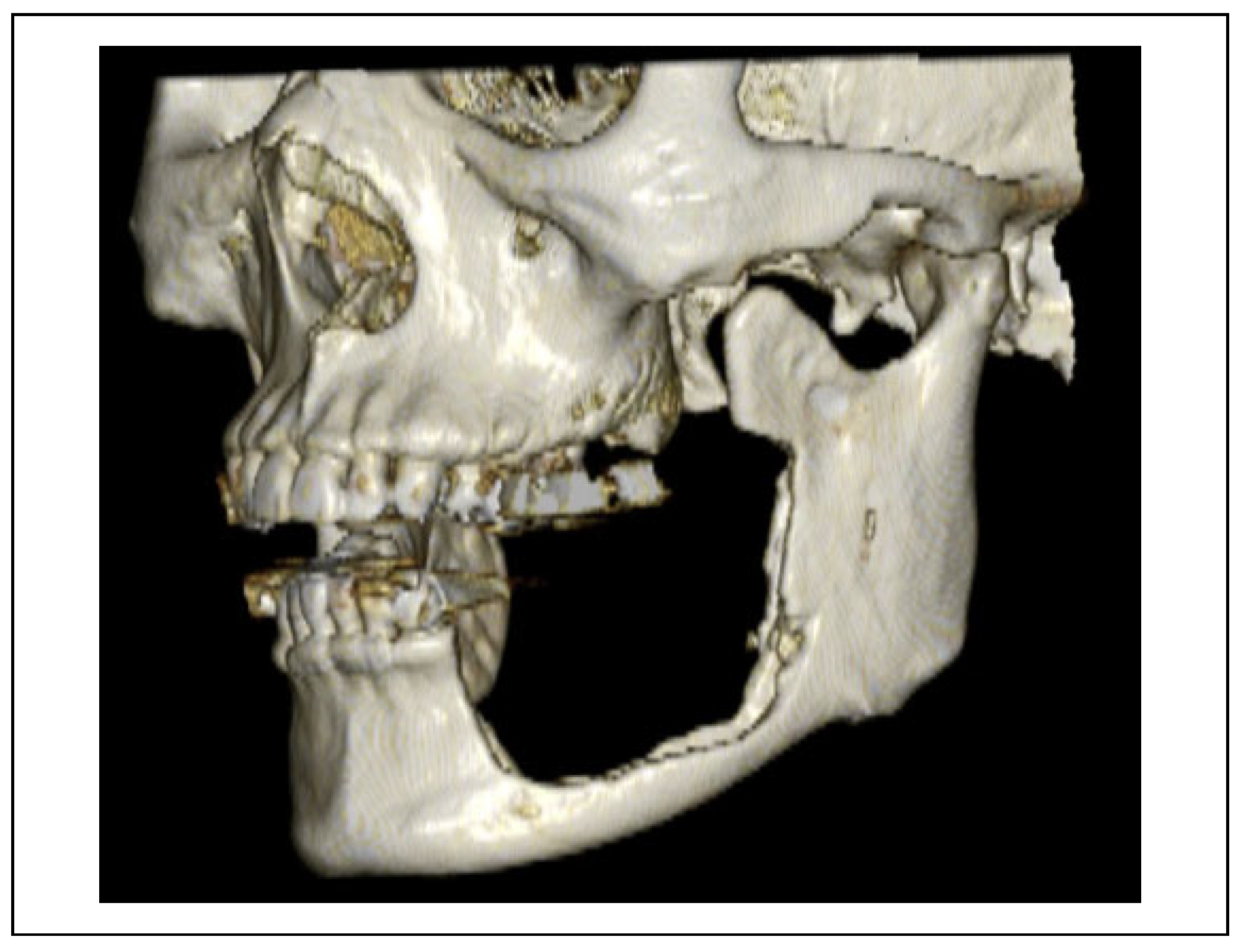
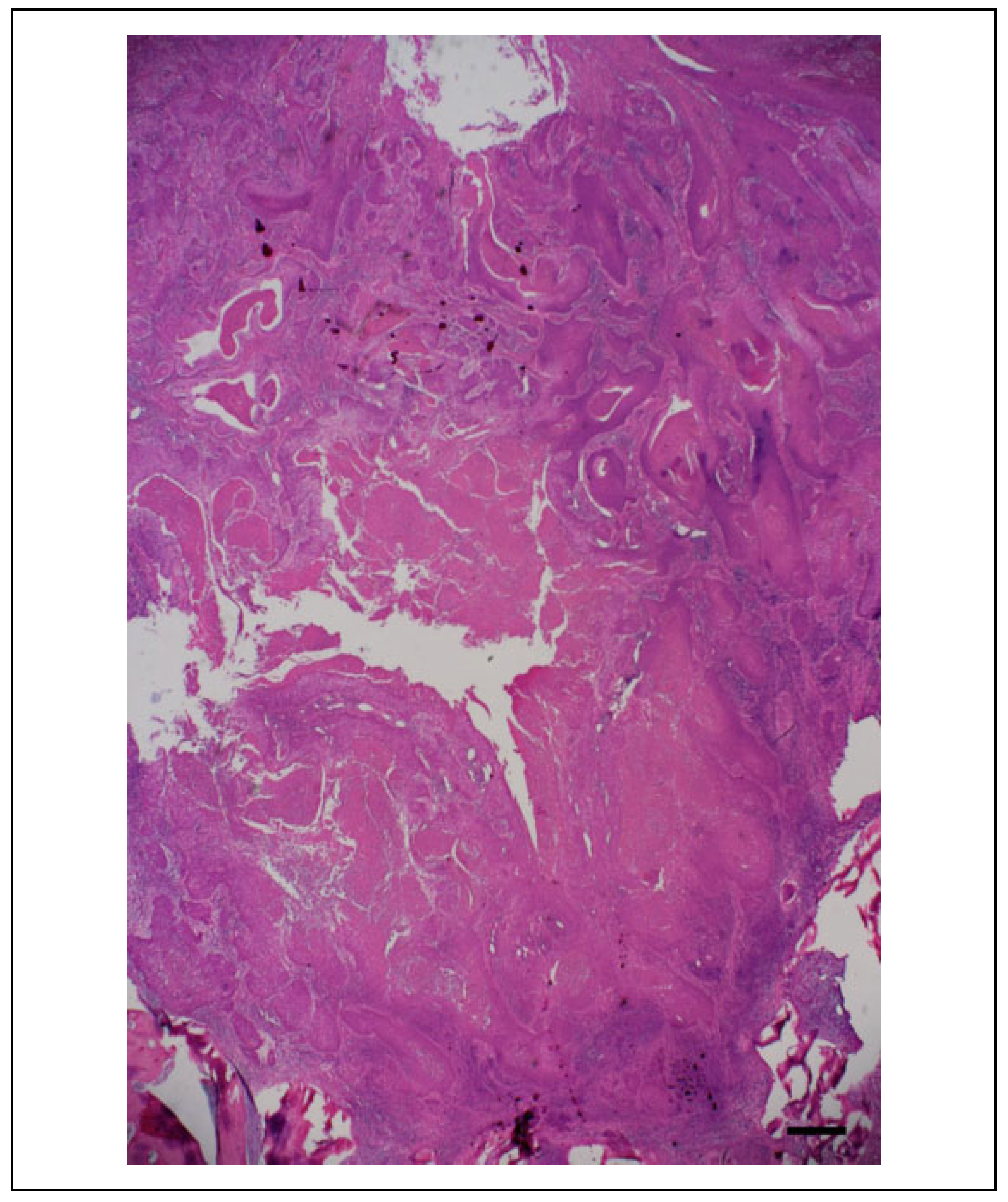
The patient began oral intake of soft food several hours postoperatively. No postoperative fracture occurred, although the mandible became thinner (Figure 12); the patient was discharged 14 days postoperatively. The tumor size was 25 × 16 × 16 mm. Histopathological examination of the resected specimen confirmed the diagnosis of gingival squamous cell carcinoma; this examination also revealed the absence of tumor cells at the surgical margin and showed that no cancer had invaded the mandibular neurovascular bundle (Figure 13). Additionally, the depth of invasion was 8 mm (Figure 14). The definitive diagnosis of mandibular gingival squamous cell carcinoma pT2 was confirmed histopathologically. Postoperative adjuvant chemoradiotherapy was not administered. The patient’s postoperative course was uneventful without major complications other than paresthesia due to mandibular nerve transection (Figure 15). There has been no evidence of recurrence or metastasis during 1 year of follow-up.

Discussion

Surgical treatment for mandibular gingival squamous cell carcinoma is determined on the basis of histological type and cancer progression (location and size of primary lesion, as well as existence of bone invasion or distant metastasis); this treatment includes MM, segmental mandibulectomy, and hemimandibulectomy.[11,12] Based on their abilities to preserve mandibular continuity, there is a substantial difference between MM and the latter 2 methods in terms of the postoperative course and patient’s quality of life.[3] In properly selected patients, MM is sufficient for achieving a safe resection margin and can ensure satisfactory tumor control that is comparable to the control following segmental mandibulectomy.[13,14,15] MM can provide functionally, aesthetically, and therapeutically satisfactory outcomes.
Notably, an intraoral approach for MM may be difficult due to limited access and operating field visibility. Gingival tumors of the anterior mandible are preferred surgical indications for MM; however, the approach becomes challenging for lesions in the posterior region of the mandible, when a neck dissection or other extraoral approach is not used for tumor exposure. To avoid the classical submandibular approach, which requires a wide skin incision, the cheeksplitting technique and/or transbuccal technique have been described as relatively minimally invasive methods for MM.[16,17,18] However, skin incisions cannot be completely avoided using these surgical methods; intraoperative damage to surrounding organs (e.g., parotid duct or buccal branch of the facial nerve) or postoperative scarring (and scar contracture in some patients) may occur. Furthermore, although these surgical methods can allow surgery under direct view while ensuring the operating field remains on the outer (buccal) side of the mandible, it remains difficult to sufficiently visualize the inner (lingual) side of the mandible where the mandibular foramen and lingual nerve are present. The minimally invasive approach without any skin incision, as well as the superior visibility of the operating field on the lingual side, are advantages of endoscopically assisted MM that cannot be obtained using other surgical methods. In the present patient, the endoscopic technique was applied to the so-called “rim mandibulectomy” form of MM, but it may be more effective for “sagittal mandibulectomy” when performing excision on the lingual side of the mandible.
To the best of our knowledge, there have been no reports of endoscopically assisted MM using an intraoral approach alone. We suspect that such reports have not been published for the following reasons: (1) confined surgical indication (i.e., in patients who require neck dissection or other extraoral approach for tumor exposure, there is no requirement to maintain an intraoral surgery approach); (2) physical limitations (i.e., this method cannot be performed without using the endoscope and oscillating saw as dedicated surgical instruments); and (3) non-physical limitations (i.e., endoscopically assisted surgery requires certain technical skills in both the surgeon and the assistants). However, in patients for whom the cheek-splitting and/or transbuccal techniques are appropriate, endoscopically assisted MM may be useful.
We cannot make definitive statements regarding the usefulness of this approach because there have been few reports; that is an important limitation of this technical note. In particular, because of the ongoing wide usage of endoscopic instruments for conventional transnasal surgery, the existing reports comprise studies regarding the use of endoscopically assisted maxillectomy by otorhinolaryngologists.[19,20] For MM, studies with large cohorts of patients should be performed to confirm the findings in this report.

Conclusion

Endoscopically assisted MM using an intraoral approach alone may be useful because it avoids damage to facial skin and improves safety by employing an enlarged bright field.

Funding

The author(s) received no financial support for the research, authorship, and/or publication of this article.

Acknowledgments

The author thanks Tomoko Oonaka, a cytotechnologist in our hospital, for specimen preparation and staining. The author also thanks Ryan Chastain-Gross, PhD, from Edanz Group (https://enauthor-services.edanzgroup.com) for editing a draft of this manuscript.

Conflicts of Interest

The author(s) declared no potential conflicts of interest with respect to the research, authorship, and/or publication of this article.

References

  1. Pedroletti, F.; Johnson, B.S.; McCain, J.P. Endoscopic techniques in oral and maxillofacial surgery. Oral Maxillofac Surg Clin North Am. 2010, 22, 169–182. [Google Scholar] [CrossRef] [PubMed]
  2. Li, Y.; Xue, R.; Lai, Q.; et al. Endoscope-assisted resection of nonneoplastic space-occupying lesion in oral and maxillofacial areas. Sci Rep. 2017, 7, 16920. [Google Scholar] [CrossRef] [PubMed]
  3. Hyppolito, J.O.P.; Araújo, R.A.C.; Souza, M.S.M.; et al. The use of intra-oral endoscopy in oral and maxillofacial surgery: literature review. Int J Oral Maxillofac Surg. 2019, 48, 272. [Google Scholar] [CrossRef]
  4. Mohamed, A.H.; Joseph, P.M.; David, Y.A.; Maria, J.T. Minimally invasive endoscopic oral and maxillofacial surgery. Oral Maxillofac Surg Clin North Am. 2019, 31, 561–567. [Google Scholar]
  5. Víctor, B.; Ramon, F.; Wilfried, E. Endoscopic visualization of anatomic structures as a support tool in oral surgery and implantology. J Oral Maxillofac Surg. 2012, 70, e1–e6. [Google Scholar]
  6. Lydiatt, W.M.; Patel, S.G.; O’Sullivan, B.; et al. Head and neck cancers—major changes in the American Joint Committee on Cancer eighth edition cancer staging manual. CA Cancer J Clin. 2017, 67, 122–137. [Google Scholar] [CrossRef]
  7. Okura, M.; Yanamoto, S.; Umeda, M.; et al. Prognostic and staging implications of mandibular canal invasion in lower gingival squamous cell carcinoma. Cancer Med. 2016, 5, 3378–3385. [Google Scholar] [CrossRef]
  8. Ebrahimi, A.; Murali, R.; Gao, K.; Elliott, M.S.; Clark, J.R. The prognostic and staging implications of bone invasion in oral squamous cell carcinoma. Cancer 2011, 117, 4460–4467. [Google Scholar] [CrossRef] [PubMed]
  9. Sproll, C.K.; Holtmann, H.; Schorn, L.K.; et al. Mandible handling in the surgical treatment of oral squamous cell carcinoma: lessons from clinical results after marginal and segmental mandibulectomy. Oral Surg Oral Med Oral Pathol Oral Radiol. 2020, 129, 556–564. [Google Scholar] [CrossRef] [PubMed]
  10. Tei, K.; Totsuka, Y.; Iizuka, T.; Ohmori, K. Marginal resection for carcinoma of the mandibular alveolus and gingiva where radiologically detected bone defects do not extend beyond the mandibular canal. J Oral Maxillofac Surg. 2004, 62, 834–839. [Google Scholar] [CrossRef] [PubMed]
  11. Rao, L.P.; Shukla, M.; Sharma, V.; Pandey, M. Mandibular conservation in oral cancer. Surg Oncol. 2012, 21, 109–118. [Google Scholar] [CrossRef] [PubMed]
  12. Guerra, M.F.M.; Gias, L.N.; Campo, F.R.; Pérez, J.S. Marginal and segmental mandibulectomy in patients with oral cancer: a statistical analysis of 106 cases. J Oral Maxillofac Surg. 2003, 61, 1289–1296. [Google Scholar] [CrossRef] [PubMed]
  13. Qiu, Y.; Lin, L.; Shi, B.; Zhu, X. Does different mandibulectomy (marginal vs segmental) affect the prognosis in patients with oral squamous cell carcinoma? J Oral Maxillofac Surg. 2018, 76, 1117–1122. [Google Scholar] [CrossRef] [PubMed]
  14. Chen, Y.L.; Kuo, S.W.; Fang, K.H.; Hao, S.P. Prognostic impact of marginal mandibulectomy in the presence of superficial bone invasion and the nononcologic outcome. Head Neck 2011, 33, 708–713. [Google Scholar] [CrossRef] [PubMed]
  15. Politi, M.; Costa, F.; Robiony, M.; Rinaldo, A.; Ferlito, A. Review of segmental and marginal resection of the mandible in patients with oral cancer. Acta Otolaryngol. 2000, 120, 569–579. [Google Scholar] [PubMed]
  16. Hirsch, D.L.; Dierks, E.J. Use of a transbuccal technique for marginal mandibulectomy: a novel approach. J Oral Maxillofac Surg. 2007, 65, 1849–1851. [Google Scholar] [CrossRef] [PubMed]
  17. Ohba, S.; Yamashita, H.; Takashi, I.; Asahina, I. Marginal mandibulectomy for lower gingival carcinoma with a cheeksplitting transbuccal approach and reconstruction by buccal fat pad flap: a case report. J Oral Maxillofac Surg. 2013, 71, e143–e146. [Google Scholar] [CrossRef] [PubMed]
  18. Abe, A.; Kurita, K.; Hayashi, H.; Ito, Y. Cheek-splitting technique for marginal mandibulectomy: a novel approach. J Clin Exp Dent. 2019, 11, e675–e678. [Google Scholar] [CrossRef] [PubMed]
  19. Deganello, A.; Ferrari, M.; Paderno, A.; et al. Endoscopicassisted maxillectomy: operative technique and control of surgical margins. Oral Oncol. 2019, 93, 29–38. [Google Scholar] [CrossRef] [PubMed]
  20. He, S.; Bakst, R.L.; Guo, T.; Sun, J. A combination of modified transnasal endoscopic maxillectomy via transnasal prelacrimal recess approach with or without radiotherapy for selected sinonasal malignancies. Eur Arch Otorhinolaryngol. 2015, 272, 2933–2938. [Google Scholar] [CrossRef] [PubMed]
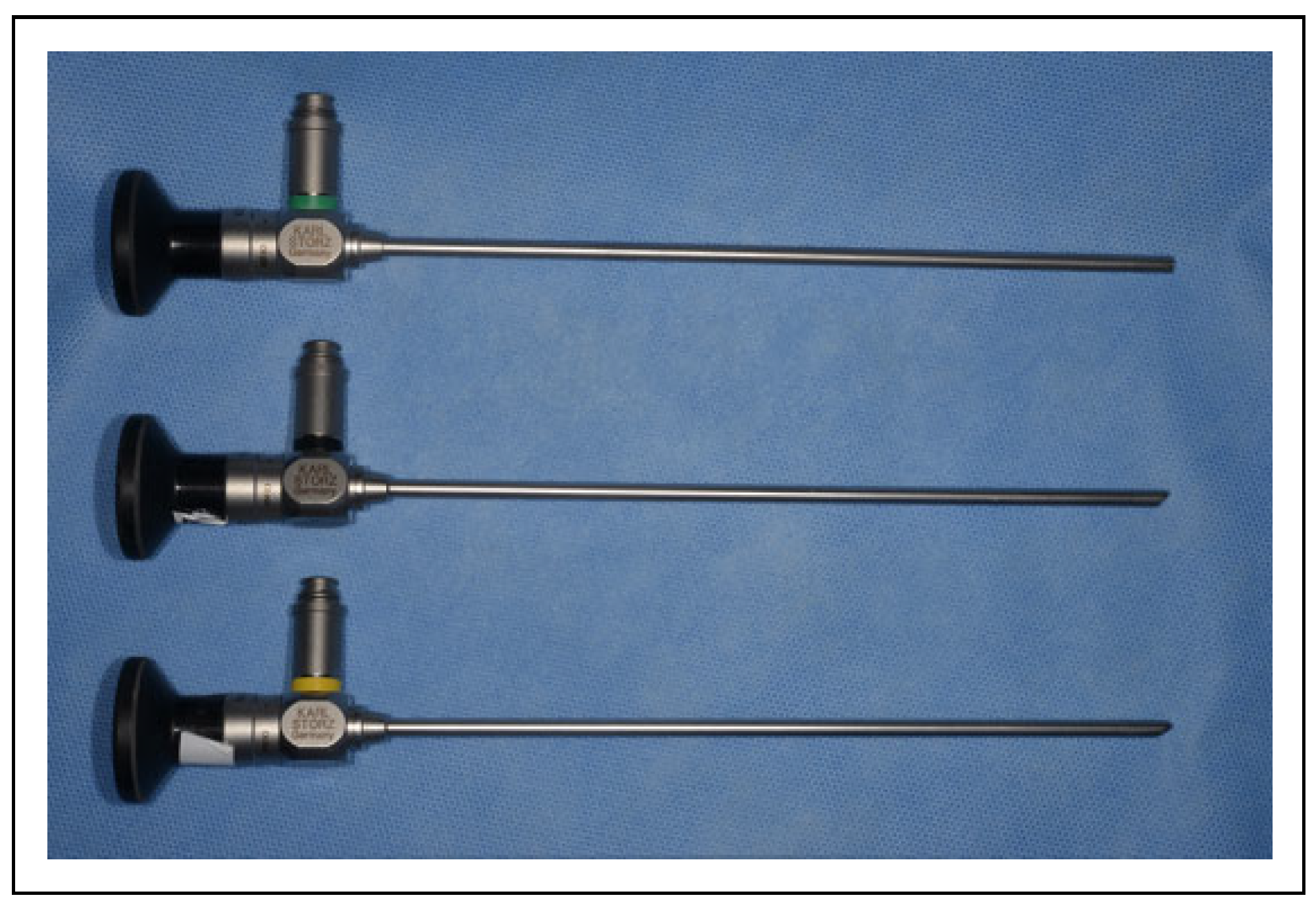
Figure 1. 4.0-mm-diameter and 18-cm-length rigid endoscopes were used (green color-coded: Straight Forward Telescope 0°, black color-coded: Forward-Oblique Telescope 45°, yellow color-coded: Lateral Telescope 70°).
Cmtr 15 00026 g001
Figure 2. Clinical presentation, intraoral view. An indurated mass (25 × 15 mm) was observed on the left mandibular gingiva.
Cmtr 15 00026 g002
Figure 3. Orthopantomography revealed erosive bone resorption of the left mandibular bone.
Cmtr 15 00026 g003
Figure 4. Plain computed tomography (coronal plane) revealed the primary lesion near the mandibular canal.
Cmtr 15 00026 g004
Figure 5. Contrast-enhanced computed tomography confirmed that the enhanced rim of the tumor was located above the mandibular canal.
Cmtr 15 00026 g005
Figure 6. 18F-fluorodeoxyglucose positron emission tomography showed no obvious findings suggestive of cervical lymph node metastasis or distant metastases.
Cmtr 15 00026 g006
Figure 7. Intraoperative endoscopic image of the ligated and incised mandibular neurovascular bundle. (A) Positional relationship when using straight endoscopy for the mandibular foramen. (B) Ligation. (C) Incision.
Cmtr 15 00026 g007
Figure 8. Intraoperative endoscopic image of bone resection. (A) Confirmation of marked bone resection line. (B) Confirmation that safety margin was secured and that there were no unsawed parts.
Cmtr 15 00026 g008
Figure 9. Progeny retractor with the 45-degree-angled endoscope. (A) Fixation using skin closure strips (3M, Steri-Strip 12-mm width). (B) Positional relationship when using angled endoscopy for the lateral mandibular body.
Cmtr 15 00026 g009
Figure 10. Intraoperative view after bone resection. The lingual nerve was exposed but kept intact.
Cmtr 15 00026 g010
Figure 11. Surgical specimen. (A) Lateral view. (B) Reverse side view.
Cmtr 15 00026 g011
Figure 12. Postoperative 3-dimensional computed tomography.
Cmtr 15 00026 g012
Figure 13. Histopathological examination of the surgical resected specimen confirmed that no cancer had invaded the mandibular neurovascular bundle. (A) Loupe image, hematoxylin and eosin staining. (B) Magnification, ×12.5; scale bar, 500 μm.
Cmtr 15 00026 g013
Figure 14. Histopathological examination showed that the depth of invasion was 8 mm (hematoxylin and eosin staining; magnification, ×12.5; scale bar, 500 μm).
Cmtr 15 00026 g014
Figure 15. Clinical examination at 3 months postoperatively. (A) Postoperative tumor site. (B) Intraoral frontal view in occlusion. (C) Extraoral view.
Cmtr 15 00026 g015