Next Article in Journal
Current Role of Radiotherapy in the Management of Oral Cavity Squamous Cell Carcinoma
Previous Article in Journal
Hemorrhage in Onodi Cell Leading to Traumatic Optic Neuropathy
 
 
Craniomaxillofacial Trauma & Reconstruction is published by MDPI from Volume 18 Issue 1 (2025). Previous articles were published by another publisher in Open Access under a CC-BY (or CC-BY-NC-ND) licence, and they are hosted by MDPI on mdpi.com as a courtesy and upon agreement with Sage.
Technical Note

The Use of a Novel CAD-CAM Splint to Simplify Open Reduction and Internal Fixation of Mandibular Angle Fracture: A Technical Note

by
Mrunalini Ramanathan
,
Elavenil Panneerselvam
*,
Sriraam Kasi Ganesh
and
Krishna Kumar Raja
Department of Oral and Maxillofacial Surgery, SRM Dental college and Hospital, Ramapuram Campus, Ramapuram, Chennai 600089, Tamil Nadu, India
*
Author to whom correspondence should be addressed.
Craniomaxillofac. Trauma Reconstr. 2021, 14(1), 74-78; https://doi.org/10.1177/1943387520904897
Submission received: 1 December 2019 / Revised: 31 December 2019 / Accepted: 1 February 2020 / Published: 12 April 2020

Abstract

:
Mandibular angle fractures are frequently encountered as they constitute an area of weakness. Complications after open reduction and internal fixation (ORIF) of angle fractures commonly arise due to improper reduction and fixation methods that fail to counteract the dynamic muscle forces present in this region. Conventional reduction methods such as digital manipulation, intermaxillary fixation, towel clip traction, and wiring are associated with various limitations. This technical note highlights the fabrication and use of a computer-aided designing/computer-aided manufacturing–generated splint for ORIF of a superiorly displaced mandibular angle fracture. The splint consisted of 2 components: (1) a distal tooth-borne component to guide the teeth into maximum intercuspation and (2) a proximal bone-borne component to reduce the angle fracture. This composite splint facilitates simultaneous restoration of occlusion as well as reduction of mandibular angle fractures. The advantages of this technique include the following: (1) easy fabrication of splint, (2) easy and precise anatomical reduction of angle fracture, and (3) less operative time.

Introduction

The surgical objectives of mandibular angle fracture include anatomical reduction and restoration of dental occlusion. This can be challenging in unfavorable fractures, where the dynamic muscle forces lead to gross displacement of fracture fragments[1] along with the resultant mal- occlusion. Improper reduction and fixation of mandibular angle fractures frequently lead to numerous complications such as malocclusion (14%); malunion, nonunion, and infection (28%); paraesthesia (6%); and hardware failure (36%).[2,3[4] Various techniques such as digital manipulation,[5] intermaxillary fixation,[5] wiring,[6] and towel clip traction[7] are practiced to achieve the abovementioned objectives[1].
Conventional interocclusal splints (manual and laboratory generated) constitute one of the frequently used methods to establish pretrauma occlusion during surgical open reduction and fixation of fractures (ORIF).[5] However, they are associated with certain limitations such as additional laboratory time and technique sensitivity in fabricating splints. Furthermore, conventional interocclusal splints offer less help in reduction of angle fracture, where the proximal fragment is edentulous. This case report demonstrates the efficacy of a novel computer-aided designing/computer-aided manufacturing (CAD/CAM) splints in achieving precise anatomical reduction of mandibular angle fractures with simultaneous restoration of occlusion.

Case Report

A 48-year-old male reported to the Department of Oral and Maxillofacial Surgery, with complaints of pain and swelling involving the left lower face. History revealed that the patient had sustained a blow to his left angle region during an assault.
Clinical examination revealed restricted mouth opening (16 mm) and posterior open bite (Figure 1) on the left side with interfragmentary mobility at the angle region. Paresthesia in relation to the left lower lip region was clinically evident. Orthopantomogram revealed unfavorable angle fracture that was displaced. Computed tomography (CT) imaging confirmed the diagnosis (Figure 2). Open reduction and internal fixation of mandibular angle fracture under general anesthesia was planned.

Preoperative Fabrication of Splint and Surgical Guide by CAD/CAM Technique

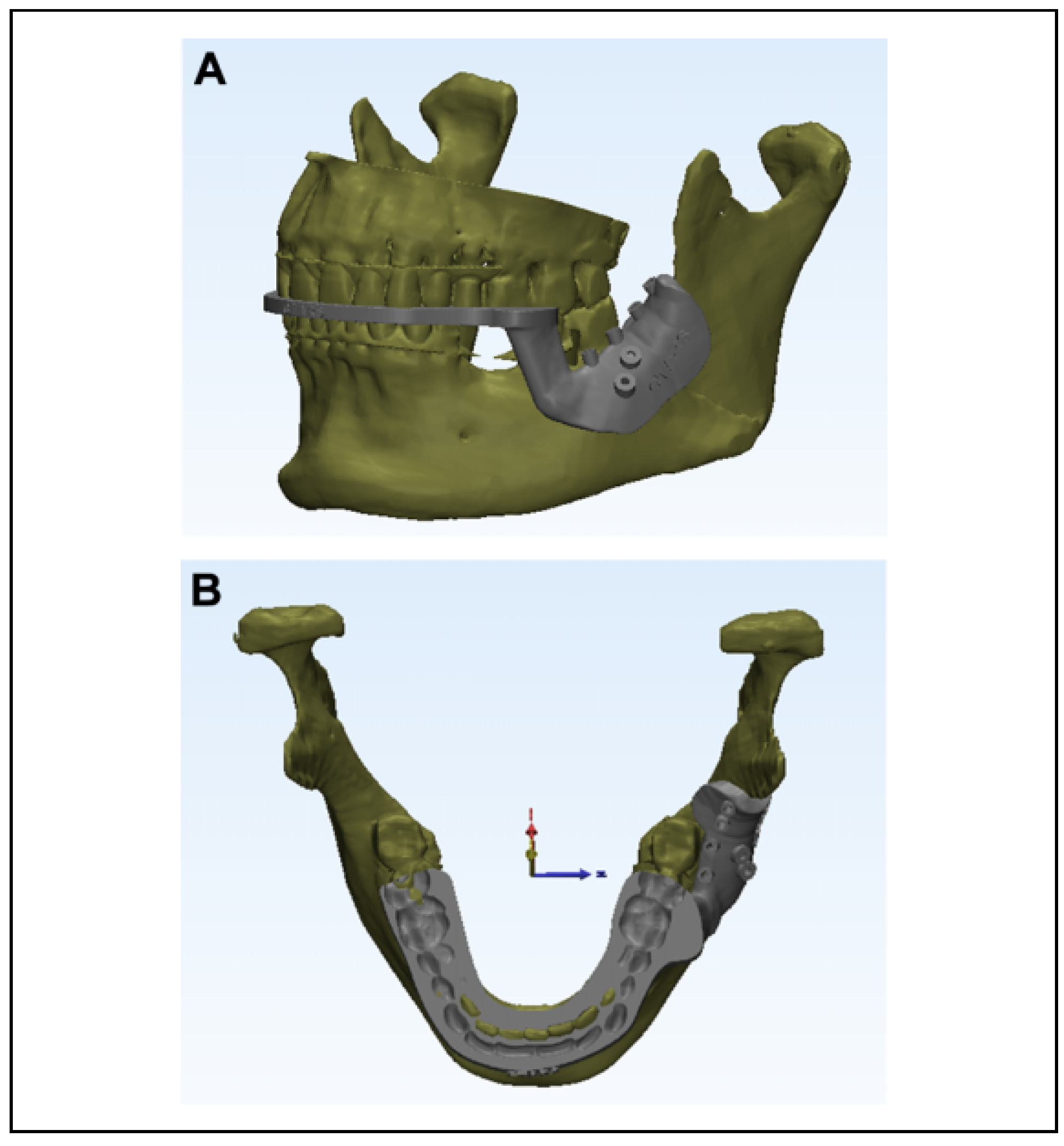
The CT scan of the patient in Digital Imaging and Communications in Medicine (DICOM) format was converted to stereolithography (STL) file using the segmentation software, Materialise Interactive Medical Image Control System “MIMICS” (Materialise NV, Leuven, Belgium). The fracture fragments were virtually reduced and repositioned to their anatomical position. The teeth were also set in maximum intercuspation virtually. The composite splint was designed with 2 components: (1) a distal interocclusal component to guide the teeth into maximum intercuspation (Figure 3A) and (2) a proximal bone-borne component to reposition the superiorly displaced angle (Figure 3B). The bone-borne component was also incorporated with drill holes to serve as a surgical guide to position the screws. The drill holes were positioned such that they bypassed the roots and inferior alveolar canal (Figure 4). The surgical splint and guide were planned using “GEOMAGIC” (3D systems, Morrisville, North Carolina) software and transferred to the printer for printing. The splint was fabricated with Photopolymer resin, utilizing the additive printing technique. The time taken to manufacture the splint was 8 hours (5 hours for planning and 3 hours for printing). The splint was sterilized by immersion in Cidex (2.45 w/v glutaraldehyde, CIDEX – activated glutaraldehyde solution; Dishman Pharmaceuticals, Ahmedabad, India) 12 hours before the surgery[8].

Intraoperative Procedure

Under general anesthesia, the patient was intubated via the nasoendotracheal route. Lignocaine (2%) with adrenaline infiltration was given in the surgical site. Vestibular incision was placed in relation to 38 (lower left third molar), and mucoperiosteal flap was raised to expose the fracture site. The fracture was reduced, and proper occlusion was achieved using the CAD/CAM splint.
The splint was first positioned intraorally on the teeth. The dental component of the splint was used to guide the mandible in achieving ideal occlusion. This maneuver delivered a simultaneous inferior thrust to the superiorly displaced angle through the bone-borne component to achieve good anatomic position (Figure 5). After ensuring maximum intercuspation between the teeth, intermaxillary fixation was done with the splint in position. Bone was drilled along the external oblique ridge, with the help of drill guides in the surgical template using 1.5-mm drill bit. A single miniplate of 2-mm system (4-hole miniplate with gap) was adapted in accordance with the drill holes and was fixed along the external oblique ridge, using 4 screws.

Postoperative Outcome

Examination of the patient in the postoperative period revealed minimal swelling in the angle region with good mouth opening (45 mm) and occlusion (Figure 6). Figure 7 demonstrates the accurate anatomical reduction of the fracture on postoperative CT. The neurosensory status of the inferior alveolar nerve was found to be intact.

Discussion

Reduction of angle fractures is challenging because of the alteration in equilibrium between dynamic muscle forces acting at the angle region which leads to displacement of the fracture fragments.[1] The presence of impacted third molar also complicates accurate reduction.[1] Reduction is generally achieved by digital manipulation,[5] towel clip,[7] wiring,[6] intermaxillary fixation,[5] and splints.[9] However, these may be time-consuming, cumbersome, and inaccurate leading to postoperative complications such as malocclusion and malunion.[4] The advantages of interocclusal splints in reduction of angle fractures has been discussed in detail by Elavenil et al.[1] The authors emphasize the role of interocclusal splints in achieving lingual occlusion and accurate transverse dimensions of the mandible which are crucial to postoperative stability of fixation of angle fractures.
However, the conventional laboratory-generated splints are associated with limitations; they require adequate fabrication time and are technique sensitive. Also, impressions are required for splint fabrication which are difficult to obtain in patients with post-traumatic trismus. Further, the conventional splints ensure accurate dental occlusion only. In contrast, the CAD/CAM-generated splint proposed by the author offered many advantages: (1) they do not require impressions[10] for fabrication of splints and (2) they permit simultaneous and precise reduction of the fracture fragments in all 3 planes along with dental occlusion.[11]
The use of CAD/CAM splints has been well established for orthognathic surgical procedures by various authors.[11]
The literature does not provide adequate information on the use of technology in manufacturing 3D splints for trauma patients.
Lee et al[12] demonstrated the intraoperative usage of a “Wing splint” using CAD/CAM technology for easy reduction of mandibular fracture in a pediatric patient. However, the splint did not have an interocclusal component to guide the occlusion. Further, the design of the splint does not facilitate correction of lingual splay. The CAD/CAM- generated surgical splints have also been used in conjunction with surgical guides in the reduction and fixation of mid-face fractures, intraoperatively.[13]
The use of CAD/CAM splints is very rewarding in the management of edentulous fractures whose reduction and fixation is challenging because of the gross displacement of fracture fragments and lack of dentition that are important in guiding reduction. In such clinical conditions, splints save considerable intraoperative time.[14] El-Gengehi et al[8] emphasized on incorporating a surgical guide in the form of a labial plate to aid in ORIF of edentulous mandible. However, this splint served only as a positional template and neither had a role in the correction of lingual splay nor establishing occlusion.
The fabrication of CAD/CAM splints for orthognathic surgery is easier because the process of obtaining patient impressions is simpler due to good mouth opening. However, in trauma patients, mouth opening might be painful and restricted, which complicates impression making. This difficulty was overcome in our patient by performing an intraoral scan that reduces patient discomfort. Performing an intraoral scan in such patients is a much easier method, wherein patient discomfort is relieved.
The total time required for virtual surgery and fabrication of splint was 8 hours, and the splint was found to be cost-effective. The greatest advantage of this composite CAD/CAM splint was the simultaneous reduction of fracture as well as restoration of dental occlusion, holding the fracture fragments in reduced state while plating was not necessary. Further, the surgical guides permitted convenient positioning of the screws that bypassed vital structures such as the inferior alveolar canal and the dental roots.
Our experience with this case demonstrated relative reduction in intraoperative time as well as manual effort required for fracture reduction while ensuring accuracy in restoration of mandibular anatomy. However, a randomized controlled trial involving large samples that compare conventional splints with CAD/CAM splints would provide more clinical evidence. The limitations concerning this technique were the relative cost involved as well as the availability of the intraoral scanning device and CAD/CAM technology.

Conclusion

The use of CAD/CAM splints with surgical guide is an effective method to ensure accurate fracture reduction and restoration of dental occlusion. These splints are surgeon and patient friendly.

Funding

This research received no external funding.

Acknowledgments

We wish to acknowledge CTARS—Dr. John Nesan and Mr. Iraimudi—for the fabrication of our CAD/CAM splint.

Conflicts of Interest

The authors declare no conflict of interest.

References

  1. Elavenil, P.; Mohanavalli, S.; Sasikala, B.; Prasanna, R.; Krishnakumar, R. Isolated bilateral mandibular angle fractures: An extensive literature review of the rare clinical phenomenon with presentation of a classical clinical model. Craniomaxil- lofac Trauma Reconstr. 2015, 8, 153–158. [Google Scholar] [CrossRef] [PubMed]
  2. Ellis, E.I.I.I. Treatment methods for fractures of the mandibular angle. Int J Oral Maxillofac Surg. 1999, 28, 243–252. [Google Scholar] [CrossRef] [PubMed]
  3. Wan, K.; Williamson, R.A.; Gebauer, D.; Hird, K. Open reduction and internal fixation of mandibular angle fractures: Does the transbuccal technique produce fewer complications after treatment than the transoral technique? J Oral Maxillofac Surg. 2012, 70, 2620–2628. [Google Scholar] [CrossRef] [PubMed]
  4. Seemann, R.; Schicho, K.; Wutzl, A.; et al. Complication rates in the operative treatment of mandibular angle fractures: A 10-year retrospective. J Oral Maxillofac Surg. 2010, 68, 647–650. [Google Scholar] [CrossRef] [PubMed]
  5. Hsu, E.; Crombie, A.; To, P.; Marquart, L.; Batstone, M. Manual reduction of mandibular fractures before internal fixation leads to shorter operative duration and equivalent outcomes when compared with reduction with intermaxillary fixation. J Oral Maxillofac Surg. 2012, 70, 1622–1626. [Google Scholar] [CrossRef] [PubMed]
  6. Fisher, J.; Cleaton-Jones, P.; Lownie, J. Relative efficiencies of various wiring configurations commonly used in open reduc- tions of fractures of the angle of the mandible. Oral Surg Oral Med Oral Pathol. 1990, 70, 10–17. [Google Scholar] [CrossRef] [PubMed]
  7. Batbayar, E.; Van Minnen, B.; Bos, R. Non-IMF mandibular fracture reduction techniques: A review of the literature. J Craniomaxillofac Surg. 2017, 45, 1327–1332. [Google Scholar] [CrossRef] [PubMed]
  8. el-Gengehi, M.; Seif, S. Evaluation of the accuracy of computer-guided mandibular fracture reduction. J Craniofac Surg. 2015, 26, 1587–1591. [Google Scholar] [CrossRef] [PubMed]
  9. Mimi, M.; Mizuno, A.; Nakano, Y.; Motegi, K. Reduction and fixation of jaw fractures using acrylic splints. Br J Oral Max- illofac Surg. 1989, 27, 321–328. [Google Scholar] [CrossRef] [PubMed]
  10. Sun, Y.; Luebbers, H.; Politis, C. Three-dimensional virtual model surgery to fabricate the digital intermediate splint. J Craniofac Surg. 2013, 24, 563–565. [Google Scholar] [CrossRef] [PubMed]
  11. Aboul-Hosn Centenero, S.; Herna´ndez-Alfaro, F. 3D planning in orthognathic surgery: CAD/CAM surgical splints and prediction of the soft and hard tissues results—Our experience in 16 cases. J Cranio-Maxillo-Facial Surg. 2012, 40, 162–168. [Google Scholar] [CrossRef] [PubMed]
  12. Lee, J.; Choi, B.; Nam, O.; Kwon, Y. Minimal invasive treatment using patient-specific template for mandibular fractures in children: “Wing-splint” by CAD/CAM technology. Br J Oral Maxillofac Surg. 2016, 54, 1140–1141. [Google Scholar] [CrossRef] [PubMed]
  13. Levine, J.; Patel, A.; Saadeh, P.; Hirsch, D. Computer-aided design and manufacturing in craniomaxillofacial surgery. J Craniofac Surg. 2012, 23, 288–293. [Google Scholar] [CrossRef] [PubMed]
  14. Wang, H.; Frost, C.; Cusano, A.; Dorafshar, A. Management of bilateral edentulous mandible fractures through an intraoral approach using CAD/CAM technology: A case report. J Oral Maxillofac Surg. 2018, 76, e1–e1056. [Google Scholar] [CrossRef] [PubMed]
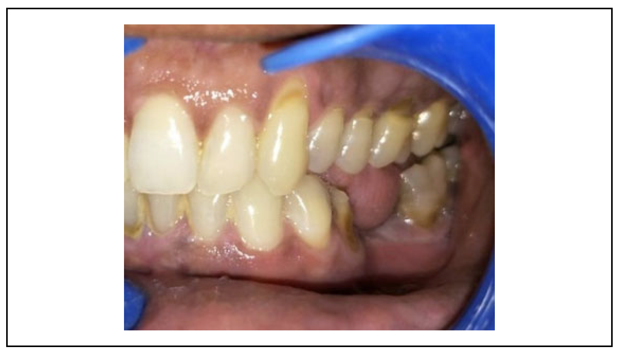
Figure 1. Preoperative image of patient demonstrating restricted mouth opening and posterior open bite.
Cmtr 14 00013 g001
Figure 2. Preoperative CT image of patient showing a superiorly displaced fracture at the left angle region. Green arrow: the direction of muscle pull; Yellow arrow: the direction of splint action. CT indicates computed tomography.
Cmtr 14 00013 g002
Figure 3. CAD/CAM splint depicting the virtual fracture reduc- tion obtained with the splint. (A) The distal interocclusal com- ponent to guide the teeth into maximum intercuspation. (B) The proximal bone-borne component to aid reduction of the super- iorly displaced left angle. CAD/CAM indicates computer-aided designing/computer-aided manufacturing.
Cmtr 14 00013 g003
Figure 4. Planning of surgical guide that bypasses vital structures.
Cmtr 14 00013 g004
Figure 5. Intraoperative image of CAD/CAM splint in position. CAD/CAM indicates Computer-Aided Designing/Computer- Aided Manufacturing.
Cmtr 14 00013 g005
Figure 6. Postoperative occlusion.
Cmtr 14 00013 g006
Figure 7. Postoperative CT image showing anatomical restoration of the left angle region. CT indicates computed tomography.
Cmtr 14 00013 g007