Next Article in Journal
A Clinical and Radiological Investigation of the Use of Dermal Fat Graft as an Interpositional Material in Temporomandibular Joint Ankylosis Surgery
Previous Article in Journal
Surgical Approach to Orbital Floor Fractures: Comparing Complication Rates Between Subciliary and Subconjunctival Approaches
 
 
Craniomaxillofacial Trauma & Reconstruction is published by MDPI from Volume 18 Issue 1 (2025). Previous articles were published by another publisher in Open Access under a CC-BY (or CC-BY-NC-ND) licence, and they are hosted by MDPI on mdpi.com as a courtesy and upon agreement with Sage.
Article

Intact Periorbita Can Prevent Post-Traumatic Enophthalmos Following a Large Orbital Blow-Out Fracture

by
Srinivas Susarla
1,2,3,*,
Richard A. Hopper
1 and
Ezgi Mercan
1
1
Division of Plastic Surgery, Department of Surgery, University of Washington School of Medicine, Seattle, WA, USA
2
Department of Oral and Maxillofacial Surgery, University of Washington School of Dentistry, Seattle, WA, USA
3
Craniofacial Center, Seattle Children’s Hospital, Seattle, WA, USA
*
Author to whom correspondence should be addressed.
Craniomaxillofac. Trauma Reconstr. 2020, 13(1), 49-52; https://doi.org/10.1177/1943387520903545
Submission received: 1 December 2019 / Revised: 31 December 2019 / Accepted: 1 February 2020 / Published: 23 March 2020

Abstract

:
Treatment of orbital floor fractures is predicated on the restoration of orbital volume to prevent enophthalmos or hypoglobus. Globe position is the result of a complex interplay between the bony orbital anatomy and the soft tissue envelope. Studies on orbital fractures have frequently suggested criteria for repair on the basis of bony defect size or volume change. In this report, we describe a case of a large orbital floor defect (4.8 cm2) with intact periorbita and no herniation of soft tissue contents in a young male following facial trauma. The patient was followed for 1-year clinically and did not develop enophthalmos. This case demonstrates that bony injury alone is not sufficient to produce enophthalmos, and that the interplay between the soft tissue and bony anatomy is a critical determinant of globe position following orbital trauma.

Introduction

Fractures of the orbital floor are among the most common injuries of the facial skeleton and have been studied extensively.[1,2,3,4,5,6,7,8] Management of orbital floor defects, however, remains controversial. While there are generally agreed upon indications for surgery (eg, acute enophthalmos, entrapment), in the absence of these clinical scenarios, decisions are frequently made on the basis of size of the orbital floor defect, with repair recommended for fractures at least of 1 to 2 cm2 in size.4 Herniation of orbital soft tissue contents into the maxillary sinus is thought to play a role in the development of enophthalmos and is potentially correlated with the size of the floor defect and orbital volume changes.[9,10,11,12,13,14,15] The role of the inferior periorbital sling in preventing enophthalmos has also been described, with the shape of the inferior rectus being an important predictor of enophthalmos.[16,17]
In this report, we describe a case of a large orbital floor defect with intact periorbita and no herniation of soft tissue contents in a young male following facial trauma. The patient was treated nonoperatively and followed for 1-year clinically and did not develop enophthalmos.

Clinical Report

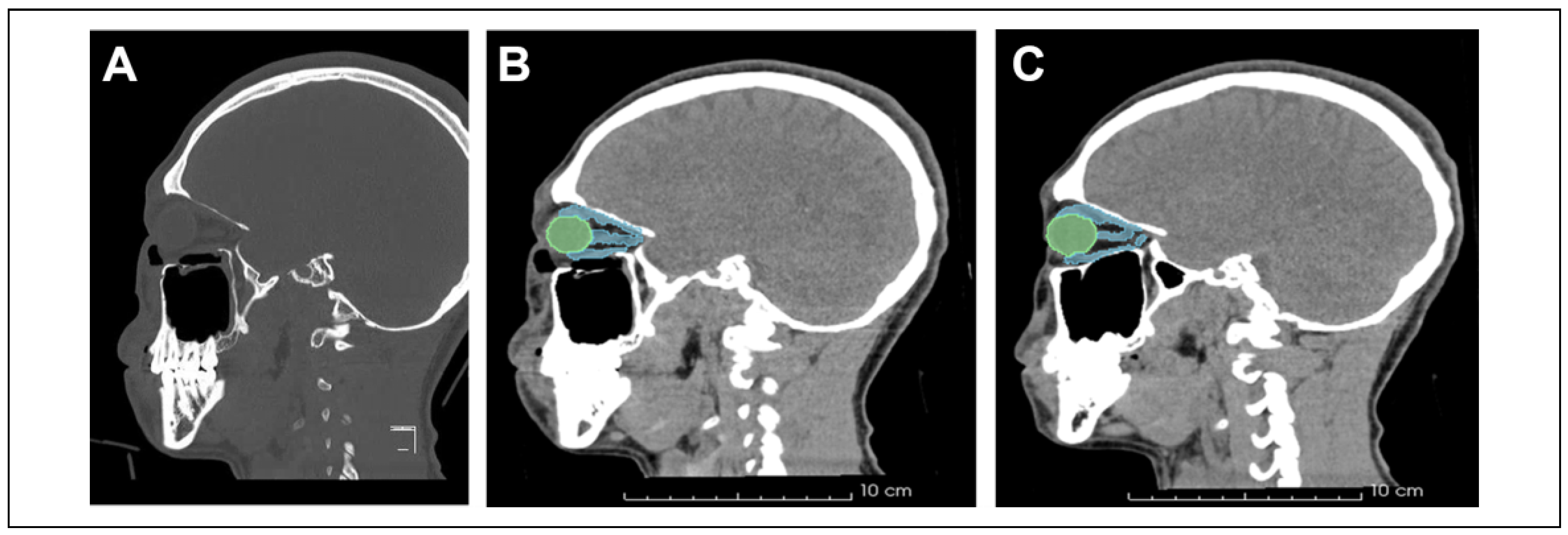
A 24-year-old man was involved in a motor vehicle accident as a restrained passenger. A clinical evaluation was notable for periorbital swelling and diplopia. Imaging (Figure 1A to C) was notable for a right orbital floor fracture with an associated large defect of 20 mm × 24 mm (4.8 cm2), without herniation of soft tissue or change in the inferior rectus morphology (height to width ratio 0.5, Figure 2A to E). The orbital volume measured to the intact soft tissue margin was 22.98 cm3; the unaffected orbital volume was 23.06 cm3. The patient was evaluated at 10 days post-injury and had improved vision without demonstrable enophthalmos (Figure 3A to B). Hertel exophthalmometry measurements were 17.2 mm OD and 16.7 mm OS. The patient was followed clinically and his diplopia resolved at 6 weeks post-injury. At 1-year post-injury (Figure 3C to D), he was reevaluated and noted to have no diplopia or clinically significant enophthalmos (Hertel 16.5 mm OD and 16.9 mm OS).

Discussion

Orbital fractures remain among the most common skeletal injuries in patients with facial trauma.[1,2,3,4] The paper thin bones of the floor and medial wall are particularly susceptible to injury and fractures of the anterior and middle portions of the bony orbit in these regions are common.[4] It is thought that traumatic expansion of the bony orbit alleviates acute increases in intraorbital pressure, resulting in a decreased propensity for globe or intracranial injury.[4,5,6] While there is an extensive body of literature on management of orbital floor fractures, the remains significant controversy regarding the decision for operative intervention.[4]
The term “blow-out” fracture was first coined by Smith and Regan in 1957; these authors advocated for early exploration and repair.[1] Converse subsequently added to the literature on repair and recommended repair within 3 weeks of injury to prevent secondary enophthalmos.[2] Dulley and Fells subsequently defined criteria for repair, many of which are still used today: enophthalmos greater than 3 mm, extensive soft tissue herniation into the maxillary sinus, diplopia, and limitation of upward gaze.[18] Subsequent investigations in the era of advanced imaging techniques have yielded guidelines on the basis of defect size (1-2 cm2) or involvement of >50% of the orbital floor.[4,7,8,9] Recent large-scale studies with regression analyses have reported that patients with defects 1.5 cm2 in size were 3 times more likely to have posttraumatic enophthalmos, when compared to patients with smaller defects.[19] Studies of 3-D volumetric changes in the traumatized orbit have suggested a relationship between orbital volume changes and enophthalmos, with 0.6-0.7 mm of enophthalmos for every 1 cm3 volume change with an orbital volume change as little as 7% necessary to create clinically significant enophthalmos.[9,10,11,12,13,14]
These data are helpful in guiding management, but the importance of the periorbital soft tissue sling is an additional critical component.[16,17] Investigations regarding the morphology of the inferior rectus muscle, whose shape will be dictated by the integrity of the periorbital-fascial support system (ie, Lockwood’s ligament), have demonstrated that rounding of the inferior rectus is associated with an increased likelihood of enophthalmos. A height to width ratio exceeding 1.0 on coronal imaging has been suggested to be indicative of late enophthalmos.[17]
This clinical case demonstrates the importance of the interplay between the bony orbital morphology and the overlying soft tissue support system. This patient had a significantly sized orbital floor defect (>4 cm2), but without significant orbital volume change or herniation of soft tissue contents, due to an intact periorbital soft tissue sling. This was only noted on close examination of fine cut CT scan images to distinguish periorbital contents from mucosal thickening and blood. As a result, no surgery was required and no significant enophthalmos was noted at long term follow-up.

Funding

The author(s) received no financial support for the research, authorship, and/or publication of this article.

Institutional Review Board Statement

The guidelines of the Declaration of Helsinki were followed at all times during this work.

Conflicts of Interest

The author(s) declared no potential conflicts of interest with respect to the research, authorship, and/or publication of this article.

References

  1. Smith, B.; Regan, W.F., Jr. Blow-out fracture of the orbit; mechanism and correction of internal orbital fracture. Am J Ophthalmol. 1957, 44, 733–739. [Google Scholar] [CrossRef] [PubMed]
  2. Converse, J.M.; Smith, B. Blowout fracture of the floor of the orbit. Trans Am Acad Ophthalmol Otolaryngol. 1960, 64, 676–688. [Google Scholar] [PubMed]
  3. Converse, J.M.; Smith, B. Blowout fracture of the floor of the orbit. J Fla Med Assoc. 1961, 47, 1337–1342. [Google Scholar]
  4. Gart, M.S.; Gosain, A.K. Evidence-based medicine: orbital floor fractures. Plast Reconstr Surg. 2014, 134, 1345–1355. [Google Scholar] [CrossRef] [PubMed]
  5. Green, R.P. , Jr, Peters, D.R.; Shore, J.W.; Fanton, J.W.; Davis, H. Force necessary to fracture the orbital floor. Ophthal Plast Reconstr Surg. 1990, 6, 211–217. [Google Scholar] [CrossRef] [PubMed]
  6. Jones, D.E.; Evans, J.N. “Blowout” fractures of the orbit: an investigation into their anatomical basis. J Laryngol Otol. 1967, 81, 1109–1120. [Google Scholar] [CrossRef] [PubMed]
  7. Manson, P.N.; Grivas, A.; Rosenbaum, A.; Vannier, M.; Zinreich, J.; Iliff, N. Studies on enophthalmos: II. The measurement of orbital injuries and their treatment by quantitative computed tomography. Plast Reconstr Surg. 1986, 77, 203–214. [Google Scholar] [CrossRef] [PubMed]
  8. Manson, P.N.; Iliff, N. Management of blowout fractures of the orbital floor: II. Early repair for selected injuries. Surv Ophthalmol. 1991, 35, 280–292. [Google Scholar] [CrossRef]
  9. Zhang, Z.; Zhang, Y.; He, Y.; An, J.; Zwahlen, R.A. Correlation between volume of herniated orbital contents and the amount of enophthalmos in orbital floor and wall fractures. J Oral Maxillofac Surg. 2012, 70, 68–73. [Google Scholar] [CrossRef]
  10. Yang, J.H.; Hwang, S.B.; Shin, J.Y.; Roh, S.G.; Chang, S.C.; Lee, N.H. 3-Dimensional volumetric analysis of relationship between the orbital volume ratio and enophthalmos in unoperated blowout fractures. J Oral Maxillofac Surg. 2019, 77, 1847–1854. [Google Scholar] [CrossRef]
  11. Ebrahimi, A.; Kalantar Motamedi, M.H.; Rasouli, H.R.; Naghdi, N. Enophthalmos and orbital volume changes in zygomaticomaxillary complex fractures: Is there a correlation between them? J Oral Maxillofac Surg. 2019, 77, 134.e1–134.e9. [Google Scholar] [PubMed]
  12. Schönegg, D.; Wagner, M.; Schumann, P.; et al. Correlation between increased orbital volume and enophthalmos and diplopia in patients with fractures of the orbital floor or the medial orbital wall. J Craniomaxillofac Surg. 2018, 46, 1544–1549. [Google Scholar] [PubMed]
  13. Sugiura, K.; Yamada, H.; Okumoto, T.; Inoue, Y.; Onishi, S. Quantitative assessment of orbital fractures in Asian patients: CT measurement of orbital volume. J Craniomaxillofac Surg. 2017, 45, 1944–1947. [Google Scholar] [CrossRef] [PubMed]
  14. Choi, S.H.; Kang, D.H. Prediction of late enophthalmos using preoperative orbital volume and fracture area measurements in blowout fracture. J Craniofac Surg. 2017, 28, 1717–1720. [Google Scholar] [CrossRef] [PubMed]
  15. Bite, U.; Jackson, I.T.; Forbes, G.S.; Gehring, D.G. Orbital volume measurements in enophthalmos using threedimensional CT imaging. Plast Reconstr Surg. 1985, 75, 502–508. [Google Scholar] [CrossRef] [PubMed]
  16. Manson, P.N.; Clifford, C.M.; Su, C.T.; Iliff, N.T.; Morgan, R. Mechanisms of global support and posttraumatic enophthalmos: I. The anatomy of the ligament sling and its relation to intramuscular cone orbital fat. Plast Reconstr Surg. 1986, 77, 193–202. [Google Scholar] [CrossRef] [PubMed]
  17. Matic, D.B.; Tse, R.; Banerjee, A.; Moore, C.C. Rounding of the inferior rectus muscle as a predictor of enophthalmos in orbital floor fractures. J Craniofac Surg. 2007, 18, 127–132. [Google Scholar] [CrossRef] [PubMed]
  18. Dulley, B.; Fells, P. Long-term follow-up of orbital blow-out fractures with and without surgery. Mod Probl Ophthalmol. 1975, 14, 467–470. [Google Scholar] [PubMed]
  19. Ahmad Nasir, S.; Ramli, R.; Abd Jabar, N. Predictors of enophthalmos among adult patients with pure orbital blowout fractures. PLoS One. 2018, 13, e0204946. [Google Scholar]
Figure 1. Multidetector thin slice CT images demonstrating large orbital floor defect. On the coronal views (A, B), the width of the defect can be seen (maximum width 20 mm). Soft tissue views (C, D) demonstrate no rounding of the inferior rectus and no herniation of soft tissue contents into the maxillary antrum. The orbital margin at the level of the soft tissue is preserved (E), without resultant increase in orbital volume (affected orbit volume = 22.98 cm3, unaffected orbit volume = 23.06 cm3). CT indicates computed tomography.
Cmtr 13 00009 g001
Figure 2. On the sagittal view (A), the floor defect measures 24 mm. There is no significant herniation of the soft tissue or bowing of the inferior rectus (blue) between the affected (B) and unaffected sides (C).
Cmtr 13 00009 g002
Figure 3. Clinical photos at 10-days post-injury (A, B) and 1-year post-injury (C, D). There is no significant enophthalmos, hypoglobus, or asymmetry.
Cmtr 13 00009 g003