Next Article in Journal
COVID-19 and the Craniomaxillofacial Surgical Community
Previous Article in Journal
Total Temporomandibular Joint Replacement: Stick to Stock or Optimization by Customization?
 
 
Craniomaxillofacial Trauma & Reconstruction is published by MDPI from Volume 18 Issue 1 (2025). Previous articles were published by another publisher in Open Access under a CC-BY (or CC-BY-NC-ND) licence, and they are hosted by MDPI on mdpi.com as a courtesy and upon agreement with Sage.
Article

Assessment of Functional Recovery and Subjective Donor-Site Morbidity Following Radial Forearm Flap Reconstruction in Small- to Moderate-Sized Palatal Defects

by
Ashok B. Chandrappa
1,
Ritu S. Batth
1,*,
Srikanth Vasudevan
1,
Anantheswar N. R. Yellambalase
1,
Pradeep N. Kumar
1,
Sudarshan Reddy
1 and
J. Nidya Seles
2
1
Department of Plastic and Reconstructive Surgery, Manipal Hospital, Bangalore, Karnataka, India
2
Department of Speech and Language Pathology, Manipal Hospital, Bangalore, Karnataka, India
*
Author to whom correspondence should be addressed.
Craniomaxillofac. Trauma Reconstr. 2020, 13(1), 71-77; https://doi.org/10.1177/1943387520904879
Submission received: 1 December 2019 / Revised: 31 December 2019 / Accepted: 1 February 2020 / Published: 16 March 2020

Abstract

:
Context: Palatal defects are encountered following tumor extirpation, trauma, or congenitally. Among the known alternatives, radial artery free forearm flap (RAFF) is a versatile flap that confers good results in head and neck reconstruction, but donor-site morbidity has been an issue of discontent among the plastic surgeons. This limitation needs to be studied further and addressed considering the unmatched quality of this tissue. Aims: This study aims to weigh the impact of the functional edge of this flap against the unpopular donor-site morbidity on a group of patients. Settings and Design: This is a retrospective analysis of recuperation of palatal function and patient concerns with the donor-site function and cosmesis on 7 consecutive patients with small-to-moderate palatal defects reconstructed with RAFF. Methods and Materials: Postoperative recovery of speech, palatal movement, and restoration of oromaxillary interface was assessed using objective tests, such as speech intelligibility testing and articulation studies. Simultaneously, subjective donor-site function and cosmesis were assessed using Patient Scar Assessment Scale (PSAS), Upper Extremity Functional Index (UEFI), and donor limb sensory testing. Results and Conclusion: Mean PSAS score was 8.28/60, and UEFI score reported was 77/80, which reflect high patient satisfaction with the donor site. Nasoendoscopy shows remarkable restoration of palate anatomy. Intelligibility testing depicts near-normal speech understandability, whereas articulation studies revealed distortions post-palatal reconstruction with RAFF. Radial artery free forearm flap should be considered as the forerunner of reconstruction in palatal defects involving less than 50%.

Introduction

Palatal defects pose a challenge to reconstructive surgeons, as they are customarily composite in nature. These types of defects are challenging to reconstruct due to lack of availability of the local tissue that can be advanced to envelope the defect. The goal of treatment is to restore the lost interface between the oral and the maxillary cavities and the palatal function of mastication and articulation and to enable a successful dental rehabilitation, wherever feasible, with minimal donor-site compromise. Maxillary defects are not as well studied as compared to oromandibular defects, hence lack a general consensus on the protocol of treatment. Obturators have always been considered the easier and readily available modality for filling small- to moderate-sized palatal defects, but the resulting halitosis and tiring maintenance of the apparatus have a much-underrated impact on the lives of patients. Local flaps, for example, buccal myomucosal flap, facial artery musculomucosal flap, tongue flap, nasolabial flap, to mention a few, are attempted with the purpose of achieving the results desired with marginally invasive and time-friendly techniques but are majorly restricted by the size and reach of the tissue. Hence the obligation to look at distant options emerges, more commonly RAFF, anterolateral thigh perforator flap (ALT), free fibula flap (FFF), pectoralis major flap (PMMC), and medial sural artery perforator flap (MSAP).[1,2,3,4,5,6]
There are diverse opinions regarding the balance between the resultant donor-site scarring and the functional rehabilitation attained with RAFF which invokes a sense of disinclination in the operative surgeon. This study seeks to analyze the patient’s perspective of radial forearm flap donor-site compromise and the level of functional competency achieved with this reconstruction in a specific subgroup of patients and indications.

Patients and Methods

This study includes patients with type II (A/B) midface defects per the Santamaria and Cordeirro et al classification that implies involvement of less than 50% of the palate and are too substantial to be reconstructed with local flaps.[7] Dental rehabilitation is seldom required, as ample dental arch is left behind. Seven consecutive patients operated in the Department of Plastic Surgery after tumor extirpation over a span of 3 years (2015-2017) were subjected to assessment. All patients were well educated about other alternatives of reconstruction such as obturators and other free flaps before they committed to the RAFF. All patients were subjected to primary reconstruction immediately following tumor resection and received postoperative radiotherapy. Donor-site defect was covered with split thickness skin graft. Donor-site dressing and splinting were continued for 7 to 10 days. The patients were advised oil massage and compression garments for the donor limb graft in the third postoperative week for 4 to 6 months. Early donor limb utilization was encouraged after 10 days. The study population was followed up for a period of 6 months postoperatively and engaged in the feedback exercise. Palatal functional status was graded according to articulation test and speech intelligibility rating scale (AYJNIHH) with the assistance of a speech language pathologist. Subjective donor-site assessment was done using Upper Extremity Functional Index (UEFI), Patient Scar Assessment Scale (PSAS), and sensory testing in the autonomous zone of superficial radial nerve. Two-point discrimination was checked using a bent paper pin readily available in the Out Patient Department (OPD), and temperature was tested using 2 glasses, one filled with warm water and the other with crushed ice. Touch sensation was tested using a cotton tip applicator for light as well as deep touch.

Surgical Technique

Flap was marked per the dimensions of the palatal defect in the distal most part of the forearm to equip the tissue with a sufficiently long pedicle and include the thin and pliable tissue of the distal forearm into the harvest. Subfascial nerve sparing dissection was done, carefully preserving the superficial branch of radial nerve (SRN). Excess adipofascial tissue was harvested on either sides of the flap pedicle to wrap the pedicle from all sides (Figure 1 and Figure 2).[8] This technique provides a protective covering to the radial artery pedicle that is destined to face discharge collection initially and later the turbulent air currents of nasal cavity to avoid complications such as air desiccation, thrombosis, and blow out of the pedicle. This technique also compensates the use of double paddled or folded bulky flaps that can possibly cause obliteration of the nasal cavity, intranasal hair growth, and halitosis. Donor site was resurfaced with an intermediate thickness split thickness skin graft harvested from the thigh. In cases with intact alveolar arch, the pedicle was passed posterior to the upper third molar to pass through a subcutaneous tunnel created superficial to the mandible to reach the neck (Figure 3). Postoperatively, wet antibiotic nasal packing was kept for a period of 48 to 72 hours to absorb any ooze from the fascial surface of the flap and avoid aspiration. All patients were directed to use saline nasal drops for a period of minimum 2 to 3 weeks to avoid crusting and related trauma in the immediate postoperative period. Patients were kept on Ryle’s tube feeding for 5 days followed by clear liquids and subsequently liquid diet and soft diet.

Results

The study included 7 patients who underwent infrastructural maxillectomy by the oncosurgical team over a period of 3 years. Two of them were diagnosed with mucoepidermoid carcinoma and 5 with squamous cell carcinoma. The age-group varied from 14 to 76 years, all of them males except one.
Weber Ferguson incision with lip split was utilized in 2 patients and intraoral approach for the rest 5. The resultant palatal defect in all of the patients was categorized type IIa per “Santamaria Cordeirro et al classification of midface defects,” which implies <50% palatal defect with intact orbital floor.[1] Two of them had alveolar arch defect, while the rest had central defects involving the hard as well as soft palate (Figure 4 and Figure 5). These defects were reconstructed with nerve sparing subfascial radial artery free forearm flap from the non-dominant limb (Figure 6 and Figure 7). Average donor-site skin graft healing time was 14 days, with no secondary procedures for any patient.
Donor-site assessment at 6 months postoperative time was done using PSAS, UEFI, and hand sensory testing (Figure 8 and Figure 9). Mean PSAS score was 8.28 with average overall patient scar satisfaction being 7+ (Figure 10). Mean UEFI score calculated was 77 (Figure 11).
Sensory testing was carried out to assess 2-point discrimination, touch and temperature, in the distribution of superficial radial nerve and was found to be bilaterally symmetrical in the donor as well as the contralateral limb.
Palatal function was assessed using articulation test and speech intelligibility rating scale. None of the patients complained of nasal regurgitation while feeding and were comfortable consuming solids as well as liquids. Intelligibility scale (Figure 12) reading was 1 for 5 patients, which implies “can be understood without difficulty, however feel the speech is not normal” and 2 for 2 patients, which implies “can be understood with a little effort, occasionally need to ask for repetition” (Figure 13). Articulation studies showed distortions for both voiced and voiceless velar sounds and at initial, medial, and final position of the words.

Discussion

Palatal defects need a thin and pliable cover that does not add to the bulk of the palate and provides a taut and well-contoured palate suitable for satisfactory articulation and mastication. Also the borrowed tissue should be alike to the native tissue to ensure an airtight seal between the 2 cavities. Generally, covering the defects of the palate becomes a challenge when the defect is too large to close with a local flap or the patient is not persuaded with the long-term maintenance required with an obturator, which is when a distant flap comes into play. Ideally, this situation warrants a flap that has suffice quantity and quality of tissue available, adequate pedicle length with safe vessel caliber, and leaves acceptable donor-site morbidity.
The mode of treatment chosen depends on a spectrum of patient and clinician-dependent factors, namely, age and comorbidities of the patient, prognosis of the disease stage, extent of palatal involvement and bone stock remaining, patient’s expectations and priorities, availability of desirable donor site in recurrent cases, and clinical expertise and infrastructure available.
To highlight, obturators are easier to use and readily available but, on the other hand, need a long-term maintenance, periodic modifications, add to the bulk, and may cause residual oromaxillary regurgitation and hypernasality.[9,10]
The superiority of free flaps in improving the quality of life even in small and medium defects of maxilla was reported by Genden et al. They also noted that patients undergoing radiotherapy had pain and difficulty tolerating the prosthesis.[11,12] Local flaps feasible are limited by their size and reach in case of larger defects crossing the midline. Flaps such as PMMC and ALT are good to plug the cavity in case of volume-extensive defects of the maxilla involving the palate but are too bulky for small–medium palatal defects. Free fibula flap is one of the best suited methods of palatal reconstruction, with dental rehabilitation in patients with larger palatal defects involving more than 50% of the palate and inadequate residual bone stock. Again the problem arises in moderately sized defects with adequate bone stock, where orienting a small FFF skin paddle becomes arduous. Adding to the turmoil is the need for finding an appropriately placed perforator feeding the skin paddle desired.
So in cases with small to moderately sized palatal defects, RAFF comes into consideration because of its favorable tissue quality, reliable anatomy, long pedicle length, and flexibility in terms of composition. Lately, donor-site morbidity following RAFF harvest has demerited the utility of this highly compliant flap and affected the armor of a plastic surgeon. Prominently, RAFF donor-site compromise is reported as delayed graft uptake,[13] poor cosmesis,[14] chronic pain,[15] numbness along the distribution of SRN,[13] reduced pinch strength,[14] and wrist extension.[16] These issues have instilled an uncertainty in context to RAFF being the first choice for such defects. But most of the studies could not emphasize on the subjective aspect of the donor-site morbidity and its significance in a patient’s routine life. All the findings reported were based on an array of mostly objective tests and were surgeon oriented. Inference derived from this study depicts average graft healing time to be 14 days, high patient scar satisfaction scores, no complaints of pain or numbness, and no wrist/ hand functional compromise in routine activities (Figure 14).
The skin graft was found to be soft and supple with no complaints of painful or diminished wrist movements. Early wrist mobilization and scar management protocols such as compression garment and graft massage, if instituted early, can have a huge impact on the patient’s perception of the secondary deformity caused. The PSAS analysis reflected that patient-perceived morbidity in the context of the donor forearm is significantly milder than expected. Also, high UEFI scores suggest that there is no substantial alteration in the way the patients used the limb in day-today tasks pre- and post-harvest.
With the careful preservation of SRN, sensory tests prove that none of the patients observed paresthesias in the donor extremity
Postoperative nasoendoscopy done at 6 months revealed mucosalization of the fascial surface of the flap carrying the pedicle, as opposed to the expected period of 11 to 12 months without an adipofascial wrap and a roomy patent nasal cavity[17] (Figure 15).
Figure 8. Patient and observer scar assessment scale.[18].
Cmtr 13 00012 g008
Figure 9. Upper extremity functional index.[19]
Cmtr 13 00012 g009
Figure 10. Patient scar assessment scores.
Cmtr 13 00012 g010
Figure 11. Line graph depicting the Upper Extremity Functional Index scores.
Cmtr 13 00012 g011
Figure 12. Speech intelligibility rating.
Cmtr 13 00012 g012
Figure 13. AYJNIHH intelligibility rating scale. Adapted from Ali Yavar Jung National Institute of Speech and Hearing Disabilities, Mumbai, Maharashtra.
Cmtr 13 00012 g013
Figure 14. Donor site at 6 months postoperatively.
Cmtr 13 00012 g014
Figure 15. Nasoendoscopy showing mucosalization of the nasal surface of the flap with a roomy nasal cavity at 6 months.
Cmtr 13 00012 g015
Functional studies divulged complete restoration of oromaxillary and oronasal interface with no regurgitation of fluids and early return to presurgical oral intake. Encouraging postoperative speech understandibility achieved with RAFF plays a vital role in postoperative rehabilitation of this subgroup of patients. The prime downside of this flap was found in articulation studies that conveyed noticeable deflections from the normal. All patients undergoing palatal reconstruction should thus be enrolled in postoperative speech therapy for 3 to 6 months depending on the type and size of the defect. The postoperative speech protocol should include a periodic oral peripheral motor examination by a qualified speech language pathologist, followed by articulation and resonance therapy with customized oromotor exercises.
Intraoral hair following RAFF reconstruction in hairy individuals is another pitfall that should be considered, although the growth decreases significantly as the flap mucosalizes, especially if the patient is planned for postoperative radiotherapy.

Conclusion

Radial artery free forearm flap is a compliant composite tissue flap that resurfaces palatal defects in complementary proportions and volume as shown by the nasoendoscopy. These results are onerous to engineer with other flaps. The RAFF donor-site morbidity is substantially acceptable to the patient population and does not hinder patient’s daily activities in any way reflected by favorable scar and extremity functional assessment scores. The oromaxillary seal obtained after reconstruction with RAFF is exemplary. The speech articulation is altered, but the restored intelligibility is good. The authors advocate early initiation of tailored postoperative speech therapy and donor-site scar management protocols with prompt donor hand mobilization. The results of this study promote RAFF for reconstructing moderate-sized palatal defects not crossing the midline and not amenable to local flaps.

Funding

The author(s) received no financial support for the research, authorship, and/or publication of this article.

Conflicts of Interest

The author(s) declared no potential conflicts of interest with respect to the research, authorship, and/or publication of this article.

References

  1. Lissaois, B. Immediate reconstruction of the hard palate following maxillectomy for carcinoma. Int Surg. 1975, 60, 477–478. [Google Scholar] [PubMed]
  2. Borges, A.F. Palatal defect reconstruction with hinged nasal septum flap. Ann Plast Surg. 1983, 10, 167–171. [Google Scholar] [CrossRef] [PubMed]
  3. Biorklund, A.; Koch, H.; Pettersson, K.I. A new method for strengthening palatal closure defects of the hard palate. Acta Otolaryngol. 1976, 82, 147–150. [Google Scholar] [PubMed]
  4. Carreirao, S.; Lessa, S. Tongue flaps and the closing of large fistulas of the hard palate. Ann Plast Surg. 1980, 4, 182–190. [Google Scholar] [PubMed]
  5. Konno, A.; Togawa, K.; Iizuka, K. Primary reconstruction after total or extended total maxillectomy for maxillary cancer. Plast Reconstr Surg. 1981, 67, 440–448. [Google Scholar] [CrossRef] [PubMed]
  6. Colmenero, C.; Martorell, V.; Colmenero, B.; Sierra, I. Temporalis myofascial flap for maxillofacial reconstruction. J Oral Maxillofac Surg. 1991, 49, 1067–1073. [Google Scholar] [CrossRef] [PubMed]
  7. Cordeiro, P.G.; Santamaria, E. A classification system and algorithm for reconstruction of maxillectomy and midfacial defects. Plast Reconstr Surg. 2000, 105, 2331–2346; discussion 2347–2348. [Google Scholar] [CrossRef] [PubMed]
  8. Ashok, B.C.; Nagaraj, P.K.; Vasudevan, S.; Rao, A.Y.; Nagireddy, S.R.; Batth, R.S. Extended adipofascial wrap around radial forearm flap for hard palate reconstruction. Indian J Plast Surg. 2018, 51, 306–308. [Google Scholar] [PubMed]
  9. Gillespie, C.A.; Kenan, P.D.; Ferguson, B.J. Hard palate reconstruction in maxillectomy. Laryngoscope. 1986, 96, 443–444. [Google Scholar] [PubMed]
  10. Shestak, K.C.; Schusterman, M.A.; Jones, N.F.; Johnson, J.T. Immediate microvascular reconstruction of combined palatal and midfacial defects using soft tissue only. Microsurgery. 1988, 9, 128–131. [Google Scholar] [PubMed]
  11. Genden, E.M.; Wallace, D.I.; Okay, D.; Urken, M.L. Reconstruction of the hard palate using the radial forearm free flap: Indications and outcomes. Head Neck. 2004, 26, 808–814. [Google Scholar] [PubMed]
  12. Genden, E.M.; Okay, D.; Stepp, M.T.; et al. Comparison of functional and quality of life outcomes in patients with and without palatomaxillary reconstruction: A preliminary report. Arch Otolaryngol Head Neck Surg. 2003, 129, 775–780. [Google Scholar] [CrossRef] [PubMed]
  13. Lutz, B.S.; Wei, F.C.; Chang, S.C.; Yang, K.H.; Chen, I.H. Donor site morbidity after Suprafascial elevation of the radial forearm flap: A prospective study in 95 consecutive cases. Plast Reconstr Surg. 1999, 103, 132–137. [Google Scholar] [PubMed]
  14. De Witt, C.A.; de Bree, R.; Verdonck-de Leeuw, I.M.; Quak, J.J.; Leemans, C.R. Donor site morbidity of the fasciocutaneous radial forearm flap: What does the patient really bother? Eur Arch Otorhinolaryngol. 2007, 264, 929–934. [Google Scholar] [CrossRef] [PubMed]
  15. Huang, C.H.; Chen, H.C.; Huang, Y.L.; Mardini, S.; Feng, G.M. Comparison of the radial forearm flap and the thinned anterolateral thigh cutaneous flap for reconstruction of tongue defects: An evaluation of donor-site morbidity. Plast Reconstr Surg. 2004, 114, 1704–1710. [Google Scholar] [CrossRef] [PubMed]
  16. Jaquet, Y.; Enepekides, D.J.; Torgerson, C.; Higgins, K.M. Radial forearm free flap donor site morbidityulnar-based transposition flap vs split-thickness skin graft. Arch Otolaryngol Head Neck Surg. 2012, 138, 38–43. [Google Scholar] [CrossRef] [PubMed]
  17. Shibahara, T.; Noma, H.; Takeda, E.; Hashimoto, S. Morphologic changes in forearm flaps of the oral cavity. J Oral Maxillofac Surg. 2000, 58, 495–499. [Google Scholar] [PubMed]
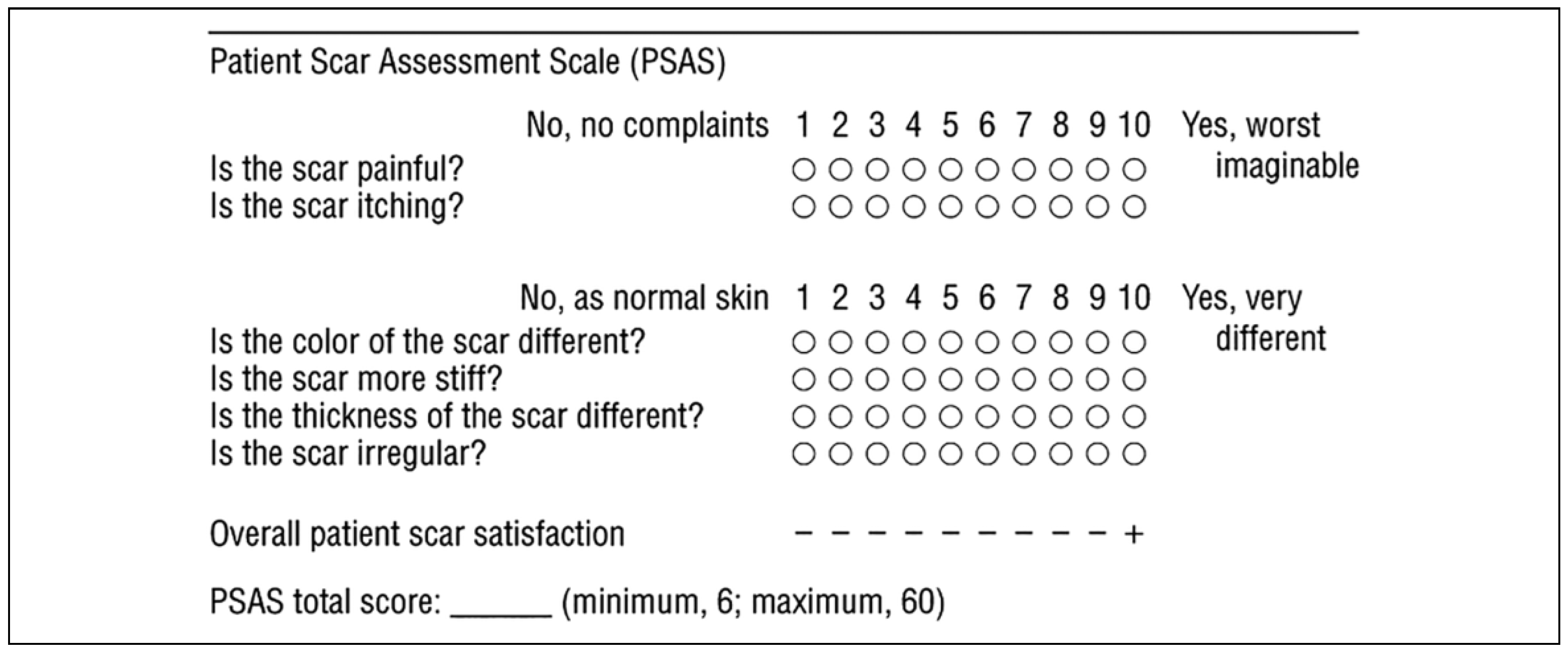
  18. Draaijers, L.J.; Tempelman, F.R.; Botman, Y.A.; et al. The patient and observer scar assessment scale: A reliable and feasible tool for scar evaluation. Plast Reconstr Surg. 2004, 113, 1960–1965; discussion 1966–7. [Google Scholar] [PubMed]
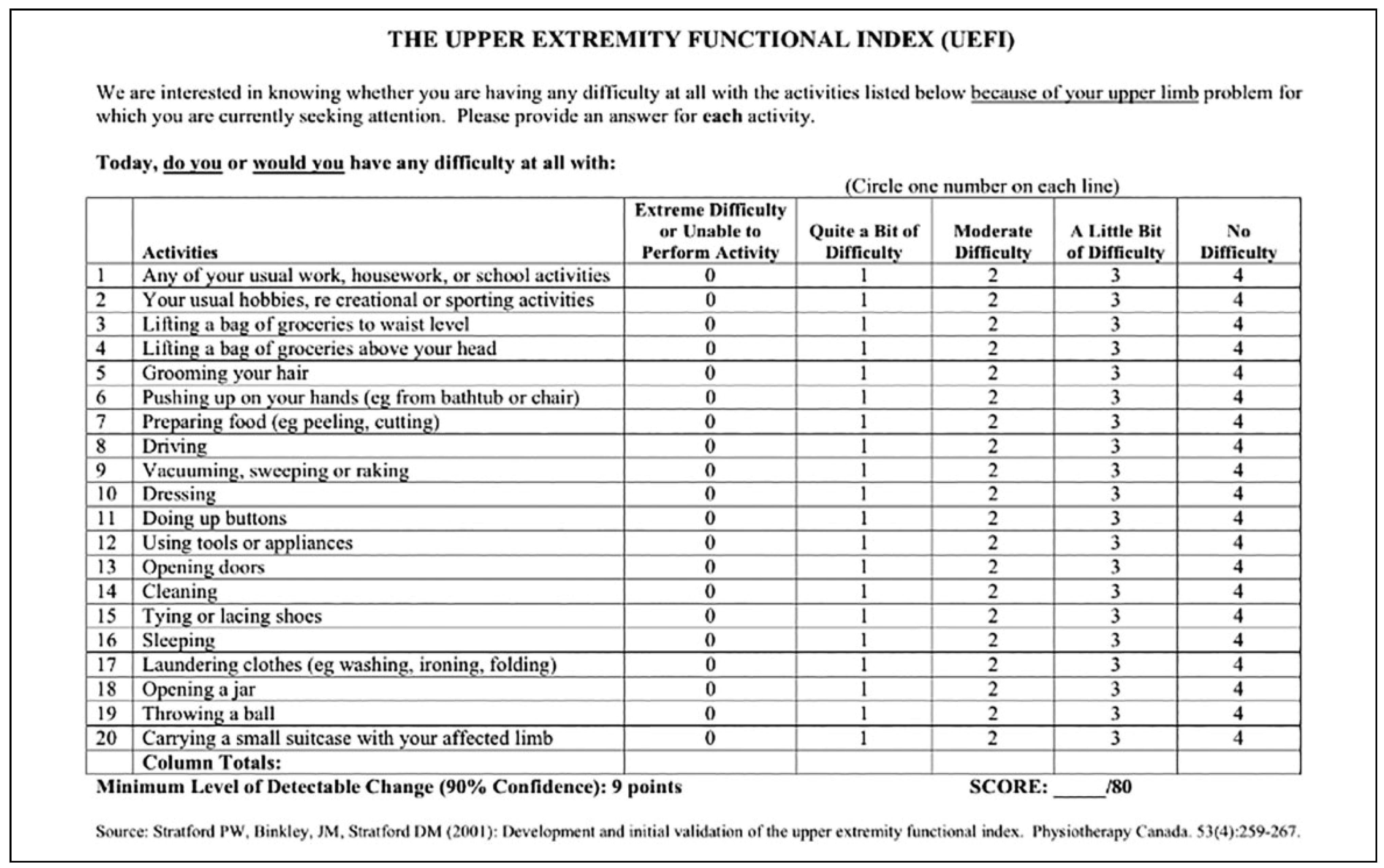
  19. Stratford, P.W.; Binkley, J.M.; Stratford, D.M. Development and initial validation of the upper extremity functional index. Physiotherapy Canada. 2001, 53, 259–267. [Google Scholar]
Figure 1. Excess adipofascial tissue harvested on either side of the pedicle.
Cmtr 13 00012 g001
Figure 2. Pedicle wrapped in the adipofascial tissue.
Cmtr 13 00012 g002
Figure 3. Retromolar route of passing the pedicle in patients with intact alveolar arch.
Cmtr 13 00012 g003
Figure 4. Central palatal defect with intact alveolar arch.
Cmtr 13 00012 g004
Figure 5. Alveolar arch defect.
Cmtr 13 00012 g005
Figure 6. A 14-year-old patient depicting the preoperative palatal lesion.
Cmtr 13 00012 g006
Figure 7. Post-reconstruction with RAFF. RAFF indicates radial artery free forearm flap.
Cmtr 13 00012 g007

