Next Article in Journal
The Comprehensive AO CMF Classification System for Mandibular Fractures: A Multicenter Validation Study
Previous Article in Journal
Frontal Sinus Fractures: Management and Complications
 
 
Craniomaxillofacial Trauma & Reconstruction is published by MDPI from Volume 18 Issue 1 (2025). Previous articles were published by another publisher in Open Access under a CC-BY (or CC-BY-NC-ND) licence, and they are hosted by MDPI on mdpi.com as a courtesy and upon agreement with Sage.
Article

Posttraumatic Lateral Dislocation of Mandibular Condyle: A Proposed New Classification with Report of 14 Dislocated Condyles

by
Tabishur Rahman
*,
Ghulam Sarwar Hashmi
,
Syed Saeed Ahmed
and
Sajjad Abdur Rahman
Department of Oral and Maxillofacial Surgery, Faculty of Medicine, Aligarh Muslim University, Aligarh 202002, Uttar Pradesh, India
*
Author to whom correspondence should be addressed.
Craniomaxillofac. Trauma Reconstr. 2019, 12(4), 249-253; https://doi.org/10.1055/s-0039-1677725
Submission received: 10 July 2018 / Revised: 1 October 2018 / Accepted: 19 October 2018 / Published: 16 January 2019

Abstract

:
Lateral dislocation of the intact mandibular condyle is a relatively uncommon clinical condition. Since the first description and classification of these dislocations given by Allen and Young, few classification systems have been proposed in literature with incorporation of different patterns of dislocations identified over the years. We share our clinical experience of nine cases of such dislocations with 14 dislocated condyles, and on the basis of clinical and radiological findings coupled with the review of existing classification systems, we propose a new classification system which includes all the possible patterns of such dislocations overcoming the major shortcomings of preexisting classification systems identified by the authors.

Lateral dislocation of the intact mandibular condyle is a relatively uncommon clinical condition. The pattern of such condylar dislocations is influenced by certain factors such as direction and amount of impact, the position of jaw during impact along with anatomical features of the joint, and the surrounding muscles which need to be favorable to push the condyle laterally and then superiorly. [1,2] Since the first description and classification of these dislocations given by Allen and Young, [3] few classification systems have been proposed in literature with incorporation of different patterns of dislocations identified over the years. The authors share their clinical experience of nine cases of such dislocations with 14 dislocated condyles, and on the basis of clinical and radiological findings coupled with the review of existing classification systems, they propose a new classification system which includes all the possible patterns of such dislocations overcoming the shortcomings of preexisting classification systems identified by the authors.

Materials and Methods

We summarized the data of nine patients with 14 laterally (superolaterally or anterosuperiorly) dislocated condyles who were operated by us. The data included age, gender, type of trauma, side of condylar involvement, type of dislocation, associated other mandibular fractures, method of reduction of condyle, treatment procedure, outcome, follow-up period, and maximal interincisal mouth opening at the last follow-up. A systematic literature search was performed based on two different methods: (1) main search—made in PubMed, Embase, and ScienceDirect databases; (2) search handling—all references of the included studies were read to find articles not found on PubMed, Embase, or ScienceDirect. Papers proposing classification of cases of lateral dislocation of mandibular condyle from 1969 to 2017 were identified and reviewed.

Results

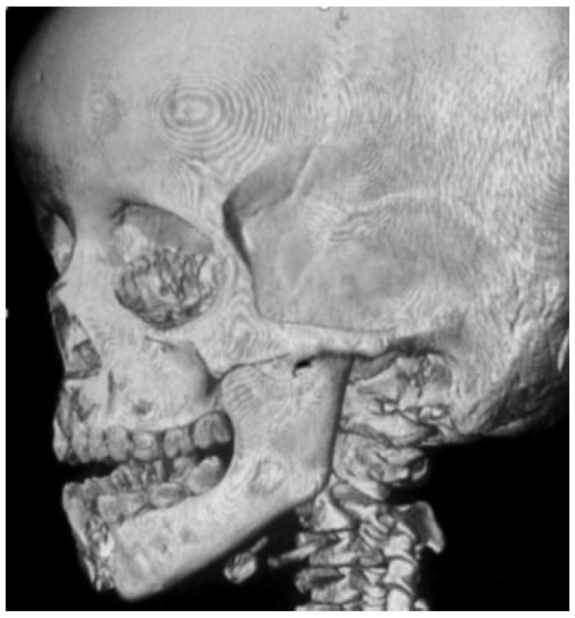
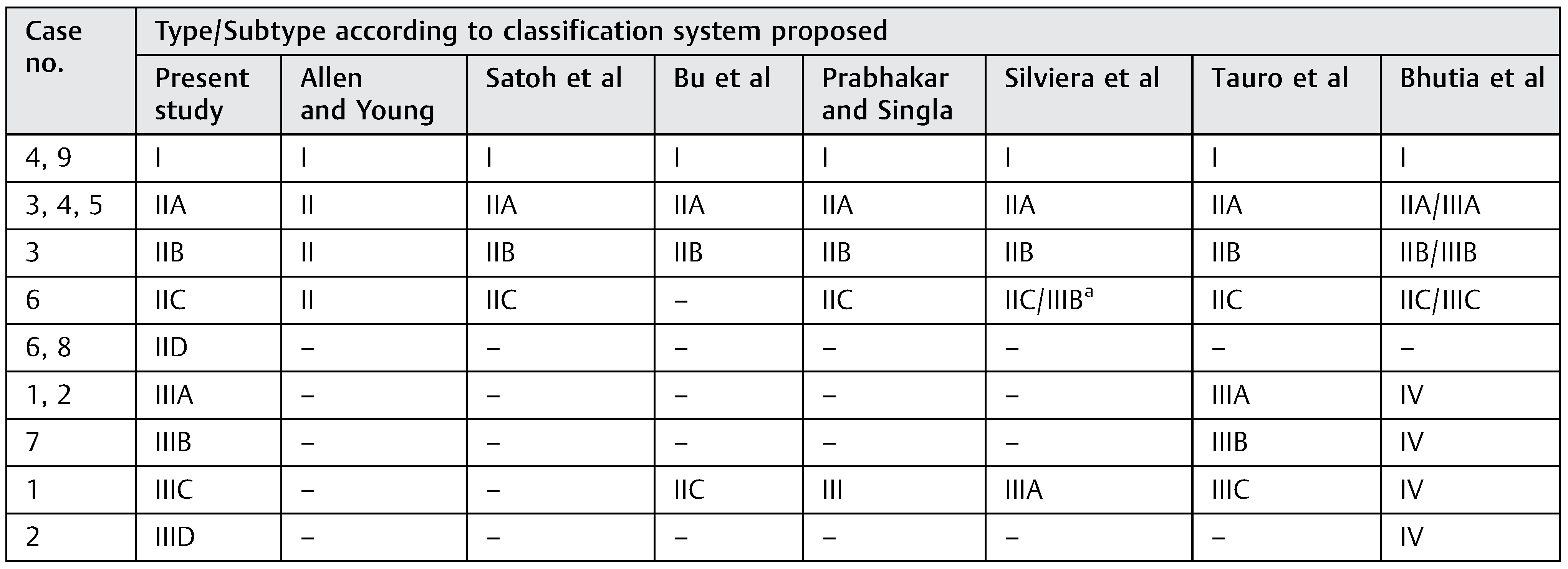
The age of our study patients ranged from 6 to 55 years with mean age of 29.4 years. There were three female patients and six male patients. Five patients were involved in road traffic accidents, three patients were victims of fall from height, and one case was of assault. Five patients had bilateral dislocations, whereas four patients suffered unilateral dislocation. Intact mandible was present in four cases, symphyseal/parasymphyseal fracture was found in four cases, and one had fracture of mandibular body. Three condyles required open reduction and nine patients were treated by closed methods. One patient with bilateral dislocation was mentally unstable and his guardian denied any treatment at our center. Eleven condyles were successfully reduced and in one case, the reduction was imperfect due to associated skull base fracture which demorphed the anatomy of the glenoid fossa (Figure 1). The follow-up period ranged from 21 to 180 days with the mean period of follow-up of 77.3 days. Maximum interincisal mouth opening at last follow-up appointment ranged from 21 to 33 mm with the mean being 25.5 mm. The findings of our cases have been summarized in Table 1. For the sake of argument, we have not mentioned the type of condylar dislocation in Table 1 but mentioned them in Table 2 after proposing our classification system.
The literature search yielded only 44 publications describing the condition in question. Out of these we narrowed down to seven articles which proposed a different classification system for these types of dislocations.
Allen and Young [3] divided such dislocations into two types on the basis of relation of the condyle with the glenoid fossa: type I (lateral subluxation), in which the condyle is laterally dislocated out of the fossa, and type II (complete dislocation), in which the condyle passes laterally and above the zygomatic arch, before actually entering the temporal fossa. Satoh et al. [4] reported one case, and further subclassified type II dislocation into type IIA (the condyle is not hooked above the zygomatic arch); type IIB (the condyle is hooked above the zygomatic arch); and type IIC (the condyle is lodged inside the zygomatic arch, which is fractured). Bu et al. [5] reported a unique case and suggested type IIC to be considered “when the condyle has passed laterally, and then superiorly to enter the temporal fossa and is lodged inside the zygomatic arch which is not fractured.” Prabhakar and Singla [6] reported a case similar to that reported by Bu et al., [5] but they classified it into type III dislocation, which described the dislocation of condyle inside the intact zygomatic arch with no mandible fracture. Silviera et al., [7] on the basis of a single unique presentation of their case, proposed a new type III classification by dividing it into two categories: type IIIA, as described by Prabhakar and Singla, [6] and type IIIB, which could be described as a “clinical condition when the condyle has passed anteriorly and then superiorly to enter the temporal fossa and is between the zygomatic arch fracture but without displacement to the lateral side and the temporal fossa and its contents on the medial side are associated with a mandible fracture.”
Tauro et al., [8] on the basis of their one case report and review of literature, proposed a change in existing classification by introducing new type II and type III dislocations where type II dislocations were complete dislocations associated with fracture of anterior mandible and type III dislocations were complete dislocations without associated anterior mandible fracture. They further gave three similar subtypes in both type II and type III with subtypes A, B, and C corresponding to type II A, II B, and II C given by Satoh et al. [4] Bhutia et al., [9] on the basis of their report of 11 cases and literature review, classified the dislocations taking into account the location of associated mandibular fracture, that is, anterior or posterior mandible. The type I dislocations were similar to that described by Allen and Young. [3] The type II dislocations were associated with fracture of anterior mandible and type III dislocations were associated with fracture of posterior mandible. They also subclassified type II and III dislocations into subtypes A, B, and C similar to those described by Tauro et al. [8] However, they further added a type IV dislocation which included complete dislocations without any associated mandibular fracture.

Discussion

Lateral dislocations of intact mandibular condyles are a rare clinical presentation. In the literature, the terms “superolateral” and “anterosuperior” dislocations have been interchangeably used for lateral dislocations of intact mandibular condyle. However, we are of the opinion that the correct nomenclature would be anterosuperior dislocation in agreement with Worthington’s [10] explanation of the mechanism of dislocation (which took into account the dislocations into the temporal fossa, i.e., anterosuperior, as well as the dislocations lateral to the zygomatic arch, i.e., superolateral). But for the sake of our study, we have reviewed the reports of anterosuperiorly as well superolaterally dislocated intact mandibular condyles, as strictly speaking both the types of dislocations are lateral dislocations of the condyle. The mechanism of lateral dislocations was suggested by Worthington [10] who stated that two obstacles need to be overcome to achieve such a condition. First, the capsular and ligamentous attachments to the condyle should be ruptured and second, the transverse dimension of the condylar head (from lateral pole to medial pole) should exceed the lateral dimensions of the space between the zygomatic arch and the medial bony wall of temporal fossa. For the latter to occur, he further stated that at least one of the following events must occur: the zygomatic arch may fracture, affording more room for the condyle to pass; the condylar head may fracture, decreasing the bulk; and the condyle head may rotate about a vertical axis, which would be favored when a mandibular fracture is present. The classifications given by Allen and Young [3] and Satoh et al. [4] supported this mechanism. Bu et al. [5] suggested that the dislocation of intact condyle medial to intact zygomatic arch could be attributed to the round shape of condyle and elasticity of zygomatic arch. Prabhakar and Singla [6] further concluded that such types of dislocations are favored by the presence of deep glenoid fossa and a steep articular eminence resulting from deep overbite which also causes the rounding of condyle induced by specific loading pattern. This formed their basis of introducing type III dislocations in the preexisting classification of Satoh et al. [4] Silviera et al. [7] subtyped type III on the basis of presentation of their case which had the intact condyle medial to a fractured but undisplaced zygomatic arch and associated mandibular fracture. However, if we take a closer look, we find that type III B as suggested by Silviera et al. [7] is similar to type IIC described by Satoh et al. [4] with the only difference being the posterior mandible fracture rather than anterior. Tauro et al. [8] emphasized that the dislocations corresponded to the size and direction of applied force on impact, position of the jaw during the impact, and anatomic variations of the joint. They further modified type II and type III dislocations as they were of the opinion that it was not just the presence or absence of anterior mandible fracture that influenced the type of dislocation but multiple impacts at the time of injury that led to the variable presentation.
Bhutia et al. [9] modified type II and type III dislocations on the basis of location of fracture of mandible—anterior or posterior. They also suggested a type IV which was not associated with fracture mandible. In this classification, type IV might include four types of presentation as suggested by us in our proposed classification, thus creating confusion when it comes to interclinician communication.
If we take a closer look, we can arrive on the following conclusions pertaining to the classification systems proposed in the literature:
  • None of the classification system includes all possible patterns of lateral dislocation (Table 2) (Figure 2).
  • There is an overlapping between the various types proposed in different classification systems. For example, type IV of Bhutia et al. [9] could be type IIIA of Silviera et al.’s [7] classification, type III of Prabhakar’s and Singla’s [6] classification, and type IIIA/B/C of Tauro et al.’s [8] classification (Table 2).
  • One given type in some classification systems accounts for many variable clinical presentations. For example, in the classification system proposed by Bhutia et al., [9] type IV accounts for complete dislocations with an intact mandible. This may include type IIIA, B, and C of Tauro et al.’s [8] classification; each of which corresponds to a different type of presentation (Table 2).
  • Additional “types” apart from those given by Allen and Young [3] were proposed by clinicians after the identification of a new clinical presentation in an attempt to classify them but were mostly based on report of a single case. [4,5,6,7]
The identification of different anatomical presentations of these dislocations could be attributed to the increase in traffic density and introduction of motor vehicles capable of achieving high speeds coupled with advancements made in the field of diagnostic radiography over the years since Allen and Young [3] first reported such dislocations. We also agree with Li et al.’s [1] explanation that the position of jaw during impact (wide open) along with anatomical features of the joint and the surrounding muscles influence the clinical presentation. Similarly, Tauro et al.’s [8] emphasis on multiple impacts producing the variable types of such dislocations cannot be overlooked. With only 44 publications reported in literature related to the type of injury in questions, we identified seven different classification systems utilizing similar terminology. This may lead to confusion when classifying these injuries for the sake of communication among clinicians. Also, a uniform consensus cannot be arrived among clinicians regarding management of these dislocations. Therefore, we suggest the following classification system.
  • Type I: Lateral subluxation.
  • Type II: Complete dislocation with fracture of anterior/posterior mandible.
    • IIA: Condyle not hooked above zygomatic arch.
    • IIB: Condyle hooked above zygomatic arch.
    • IIC: Condyle medial to fractured zygomatic arch.
    • IID: Condyle medial to intact zygomatic arch.
  • Type III: Complete dislocation without fracture of anterior/posterior mandible.
    • IIIA: Condyle not hooked above zygomatic arch.
    • IIIB: Condyle hooked above zygomatic arch.
    • IIIC: Condyle medial to fractured zygomatic arch.
    • IIID: Condyle medial to intact zygomatic arch.
To stress on our idea, we made a comparison of our described types of dislocations with those in other classification systems (Table 2) and concluded that we were correct in assuming that other classification systems did not take into account all possible variable clinical presentations of lateral condylar dislocation.
In summary, the merits of this classification system are as follows: (1) it is systematic, (2) it includes all possible patterns of lateral dislocations, and (3) it is easy to remember, thus offering ease of communication among clinicians. However, it is still very early to predict treatment modality to be adopted for each type of dislocation, as the number of cases reported till date are scant and clinicians who have reported such cases have attempted reduction of such condyles not on the basis of a set protocol but on the basis of the presentation of the case and their expertise.

Funding

None.

Acknowledgments

All the authors have viewed and agreed to the submission of the manuscript.

Conflicts of Interest

None.

References

  1. Li, Z.; Li, Z.B.; Shang, Z.J.; Wu, Z.X. An unusual type of superolateral dislocation of mandibular condyle: Discussion of the causative mechanisms and clinical characteristics. J Oral Maxillofac Surg 2009, 67, 431–435. [Google Scholar] [CrossRef]
  2. Rahman, T. Unusual superolateral dislocation of mandibular condyle. Craniomaxillofac Trauma Reconstr 2018, 11, 142–144. [Google Scholar] [CrossRef] [PubMed]
  3. Allen, F.J.; Young, A.H. Lateral displacement of the intact mandibular condyle. A report of five cases. Br J Oral Surg 1969, 7, 24–30. [Google Scholar] [CrossRef] [PubMed]
  4. Satoh, K.; Suzuki, H.; Matsuzaki, S. A type II lateral dislocation of bilateral intact mandibular condyles with a proposed new classification. Plast Reconstr Surg 1994, 93, 598–602. [Google Scholar] [CrossRef] [PubMed]
  5. Bu, S.S.; Jin, S.L.; Yin, L. Superolateral dislocation of the intact mandibular condyle into the temporal fossa: Review of the literature and report of a case. Oral Surg Oral Med Oral Pathol Oral Radiol Endod 2007, 103, 185–189. [Google Scholar] [CrossRef] [PubMed]
  6. Prabhakar, V.; Singla, S. Bilateral anterosuperior dislocation of intact mandibular condyles in the temporal fossa. Int J Oral Maxillofac Surg 2011, 40, 640–643. [Google Scholar] [CrossRef] [PubMed]
  7. Silviera, R.L.; Ranuzia, I.; Melo, M.F.S.; de Oliveira, R.A.; de Brito, A.A.; Vidigal, V.L. Traumatic anterosuperior dislocation of the intact mandibular condyle into the temporal fossa. Craniomaxillofac Trauma Reconstr 2018, 11, 296–301. [Google Scholar] [CrossRef] [PubMed]
  8. Tauro, D.; Lakshmi, S.; Mishra, M. Superolateral dislocation of the mandibular condyle: Report of a case with review of literature and a proposed modification in the classification. Craniomaxillofac Trauma Reconstr 2010, 3, 119–123. [Google Scholar] [CrossRef] [PubMed]
  9. Bhutia, D.P.; Mehrotra, D.; Mahajan, N.; Howlader, D.; Gamit, J. Posttraumatic superolateral dislocation of condyle: A case series of 18 condyles with review of literature and a proposed classification. J Oral Biol Craniofac Res 2017, 7, 127–133. [Google Scholar] [CrossRef] [PubMed]
  10. Worthington, P. Dislocation of the mandibular condyle into the temporal fossa. J Maxillofac Surg 1982, 10, 24–27. [Google Scholar] [CrossRef] [PubMed]
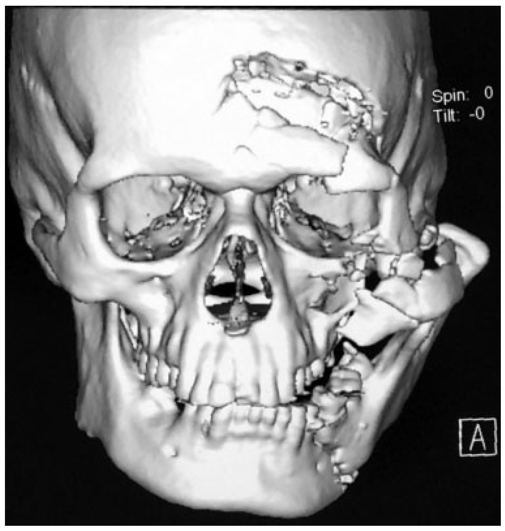
Figure 1. Imperfect reduction was achieved in this patient because of the severe comminuted fracture of the midface as well as base of the skull.
Cmtr 12 00035 g001
Figure 2. A unique type of clinical presentation which cannot be classified in any of the existing classification systems.
Cmtr 12 00035 g002
Table 1. Summary of review of records of nine patients with lateral condylar dislocations.
Cmtr 12 00035 i001
Abbreviations: FFH, fall from height; RTA, road traffic accident.
Table 2. Comparison of present classification system with existing classification systems.
Cmtr 12 00035 i002
a In both IIC and IIIB, the zygomatic arch is fractured; the only difference is that in IIIB the zygomatic arch is fractured and undisplaced, whereas in IIC the zygomatic arch is fractured and displaced.