Next Article in Journal
Psychological Impact of Facial Trauma
Previous Article in Journal
Smile Train: Making the Grade in Global Cleft Care
 
 
Craniomaxillofacial Trauma & Reconstruction is published by MDPI from Volume 18 Issue 1 (2025). Previous articles were published by another publisher in Open Access under a CC-BY (or CC-BY-NC-ND) licence, and they are hosted by MDPI on mdpi.com as a courtesy and upon agreement with Sage.
Review

Prosthetics in Facial Reconstruction

by
Jaclyn Klimczak
1,
Samuel Helman
1,
Sameep Kadakia
1,
Raja Sawhney
2,
Manoj Abraham
3,
Allison K. Vest
4 and
Yadranko Ducic
5,*
1
Department of Otolaryngology-Head and Neck Surgery, New York Eye and Ear Infirmary of Mount Sinai, New York, NY, USA
2
Department of Facial Plastic and Reconstructive Surgery, University of Florida Health Science Center, Gainesville, FL, USA
3
Department of Facial Plastic Surgery, New York Medical College, West Chester, NY, USA
4
Department of Anaplastology, Medical Arts Prosthetics LLC, Mckinney, TEX, USA
5
Otolaryngology and Facial Plastic Surgery Associates, 923 Pennsylvania Avenue, Suite 100 Fort Worth, TX 76104, USA
*
Author to whom correspondence should be addressed.
Craniomaxillofac. Trauma Reconstr. 2018, 11(1), 6-14; https://doi.org/10.1055/s-0037-1603459
Submission received: 11 January 2017 / Revised: 1 February 2017 / Accepted: 25 February 2017 / Published: 22 May 2017

Abstract

:
Reconstruction of the head and neck can be a challenging undertaking owing to numerous considerations for successful rehabilitation. Although head and neck defects were once considered irretrievably morbid and associated with a poor quality of life, advances in surgical technique has immensely contributed to the well-being of these patients. However, all patients are not suitable surgical candidates and many have sought nonsurgical options for functional and cosmetic restoration. As such, the advent of prostheses has ameliorated those concerns and provided a viable alternative for select patient populations. Prosthetic reconstruction has evolved significantly over the past decade. Advances in biocompatible materials and imaging adjuncts have spurred further discovery and forward progress. A multidisciplinary approach to head and neck reconstruction focused on appropriate expectations and patient-centered goals is most successfully coordinated by a team of head and neck surgeons, maxillofacial surgeons, and prosthetic specialists. The aim of this article is to provide a comprehensive review of the current trends for prosthetic rehabilitation of head and neck defects, and further elaborate on the limitations and advancements in the field.

Prostheses have become an integral part of head and neck restoration. With significant advancements over the past decade, postsurgical rehabilitation and quality-of-life measures have taken on paramount roles. Head and neck prostheses provide a synthetic replacement for ablative, congenital, or traumatic defects of the head and neck, and attempt to restore cosmetic appearance and functionality. The dual goals of craniofacial reconstruction, form and function, can often be harmoniously achieved. However, the challenging anatomy and physiology of the head and neck can prove daunting and time consuming to the most experienced surgeon. Therefore, a multidisciplinary approach employing the ablative, reconstructive, and maxillofacial techniques for pre- and postsurgical planning is critical to a successful patient outcome. The purpose of this article is to review the current concepts surrounding prosthetic rehabilitation of head and neck defects, with emphasis on the advancements and limitations in the field of prosthetic reconstruction.

Methods

This review seeks to collate all full-text peer-reviewed publications concerning facial prosthetic rehabilitation of patients with head and neck cancer, posttraumatic, and congenital defects. The most current prosthetic biomaterials and their indications were also identified. A comprehensive search of studies published from January 1990 to July 2016 and listed in the PubMed/Medline and Cochrane Library databases was performed. Three authors independently reviewed the titles and abstracts of all citations identified by the literature search. Special attention was placed on auricular, orbital, midface, and nasal defects; prosthetic materials; retention mechanisms; and technique for placement. Articles were reviewed, and those appropriate to the central theme of this article were included in the study.

Presurgical Planning

The multidisciplinary approach to head and neck reconstruction is critical because treatment of these defects is complex [1]. In some cases, substitutions of biologic structures with prostheses are advantageous and may be superior to surgical reconstruction both in function and aesthetics [2]. Multiple factors must be taken into account when reconstructing a head and neck defect, including the size, location, age of the defect, and amount of adjacent supporting tissue around the defect. Overly large defects [3,4], and defects adjacent to mobile structures, may be more successful with surgical versus prosthetic approach to ensure fit and survival [5]. The timing for reconstruction is critical in cases of trauma-induced defects and congenital malformations [6]. Patients’ age in congruence with their anticipated growth is also a factor in reconstructing an anatomical area that compliments their appearance. Prosthetics can be tailored to patients’ age and facial features during different growth stages [4,5,6].
Head and neck malignancy can ensue devastating aesthetic and functional consequences that patients require extensive preoperative counseling to tailor postoperative expectations [5,7,8]. Reconstruction, when needed, is informed by disease features, including the presence or absence of metastases, extent of extirpation, and patient prognosis, among others [8]. In patients in whom recurrence is more likely, prosthesis would be more favorable for long-term management and surveillance [5]. Microvascular flap reconstruction affords the best reconstructive option if infection, osteonecrosis, or pathologic fractures are anticipated [9].
Successful reconstruction in part relies on careful consideration of the patient’s demographics, functional status, and preexisting medical conditions. The psychological well-being of the patient should also be considered throughout the reconstructive process so that their goals and expectations are well communicated [10,11]. The postsurgical hospital course in patients undergoing microvascular free flap surgery is extensive and may require revision surgeries and/or procedures which predisposes the patient to a variety of possible complications [12]. Hence, in each patient, a cost–benefit analysis of their wants, surgical needs, and expected outcomes must be weighed, and clinically prostheses may provide a better functional and cosmetic result in the face of attenuated survival [13,14,15]. Therefore, elderly patients or those with significant comorbidities may benefit from prosthetic rehabilitation [16,17,18].

Imaging Modalities and Prosthesis Design

Reconstruction of head and neck defects has improved immensely over the years due to advancements in threedimensional (3D) printing technologies. These digital imaging modalities allow a surgeon to preoperatively visualize the patient’s defect and create customizable patient-tailored prosthetics. Further advancements in the computer-aided design/computer-aided manufacturing (CAD/CAM) technology allow for creating and manufacturing patient-specific custom prostheses [19,20,21]. Imaging modalities such as computed tomography (CT) and magnetic imaging resonance (MRI) can be converted to a rapid prototyping model that can be printed in wax or acrylic [22,23]. These models can be further modified, or duplicated with other prosthetic materials.

Materials

Throughout history, materials such as silver, leather, porcelain, paper mache, gelatin, latex, and acrylic were employed in prosthetic reconstruction [24,25]. Development of an ideal facial prosthetic relies on factors such as durability, flexibility, weight, color matching, longevity, biocompatibility, texture, hygiene, and thermal conductivity [26]. Modern materials used for head and neck prostheses are divided into methacrylates, polyurethane elastomers, and silicone elastomers. These clinically inert materials easily absorb pigmentation and can be designed to match the color and texture of the surrounding structures for the most natural result [24,27,28]. Silicone has become the most widely used materials in facial prosthetics. The soft and flexible texture of a silicone elastomer retains body temperature without distortion and can be stretched until transparent to blend with the adjacent skin. However, facial prosthetics are not without their disadvantages. On average, a facial prosthetic is remade or reconditioned every 2 years [18]. Environmental influences, ultraviolet light, and discoloration play important roles in the degradation of a prosthesis, which puts a considerable burden on patients [17,18,26].

Retention Systems

The success of a prosthesis and overall patient compliance relies heavily on its retention system. Anchorage relies on a range of patient-specific factors, including anatomic, mechanical, chemical, and surgical considerations [27,28,29,30]. Facial prostheses were often mechanically anchored to spectacles or secured via anatomical undercuts early in the 20th century [18]. Complications with these retention systems and advancements in prosthetic anchoring, including adhesives, have since largely replaced these older practices. While easier to use, medical adhesives often lose their bone strength as the adhesive naturally weakens, requiring reapplication every 4 to 8 h, or sooner if compromised by humidity and/or perspiration. Patients with active lifestyles are poor candidates for an adhesive-based prosthesis, as they easily dislodge with frequent movement [28,31,32]. These early methods of adhesion often resulted in skin irritation, allergic reactions, and difficulty in positioning the prosthetic [28,33,34]. Compliance and satisfaction with daily-applied prosthetic adhesive is reduced relative to surgical methods of prosthetic anchorage [35].
Implant-retained facial prosthetics has vastly improved anchorage and become a reliable practice of retention. Osseointegration gained popularity in the 1950s when Swedish physician Per-Ingvar Brånemark discovered that titanium was biocompatible with bone [36]. For a successful rigid fixation of an alloplastic material to bone, two processes play a vital role in osseointegration: osteointegration and osteoconduction [37]. Implants introduced into the bone within or adjacent to the defect triggers osteoinduction. The stimulation of preosteoblasts in the formation of new bone is speculated to be the most important factor in ensuring prosthesis survival. Once the implants are placed, osteoconduction begins a cycle of remodeling and bone formation [37,38,39]. When the bone is finally healed, it bonds with the implant. Abutments or attachments to the bone-anchored implant exit the skin and allow for external prosthesis attachment [24]. Osseointegration is a simple surgical procedure that is generally well tolerated, and holds a low risk of long-term complications [29,30]. Bone-anchored implant mechanisms have revolutionized craniofacial prosthetic rehabilitation, as they are comfortable to wear and easy to clean and self-align in an anatomically correct position [34]. While bone anchoring is an excellent modality in many respects, it is by no means a panacea. Implant-retained prostheses have a greater risk of failure in irradiated bone, and may portend an increased risk of microbial infections [18,40]. Individual assessment among patients is therefore critical when planning their prosthetic rehabilitation.
Several mechanisms exist to couple the osseointegrated implant with the external prosthesis. These include bar-clip attachments, ball attachments, and magnetic retention [41]. Bar-clip retention for facial prostheses is most widely used, and provides the strongest bond. However, adequate surface area to sustain the load is required to support the attachment [42]. Magnetic retention has strong attractive forces in small and inconspicuous sizes that are ideal for craniofacial deficits [42,43]. Nasal and orbital prostheses, for example, are almost exclusively retained by magnets [24,41]. Bone-anchored prostheses typically last between 3 and 5 years compared with 1 and 3 years for an adhesive-retained prosthesis before replacement as natural wear is needed [44].

Auricular Prostheses

Auricular defects are commonly associated with congenital malformations, ablative tumor surgery, or trauma. The size and extent of the deformity determine whether the defect can be treated with surgical reconstruction or prosthesis [45]. The best approach to surgically restore the auricle is dependent on patient preference, surgeon experience and comfort, and available tissue [46]. Larger defects are harder to reconstruct surgically and depend on multiple factors to make the reconstruction a success. The adjacent skin and subcutaneous tissue, which are frequently damaged by trauma or radiation therapy, must be intact with a rich blood supply to support an autologous graft [46,47,48]. Auricular reconstruction presents a challenge to even the most accomplished surgeon. Multiple surgeries are required for graft harvesting, tissue expansion, and cosmetic revision, increasing the morbidity and risk for complications. Bleeding, infection, and hematoma are more frequent in comparison to an auricular prosthesis at the implant and graft site [49]. Moreover, overall patient satisfaction is frequently lower due to inconsistent aesthetic outcomes [50,51,52].
Variations and inconsistencies in surgical practice paved the way for prosthetic reconstruction. In these cases, preservation of the tragus aids in prosthesis alignment and defect concealment [53]. Retention of an auricular prosthesis is achieved via chemical adhesives or more commonly with osseointegrated implants. Osseointegration for retention of auricular implants is favorable due to the high success rate in temporal bone anchoring, even in irradiated patients [54,55,56]. Implants are placed in two to three locations to lend greater infrastructure support [57].The implants are placed 20 mm from the external ear canal to correspond with the 1-, 3-, and 5-o’clock positions along the antihelix of the left ear and 7-, 9-, and 11-o’clock positions for the right ear [5,58]. Placement as such allows for a sufficiently high space for the abutment to support the prosthesis in correct anatomical position [24].The bone must be well vascularized and thick enough (>2.5 mm) to support the load of the prosthesis [25,45,59]. It is important to consider that portions of the normal anatomy, such as the tragus, can be kept intact to hide the transition from skin to prosthesis. Preoperative CT imaging is always needed for presurgical planning to review the integrity of the temporal bone and map out adequate sites for implants [59].
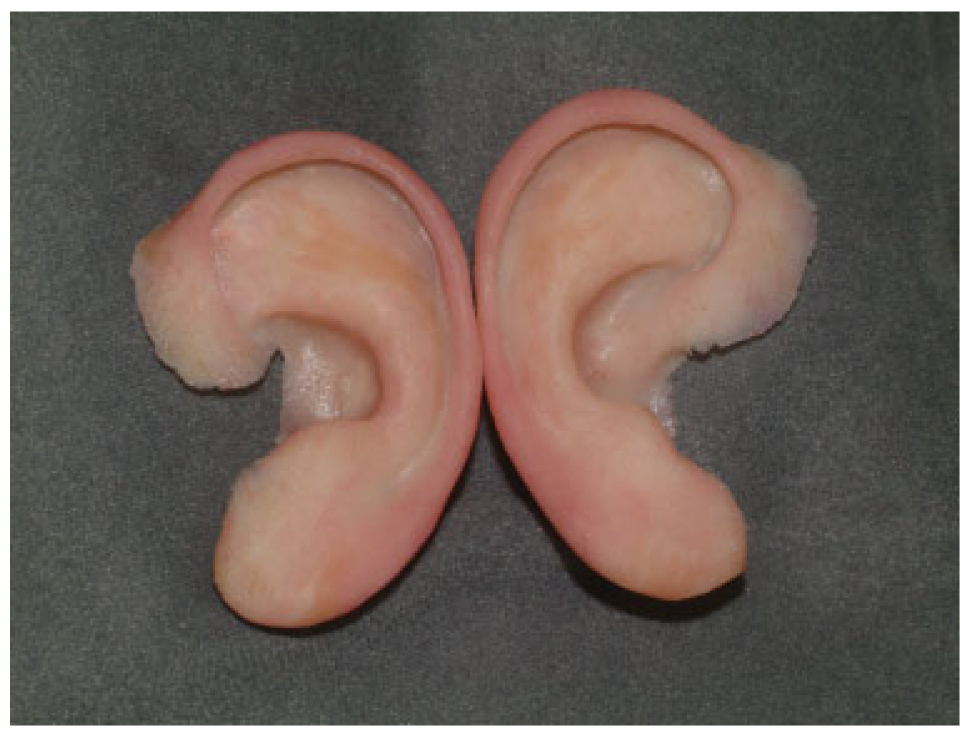
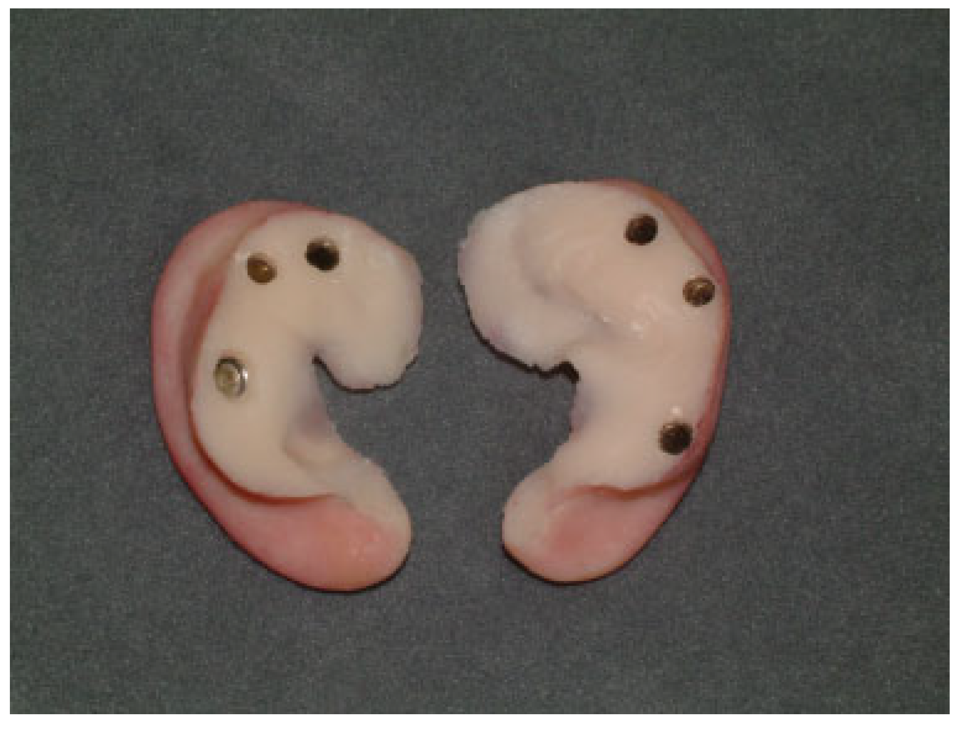
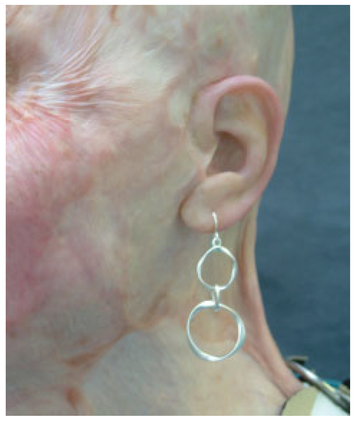
An auricular prosthesis can be easily implemented, and provides significant functional and aesthetic benefits. Sound amplification and acoustic gain are markedly attenuated in the deformed ear. Auricular prostheses provide a clinically relevant acoustic gain at certain head positions and frequencies that aid speech recognition in noise [56]. Figure 1, Figure 2, Figure 3 and Figure 4 show a patient following auriculectomy successfully restored using an auricular prosthesis.

Nasal Prostheses

Nasal lesions may require a partial or total resection, and are commonly reconstructed with the use of regional flaps. The paramedian forehead flap is the most cosmetically favorable because of the similar skin color and texture to the nasal region [60,61]. Nonetheless, color match is not always optimal, and revision surgeries come with increased morbidity and mortality [62,63]. Moreover, patients undergoing postoperative radiation have delayed wound healing and an increased risk of flap failure [64,65,66]. In some cases, prosthesis may be the best alternative to match and restore the defect with a nose similar in color, thickness, and texture to the surrounding anatomy [64,66].
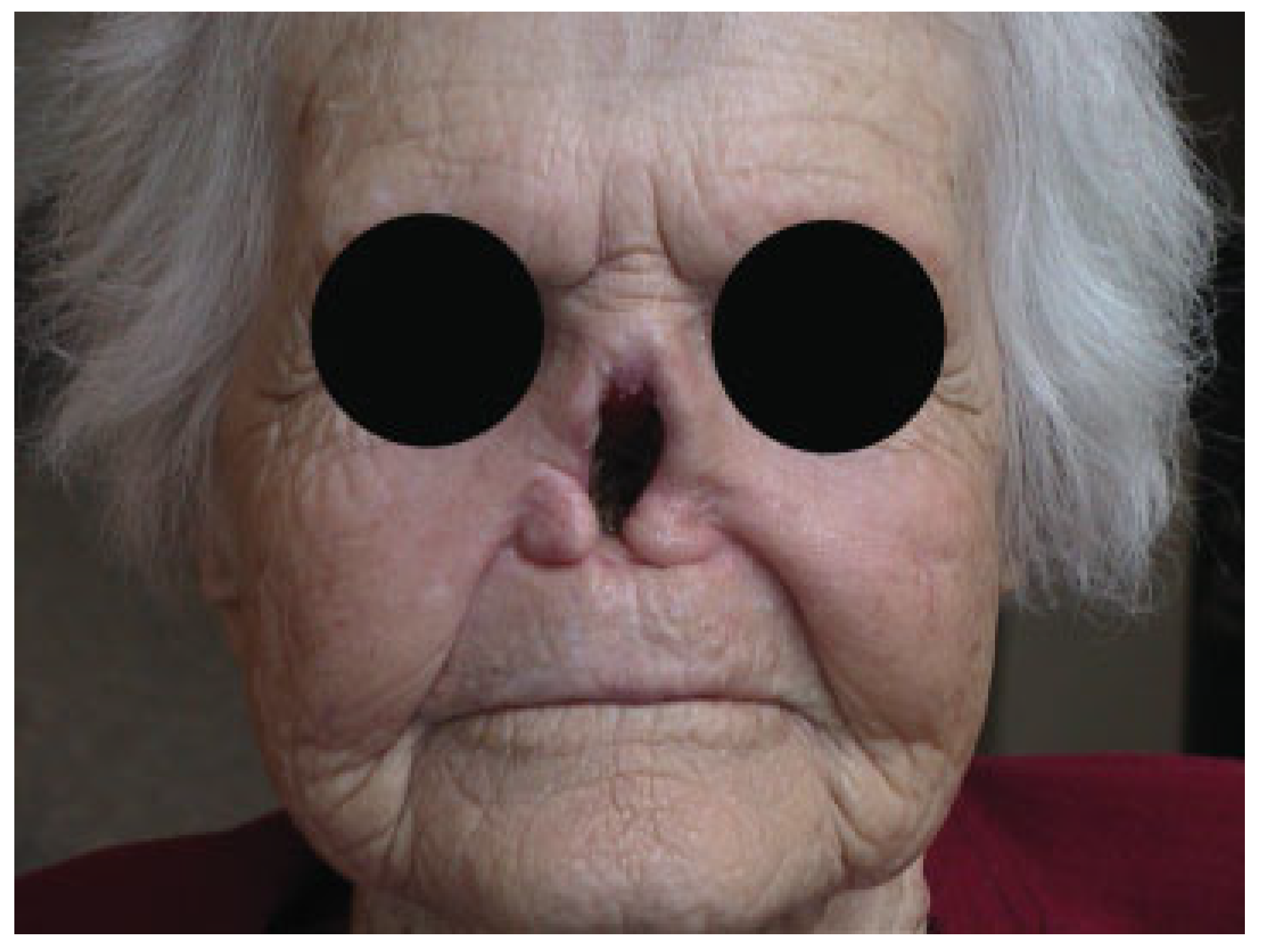
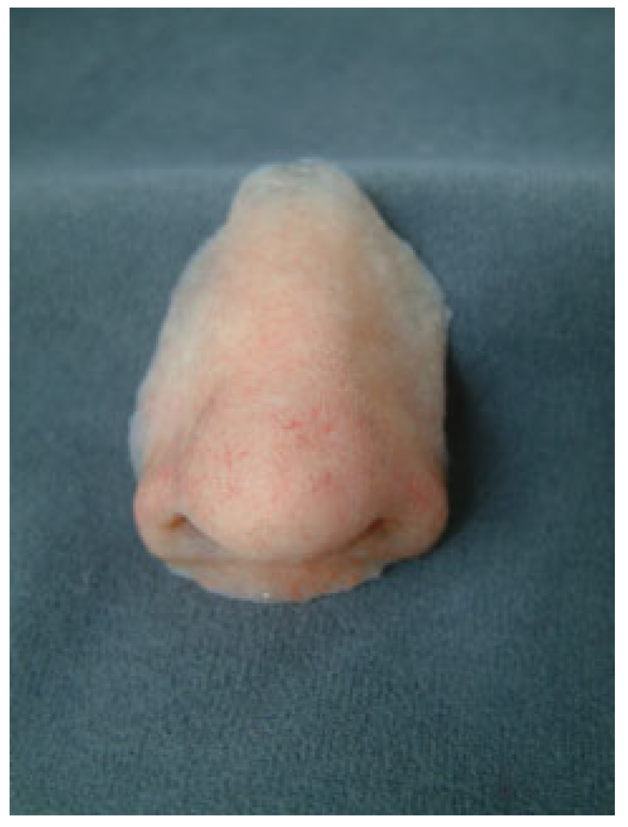
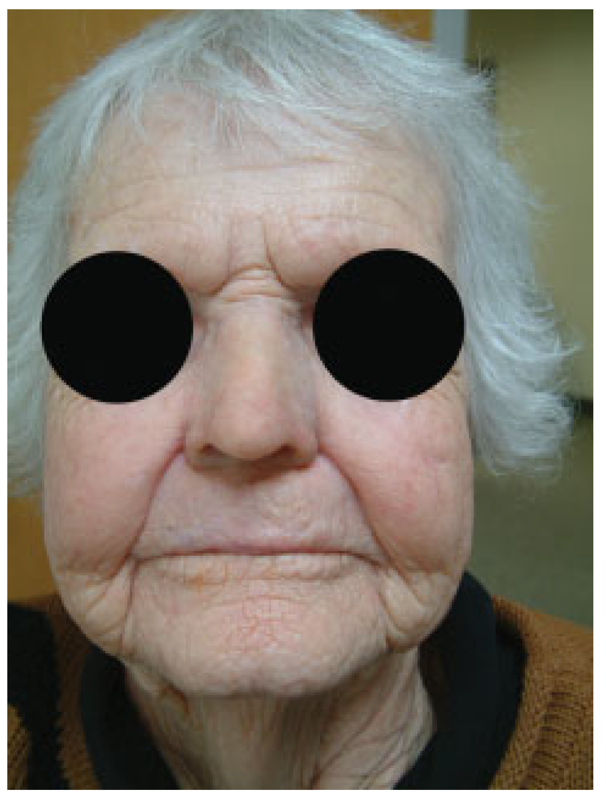
Before determining the appropriate method of reconstruction, careful attention to the retention mechanism used for a nasal prosthesis must be acknowledged. Adhesives have a short life span and often fail because of the constant air exchange, humidification, and moisture in the nasal cavity [67]. Mechanical retention may be sufficient if the appropriate surgical undercuts are made to position the implant, and enough surface area is available to achieve appropriate anchorage [68]. Osseointegration has significantly improved nasal implant retention because of its tenacity and resilience in the face of dynamic environmental conditions. This technique, however, is dependent on the quantity and quality of the available bone stock [67,68,69]. Osseointegrated implants are traditionally placed in the floor of the nasal cavity or glabella. Limitations in the amount of bony support can compromise the stability of an implant, or cause dislodgement with functional movements such as mastication [69]. Scott et al. looked into the placement of zygomatic implants to support a nasal prosthesis in patients who had undergone rhinectomy. In comparison to nasal implantation, using the zygoma provides a region with greater length and bone stock for successful osseointegration [70]. Second, the length of the zygomatic implant is often not in the field of radiation therapy, thus improving implant success [71,72]. It is again important to consider that portions of the normal anatomy may need to be removed, such as the ala, anterior septum, and columella to allow the prosthesis to fit properly without requiring a large construct. Care must also be taken to ensure that the upper lip or melolabial crease is not disturbed to prevent stark contrast between native tissue and the prosthesis. While nasal prostheses share some disadvantages, overall they provide a cosmetically advantageous result that can be achieved with a low morbidity surgical procedure. Figure 5, Figure 6 and Figure 7 show a patient following rhinectomy successfully restored using a nasal prosthesis.

Maxillary and Midface Prostheses

Reconstruction of midface and maxillary defects is a complex undertaking. The maxilla is essential for mastication, phonation, malar projection, and deglutition, The maxilla provides structural support between the skull base and occlusal plane, and supports facial structures involved in expressive movements [73]. Reconstruction of maxillary defects must accomplish closure of the oral cavity, support of orbital contents and maxillary buttresses, restoration of dentition and functionality, and restitution of midface contour and symmetry. Traditionally, maxillectomy defects were reconstructed with prosthetic obturation, with the primary goal of separating the oral cavity from the sinonasal cavity [74,75]. Obturator stability is directly dependent on the amount of tissue available for support, and extensive hard palate resections compromise the airtight fit of the obturator with remnant structures [75,76,77]. This instability leads to leak from the nasal cavity and sinuses, increasing irritation and wear on the oral cavity [77,78,79,80,81].
Maxillary and midface defects have spanned the reconstructive ladder from prosthetic rehabilitation to microvascular free flap. The size and location of the defect largely dictate the most appropriate reconstruction. Brown et al. developed a modern classification system for maxillary defects in 2000 [82], dividing defects into four vertical and three horizontal classes with associated surgical and prosthodontic approaches and reconstructive strategies. Smaller defects without alveolar involvement are most amenable to obturator rehabilitation and can be reconstructed using a local advancement or microvascular free flap. Prosthesis for smaller defects is well tolerated but maintenance is the greatest factor that drives patient preference between single and multiple surgery reconstructions [75].
Midface defects typically require three to four implants for adequate support [29]. The zygomatic buttress, supraorbital rim, vomer, and horizontal part of the hard palate are stable recipient sites [53]. The amount of remnant palate and/or the adjacent abutment tooth is critical to utilize for stability and retention of the prosthesis [83]. The integrity of the canine and molar teeth is also critical for strong prosthesis retention. Forces that influence the fit of a prosthesis need to be considered and include downward gravitational forces, upward occlusive forces, and rotational forces involved with functional speech, swallow, and mastication [81,84].
Large maxillary defects may not be amenable to prosthetic rehabilitation due to the absence in remaining structures that provide stability. In these cases, microvascular composite free flaps can achieve outstanding functional and aesthetic results. Even with complex resections, these composite flaps provide adequate bone length and tissue to fill the defect [75]. A combination of techniques using local advancement flaps or microvascular free flaps in addition to maxillofacial prostheses may achieve a more satisfactory functional and aesthetic result [73,75,85].

Orbital Prostheses

The eyes are integral to vision and an important component of self-image and expression. Orbital exenteration heavily impacts a patient’s self-confidence, thus necessitating speedy repair [86]. Orbital enucleation involves removal of the globe by severing the optic nerve and extraocular muscles, while evisceration consists of removal of the entire globe, leaving the surrounding orbital contents intact including Tenon’s capsule, extraocular muscles, and the optic nerve [87]. Exenteration is the most radical where all orbital contents including the globe are removed. Reconstruction after exenteration is patient dependent and can be done with a microvascular free flap or orbital prosthesis [88]. However, for the aforementioned lesser defects, repair with a prosthetic eye is the most common form of reconstruction. After removal of the eye, an orbital implant is placed for 4 to 6 weeks. The implant occupies the space that will eventually support the prosthesis, and prevents scar formation and tissue contraction. The bony cavity is lined with a splitthickness skin graft to create an adhesive base for the implant [53]. The orbital boundaries must be stable and healthy, the sinuses must be closed from the orbital contents, and the depth of the defect must be maintained to prevent endopthalmos [89]. The inferior margin of the resection should have solid contour and the stability to support a prosthetic load [90]. The superior portion should take careful consideration to maintain the position of the eyebrow. In cases of evisceration surgery, the extraocular muscles remain attached to the scleral envelope that lines the implant. Enucleation requires reattachment of the muscles directly onto the implant or indirectly to a wrapping material over the implant for anchoring [91,92]. The cosmetic success is largely due to the orbital implant. If properly placed, the prosthetic eye will look and move with similar behavior to the contralateral eye [93].
The orbital implant is either made from a nonporous material such as silicone and polymethyl methacrylate or a porous material such as porous polyethylene, hydroxyapatite, and aluminum oxide [91]. Nonporous materials are best used for implants that will not be integrated or pegged, and are well tolerated. Porous materials have become the mainstay in orbital implant material, as they promote a fibrovascular ingrowth of host tissue, improving stability, lowering the risk of rejection, and allowing the insertion of pegs or posts [92]. The material and size of the implant are determined by the surgeon to meet the patient’s anatomic needs. These implants achieve 65 to 75% of the volume of the original ocular globe [94], with the remaining space filled by an external prosthesis.
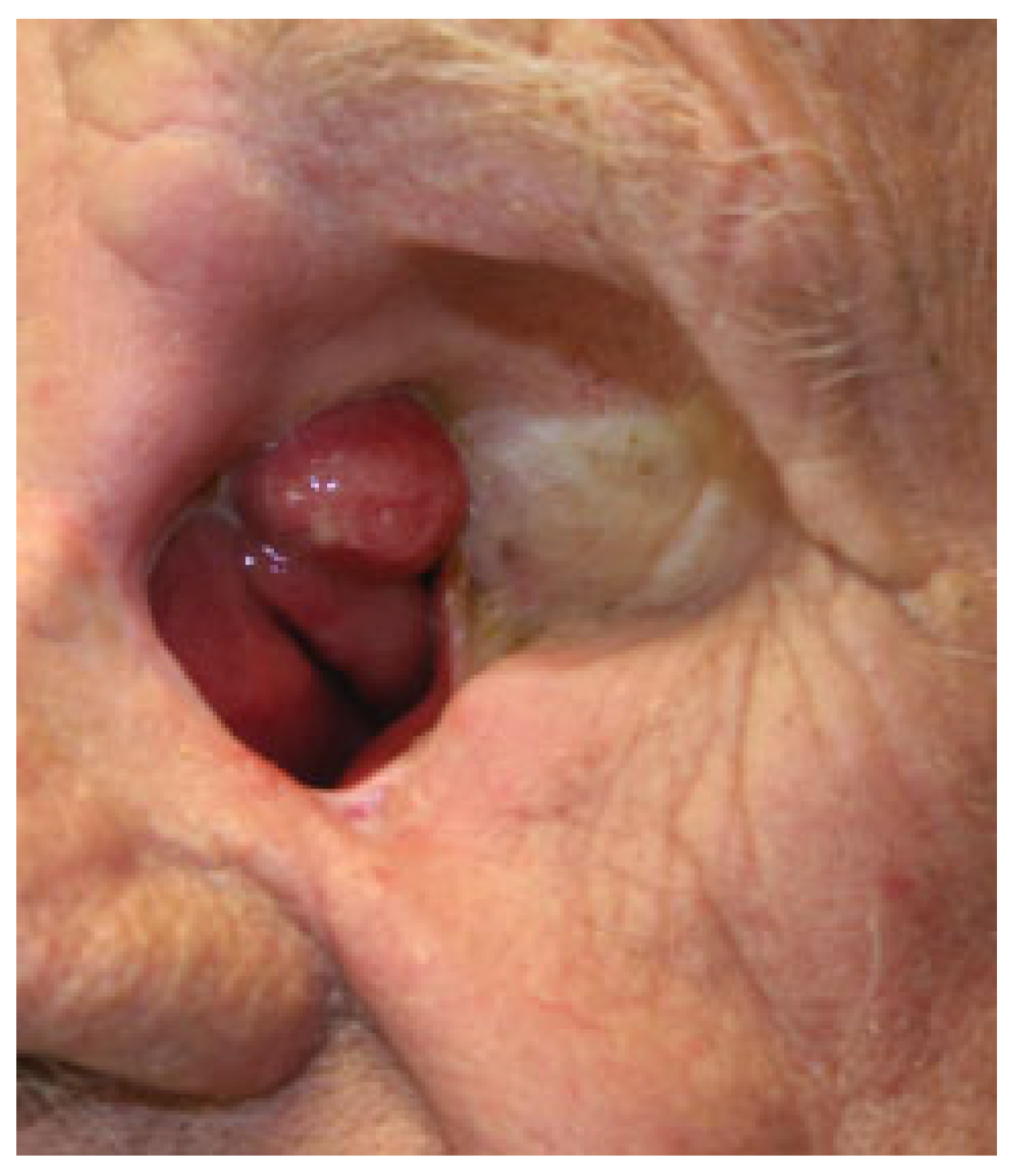
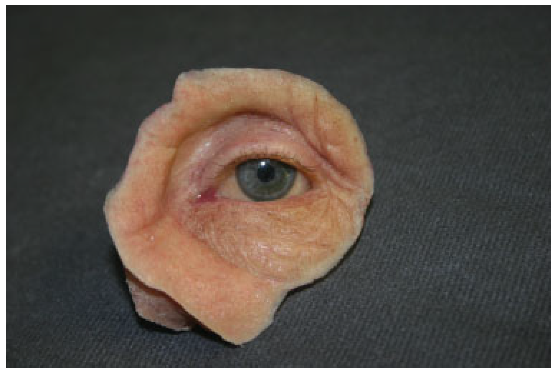
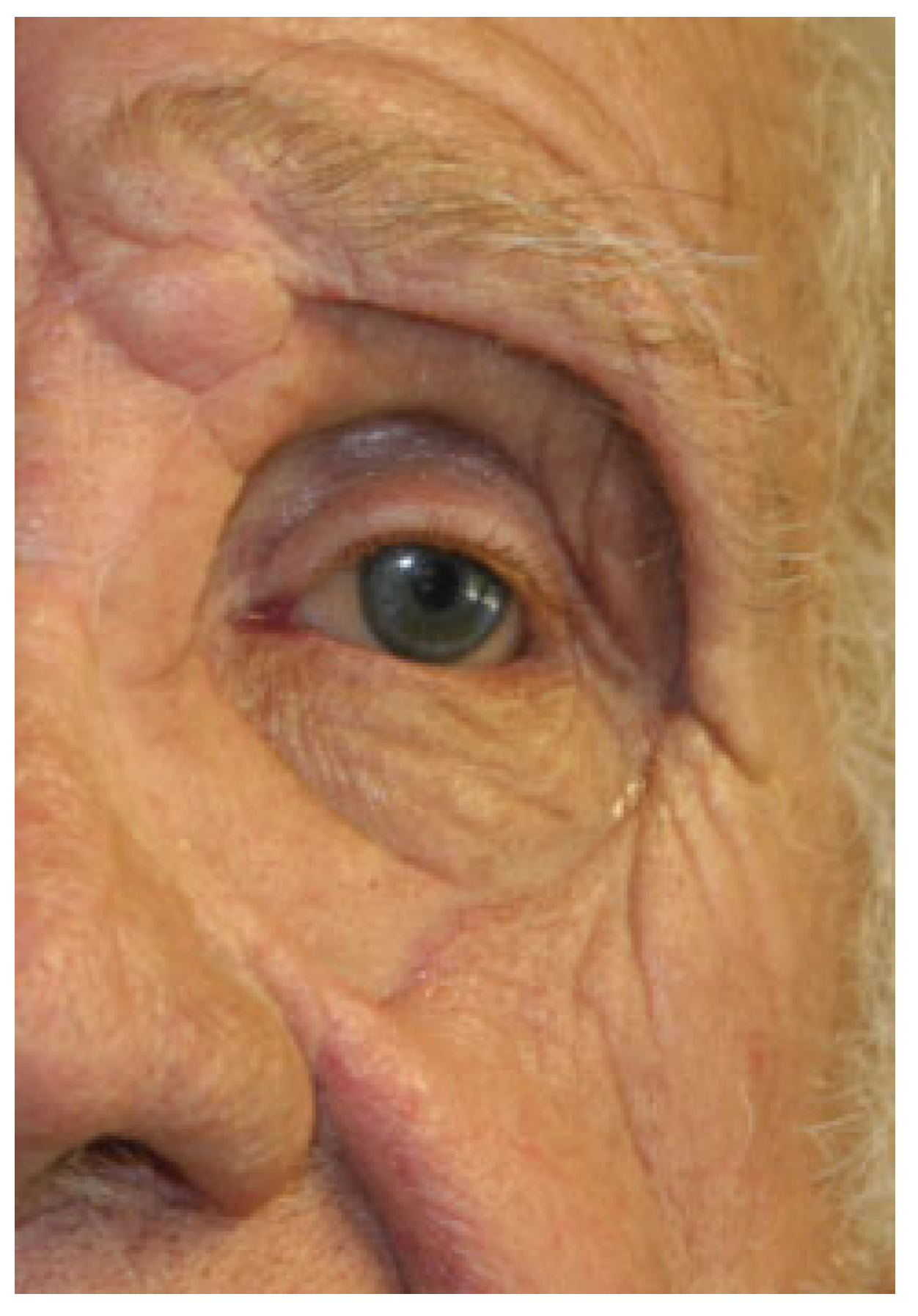
An orbital or ocular prosthesis covers the orbital cavity and underlying implant. Ocular prostheses may be either fit from a stock set of prefabricated eyes matched to the patient or custom made [95]. Custom-made prosthetics is made from an impression of the patient’s eye socket, and hand painted to match the contralateral eye [92]. Retention of an orbital prosthesis is most successful after osseointegration [96]. Adhesive retention should be used in patients with incomplete bone growth or low bone density. Orbital prostheses present a viable reconstructive option after removal of the globe. Figure 8, Figure 9 and Figure 10 show a patient following orbital exoneration successfully restored using an orbital prosthesis.

Conclusions

Head and neck prosthetic rehabilitation is an excellent alternative to surgical reconstruction for patients who have undergone treatment for malignancy, congenital deformities, or trauma. This review presents the current standards of care, limitations, and data behind the materials used for prosthetic head and neck rehabilitation.

References

  1. Gibson, M.K.; Forastiere, A.A. Multidisciplinary approaches in the management of advanced head and neck tumors: State of the art. Curr Opin Oncol 2004, 16, 220–224. [Google Scholar]
  2. Thawley, S.E.; Batsakis, J.G.; Lindberg, R.D.; et al. Comprehensive Management of Head and Neck Tumors; Elsevier: St Louis, MO, USA, 1998; pp. 526–527. [Google Scholar]
  3. Harrison, D.F. Total rhinectomy–a worthwhile operation? J Laryngol Otol 1982, 96, 1113–1123. [Google Scholar] [PubMed]
  4. Bhandari, S. Prosthetic considerations in oral and maxillofacial trauma. J Postgrad Med Educ Res 2014, 48, 87–90. [Google Scholar]
  5. Salinas, T.J. Prosthetic rehabilitation of defects of the head and neck. Semin Plast Surg 2010, 24, 299–308. [Google Scholar] [CrossRef]
  6. Thaller, S.; Bradley, J.P.; Garri, J.I. Craniofacial Surgery; CRC Press: New York, NY, USA, 2007; p. 334. [Google Scholar]
  7. Lemon, J.C.; Chambers, M.S.; Wesley, P.J.; Reece, G.P.; Martin, J.W. Rehabilitation of a midface defect with reconstructive surgery and facial prosthetics: A case report. Int J Oral Maxillofac Implant 1996, 11, 101–105. [Google Scholar]
  8. Parr, G.R.; Goldman, B.M.; Rahn, A.O. Maxillofacial prosthetic principles in the surgical planning for facial defects. J Prosthet Dent 1981, 46, 323–329. [Google Scholar]
  9. Oh, H.K.; Chambers, M.S.; Martin, J.W.; Lim, H.J.; Park, H.J. Osteoradionecrosis of the mandible: Treatment outcomes and factors influencing the progress of osteoradionecrosis. J Oral Maxillofac Surg 2009, 67, 1378–1386. [Google Scholar] [CrossRef] [PubMed]
  10. Wiens, J.P.; Wiens, R.L. Psychological management of the maxillofacial prosthetic patient. In Clinical Maxillofacial Prosthetics; Taylor, T.D., Ed.; Quintessence Publishing: Batavia, IL, USA, 2000; pp. 1–14. [Google Scholar]
  11. Raghoebar, G.M.; van Oort, R.P.; Roodenburg, J.L.; Reintsema, H.; Dikkers, F.G. Fixation of auricular prostheses by osseointegrated implants. J Invest Surg 1994, 7, 283–290. [Google Scholar]
  12. de Bree, R.; Leemans, C.R. Recent advances in surgery for head and neck cancer. Curr Opin Oncol 2010, 22, 186–193. [Google Scholar]
  13. Moreno, M.A.; Skoracki, R.J.; Hanna, E.Y.; Hanasono, M.M. Microvascular free flap reconstruction versus palatal obturation for maxillectomy defects. Head Neck 2010, 32, 860–868. [Google Scholar]
  14. Roumanas, E.D.; Freymiller, E.G.; Chang, T.L.; Aghaloo, T.; Beumer, J., III. Implant-retained prostheses for facial defects: An up to 14-year follow-up report on the survival rates of implants at UCLA. Int J Prosthodont 2002, 15, 325–332. [Google Scholar]
  15. Guttal, S.S.; Patil, N.P.; Shetye, A.D. Prosthetic rehabilitation of a midfacial defect resulting from lethal midline granuloma–a clinical report. J Oral Rehabil 2006, 33, 863–867. [Google Scholar] [CrossRef]
  16. Markt, J.C.; Lemon, J.C. Extraoral maxillofacial prosthetic rehabilitation at the M. D. Anderson Cancer Center: A survey of patient attitudes and opinions. J Prosthet Dent 2001, 85, 608–613. [Google Scholar] [CrossRef]
  17. Eleni, P.N.; Krokida, M.; Polyzois, G.; Gettleman, L.; Bisharat, G.I. Effects of outdoor weathering on facial prosthetic elastomers. Odontology 2011, 99, 68–76. [Google Scholar] [CrossRef] [PubMed]
  18. Visser, A.; Raghoebar, G.M.; van Oort, R.P.; Vissink, A. Fate of implantretained craniofacial prostheses: Life span and aftercare. Int J Oral Maxillofac Implant 2008, 23, 89–98. [Google Scholar]
  19. Feng, Z.; Dong, Y.; Zhao, Y.; et al. Computer-assisted technique for the design and manufacture of realistic facial prostheses. Br J Oral Maxillofac Surg 2010, 48, 105–109. [Google Scholar] [CrossRef] [PubMed]
  20. Verdonck, H.W.; Poukens, J.; Overveld, H.V.; Riediger, D. Computerassisted maxillofacial prosthodontics: A new treatment protocol. Int J Prosthodont 2003, 16, 326–328. [Google Scholar]
  21. Wu, G.; Zhou, B.; Bi, Y.; Zhao, Y. Selective laser sintering technology for customized fabrication of facial prostheses. J Prosthet Dent 2008, 100, 56–60. [Google Scholar] [CrossRef]
  22. Jiao, T.; Zhang, F.; Huang, X.; Wang, C. Design and fabrication of auricular prostheses by CAD/CAM system. Int J Prosthodont 2004, 17, 460–463. [Google Scholar]
  23. Subburaj, K.; Nair, C.; Rajesh, S.; Meshram, S.M.; Ravi, B. Rapid development of auricular prosthesis using CAD and rapid prototyping technologies. Int J Oral Maxillofac Surg 2007, 36, 938–943. [Google Scholar] [CrossRef]
  24. Federspil, P.A. Implant-retained craniofacial prostheses for facial defects. GMS Curr Top Otorhinolaryngol Head Neck Surg 2009, 8, Doc03. [Google Scholar] [PubMed]
  25. Giot, J.P.; Labbé, D.; Soubeyrand, E.; et al. Prosthetic reconstruction of the auricle: Indications, techniques, and results. Semin Plast Surg 2011, 25, 265–272. [Google Scholar]
  26. Montgomery, P.C.; Kiat-Amnuay, S. Survey of currently used materials for fabrication of extraoral maxillofacial prostheses in North America, Europe, Asia, and Australia. J Prosthodont 2010, 19, 482–490. [Google Scholar] [PubMed]
  27. Kiat-amnuay, S.; Jacob, R.F.; Chambers, M.S.; et al. Clinical trial of chlorinated polyethylene for facial prosthetics. Int J Prosthodont 2010, 23, 263–270. [Google Scholar]
  28. Kiat-Amnuay, S.; Gettleman, L.; Goldsmith, L.J. Effect of multi-adhesive layering on retention of extraoral maxillofacial silicone prostheses in vivo. J Prosthet Dent 2004, 92, 294–298. [Google Scholar]
  29. Wolfaardt, J.; Gehl, G.; Farmand, M.; Wilkes, G. Indications and methods of care for aspects of extraoral osseointegration. Int J Oral Maxillofac Surg 2003, 32, 124–131. [Google Scholar]
  30. Granström, G. Craniofacial osseointegration. Oral Dis 2007, 13, 261–269. [Google Scholar] [PubMed]
  31. Alvi, R.; McPhail, J.; Hancock, K. Closed-field titanium magnets for the retention of complex craniofacial prostheses. Br J Plast Surg 2002, 55, 668–670. [Google Scholar]
  32. Sencimen, M.; Bal, H.E.; Demiroğullari, M.; Kocaoglu, M.; Dogan, N. Auricular epithesis retained by an attachment system (2 case reports). Oral Surg Oral Med Oral Pathol Oral Radiol Endod 2008, 105, e28–e34. [Google Scholar]
  33. Dahl, J.E.; Polyzois, G.L. Irritation test of tissue adhesives for facial prostheses. J Prosthet Dent 2000, 84, 453–457. [Google Scholar]
  34. Huband, M. Prosthetic rehabilitation. Dermatol Clin 2011, 29, 325–330. [Google Scholar] [PubMed]
  35. Wondergem, M.; Lieben, G.; Bouman, S.; van den Brekel, M.W.; Lohuis, P.J. Patients’ satisfaction with facial prostheses. Br J Oral Maxillofac Surg 2016, 54, 394–399. [Google Scholar]
  36. Tjellström, A.; Lindström, J.; Hallén, O.; Albrektsson, T.; Brånemark, P.I. Osseointegrated titanium implants in the temporal bone. A clinical study on bone-anchored hearing aids. Am J Otol 1981, 2, 304–310. [Google Scholar] [PubMed]
  37. Albrektsson, T. Dental and maxillofacial implantology. In Principles of Osseointegration; Hobkirk, J.A., Watson, K., Eds.; Mosby-Wolfe: London, UK, 1995; pp. 9–19. [Google Scholar]
  38. Albrektsson, T.; Johansson, C. Osteoinduction, osteoconduction and osseointegration. Eur Spine J 2001, 10 (Suppl. S2), S96–S101. [Google Scholar]
  39. Frost, H.M. The biology of fracture healing. An overview for clinicians. Part I. Clin Orthop Relat Res 1989, 248, 283–293. [Google Scholar]
  40. Ihde, S.; Kopp, S.; Gundlach, K.; Konstantinović, V.S. Effects of radiation therapy on craniofacial and dental implants: A review of the literature. Oral Surg Oral Med Oral Pathol Oral Radiol Endod 2009, 107, 56–65. [Google Scholar]
  41. Voigt, A.; Christ, S.; Klein, M. Experimental analysis of retention forces of different magnetic devices for bone-anchored auricular facial prostheses. Int J Oral Maxillofac Surg 2008, 37, 664–668. [Google Scholar]
  42. de Sousa, A.A.; Mattos, B.S. Magnetic retention and bar-clip attachment for implant-retained auricular prostheses: A comparative analysis. Int J Prosthodont 2008, 21, 233–236. [Google Scholar]
  43. Ariani, N.; Visser, A.; van Oort, R.P.; et al. Current state of craniofacial prosthetic rehabilitation. Int J Prosthodont 2013, 26, 57–67. [Google Scholar]
  44. Art as Applied to Medicine. The Facial Prosthetics Clinic of Johns Hopkins University Web Site. Available online: http://hopkinsmedi-cine.org/medart/Prosthetics.htm (accessed on 2 December 2016).
  45. Mardani, M.A.; Aminian, G.; Tabatabaian, F.; Arazpour, M.; Hutchins, S.W.; Head, J.S. A novel technique for fabricating an ear prosthesis in a patient with congenital ear deformity. Prosthet Orthot Int 2013, 37, 340–343. [Google Scholar]
  46. Cox, A.; Sabbagh, W.; Gault, D. Costal cartilage or conchal cartilage for aesthetic and structural reconstruction of lower pole ear defects. Aesthet Surg J 2012, 32, 271–274. [Google Scholar] [CrossRef] [PubMed]
  47. Firmin, F. State-of-the-art autogenous ear reconstruction in cases of microtia. Adv Otorhinolaryngol 2010, 68, 25–52. [Google Scholar]
  48. Firmin, F.; Sanger, C.; O’Toole, G. Ear reconstruction following severe complications of otoplasty. J Plast Reconstr Aesthet Surg 2008, 61 (Suppl. S1), S13–S20. [Google Scholar] [CrossRef]
  49. Mevio, E.; Facca, L.; Schettini, S.; Mullace, M. Bone-anchored titanium implants in patients with auricular defects: Three years and 27 patients’ experience. Int J Otolaryngol 2016, 2016, 9872048. [Google Scholar]
  50. Arora, V.; Sahoo, N.K.; Gopi, A.; Saini, D.K. Implant-retained auricular prostheses: A clinical challenge. Int J Oral Maxillofac Surg 2016, 45, 631–635. [Google Scholar] [CrossRef] [PubMed]
  51. Taylor, T.D. Clinical Maxillofacial Prosthetics; Quintessence Publishing Co., Inc.: Chicago, IL, USA, 2000; pp. 233–276. [Google Scholar]
  52. Zhao, Y.; Wang, Y.; Zhuang, H.; et al. Clinical evaluation of three total ear reconstruction methods. J Plast Reconstr Aesthet Surg 2009, 62, 1550–1554. [Google Scholar] [PubMed]
  53. Lemon, J.C.; Kiat-amnuay, S.; Gettleman, L.; Martin, J.W.; Chambers, M.S. Facial prosthetic rehabilitation: Preprosthetic surgical techniques and biomaterials. Curr Opin Otolaryngol Head Neck Surg 2005, 13, 255–262. [Google Scholar]
  54. Eriksson, E.; Brånemark, P.I. Osseointegration from the perspective of the plastic surgeon. Plast Reconstr Surg 1994, 93, 626–637. [Google Scholar] [CrossRef]
  55. Black, B. Use of titanium in repair of external auditory canal defects. Otol Neurotol 2009, 30, 930–935. [Google Scholar] [CrossRef]
  56. Walsh, W.E.; Dougherty, B.; Reisberg, D.J.; et al. The importance of auricular prostheses for speech recognition. Arch Facial Plast Surg 2008, 10, 321–328. [Google Scholar]
  57. Wright, R.F.; Zemnick, C.; Wazen, J.J.; Asher, E. Osseointegrated implants and auricular defects: A case series study. J Prosthodont 2008, 17, 468–475. [Google Scholar] [PubMed]
  58. Subaşı, M.G.; Alnıaçık, G.; Kalaycı, A.; Akman, S.; Durmuş, E. Prosthetic rehabilitation of partial ear defect: 2 case reports. J Indian Prosthodont Soc 2014, 14 (Suppl. S1), 196–201. [Google Scholar]
  59. Badie-Modiri, B.; Kaplanski, P. Extra-oral implants: Principal areas of implantation [in French]. Rev Stomatol Chir Maxillofac 2001, 102, 229–233. [Google Scholar] [PubMed]
  60. Correa, B.J.; Weathers, W.M.; Wolfswinkel, E.M.; Thornton, J.F. The forehead flap: The gold standard of nasal soft tissue reconstruction. Semin Plast Surg 2013, 27, 96–103. [Google Scholar]
  61. Menick, F.J. A 10-year experience in nasal reconstruction with the three-stage forehead flap. Plast Reconstr Surg 2002, 109, 1839—1855, Discussion 1856—1861. [Google Scholar]
  62. Ahmed, B.; Butt, A.M.; Hussain, M.; Amin, M.; Yazdanie, N. Rehabilitation of nose using silicone based maxillofacial prosthesis. J Coll Physicians Surg Pak 2010, 20, 65–67. [Google Scholar] [PubMed]
  63. Thornton, J.F.; Griffin, J.R.; Constantine, F.C. Nasal reconstruction: An overview and nuances. Semin Plast Surg 2008, 22, 257–268. [Google Scholar]
  64. Bourget, A.; Chang, J.T.; Wu, D.B.; Chang, C.J.; Wei, F.C. Free flap reconstruction in the head and neck region following radiotherapy: A cohort study identifying negative outcome predictors. Plast Reconstr Surg 2011, 127, 1901–1908. [Google Scholar]
  65. Thankappan, K. Microvascular free tissue transfer after prior radiotherapy in head and neck reconstruction—A review. Surg Oncol 2010, 19, 227–234. [Google Scholar]
  66. Klug, C.; Berzaczy, D.; Reinbacher, H.; et al. Influence of previous radiotherapy on free tissue transfer in the head and neck region: Evaluation of 455 cases. Laryngoscope 2006, 116, 1162–1167. [Google Scholar]
  67. Ciocca, L.; Maremonti, P.; Bianchi, B.; Scotti, R. Maxillofacial rehabilitation after rhinectomy using two different treatment options: Clinical reports. J Oral Rehabil 2007, 34, 311–315. [Google Scholar] [PubMed]
  68. Seçilmiş, A.; Oztürk, A.N. Nasal prosthesis rehabilitation after partialrhinectomy: A clinical report. Eur J Dent 2007, 1, 115–118. [Google Scholar]
  69. Dimitroulis, G. Nasal implants following nasectomy. Int J Oral Maxillofac Surg 2007, 36, 447–449. [Google Scholar]
  70. Scott, N.; Kittur, M.A.; Evans, P.L.; Dovgalski, L.; Hodder, S.C. The use of zygomatic implants for the retention of nasal prosthesis following rhinectomy: The Morriston experience. Int J Oral Maxillofac Surg 2016, 45, 1044–1048. [Google Scholar]
  71. Brånemark, P.I.; Adell, R.; Albrektsson, T.; Lekholm, U.; Lindström, J.; Rockler, B. An experimental and clinical study of osseointegrated implants penetrating the nasal cavity and maxillary sinus. J Oral Maxillofac Surg 1984, 42, 497–505. [Google Scholar] [PubMed]
  72. Chrcanovic, B.R.; Albrektsson, T.; Wennerberg, A. Survival and complications of Zygomatic Implants: An updated systematic review. J Oral Maxillofac Surg 2016, 74, 1949–1964. [Google Scholar] [PubMed]
  73. Futran, N.D.; Mendez, E. Developments in reconstruction of midface and maxilla. Lancet Oncol 2006, 7, 249–258. [Google Scholar]
  74. Har-El, G.; Nathan, C.O.; Day, T.A.; Nguyen, S.A. Rehabilitation of patients with head and neck cancer: Prosthetic rehabilitation: Intraoral and extraoral prostheses. In Head and Neck Surgery; Thieme Medical Publishers: New York, NY, USA, 2013. [Google Scholar]
  75. Okay, D.J.; Genden, E.; Buchbinder, D.; Urken, M. Prosthodontic guidelines for surgical reconstruction of the maxilla: A classification system of defects. J Prosthet Dent 2001, 86, 352–363. [Google Scholar]
  76. Iyer, S.; Thankappan, K. Maxillary reconstruction: Current concepts and controversies. Indian J Plast Surg 2014, 47, 8–19. [Google Scholar]
  77. Nekora-Azak, A.; Evlioglu, G.; Ozdemir-Karataş, M.; Keskin, H. Use of biofunctional prosthetic system following partial maxillary resection: A clinical report. J Oral Rehabil 2005, 32, 693–695. [Google Scholar]
  78. Etienne, O.M.; Taddei, C.A. Use of bar-clip attachments to enhance the retention of a maxillofacial prosthetic obturator: A clinical report. J Oral Rehabil 2004, 31, 618–621. [Google Scholar] [CrossRef]
  79. Shipman, B. Evaluation of occlusal force in patients with obturator defects. J Prosthet Dent 1987, 57, 81–84. [Google Scholar] [CrossRef]
  80. Watson, R.M.; Gray, B.J. Assessing effective obturation. J Prosthet Dent 1985, 54, 88–93. [Google Scholar] [CrossRef] [PubMed]
  81. Parr, G.R.; Tharp, G.E.; Rahn, A.O. Prosthodontic principles in the framework design of maxillary obturator prostheses. 1989. J Prosthet Dent 2005, 93, 405–411. [Google Scholar] [CrossRef]
  82. Brown, J.S.; Rogers, S.N.; McNally, D.N.; Boyle, M. A modified classification for the maxillectomy defect. Head Neck 2000, 22, 17–26. [Google Scholar] [CrossRef]
  83. Murat, S.; Gurbuz, A.; Isayev, A.; Dokmez, B.; Cetin, U. Enhanced retention of a maxillofacial prosthetic obturator using precision attachments: Two case reports. Eur J Dent 2012, 6, 212–217. [Google Scholar] [CrossRef] [PubMed]
  84. Aramany, M.A. Basic principles of obturator design for partially edentulous patients. Part II: Design principles. J Prosthet Dent 2001, 86, 562–568. [Google Scholar] [PubMed]
  85. Cordeiro, P.G.; Santamaria, E. A classification system and algorithm for reconstruction of maxillectomy and midfacial defects. Plast Reconstr Surg 2000, 105, 2331—2346, Discussion 2347—2348. [Google Scholar] [CrossRef]
  86. McBain, H.B.; Ezra, D.G.; Rose, G.E.; Newman, S.P.; Appearance Research Collaboration (ARC). The psychosocial impact of living with an ocular prosthesis. Orbit 2014, 33, 39–44. [Google Scholar] [CrossRef]
  87. Baino, F.; Potestio, I. Orbital implants: State-of-the-art review with emphasis on biomaterials and recent advances. Mater Sci Eng C 2016, 69, 1410–1428. [Google Scholar] [CrossRef]
  88. Mishra, S.K.; Ramesh, C. Reproduction of custom-made eye prosthesis manoeuvre: A case report. J Dent Oral Hyg 2009, 1, 59–63. [Google Scholar]
  89. Pruthi, G.; Jain, V.; Rajendiran, S.; Jha, R. Prosthetic rehabilitation after orbital exenteration: A case series. Indian J Ophthalmol 2014, 62, 629–632. [Google Scholar] [PubMed]
  90. Parr, G.R.; Goldman, B.M.; Rahn, A.O. Surgical considerations in the prosthetic treatment of ocular and orbital defects. J Prosthet Dent 1983, 49, 379–385. [Google Scholar] [CrossRef] [PubMed]
  91. Begum, Z.; Kola, M.Z.; Joshi, P. Analysis of the properties of commercially available silicone elastomers for maxillofacial prostheses. J Dent 2011, 31, 67–74. [Google Scholar]
  92. Cafiero-Chin, M.; Marques, C.; Danz, H. Ocular prosthesis: Indications to management. Canad J Optometr 2015, 77, 24–33. [Google Scholar] [CrossRef]
  93. Sami, D.; Young, S.; Petersen, R. Perspective on orbital enucleation implants. Surv Ophthalmol 2007, 52, 244–265. [Google Scholar] [CrossRef]
  94. Hughes, M.O. A pictorial anatomy of the human eye/anophthalmic socket: A review for ocularists. J Ophthal Prosthet 2007, 12, 51–63. [Google Scholar]
  95. Gunaseelaraj, R.; Karthikeyan, S.; Kumar, M.N.; Balamurugan, T.; Jagadeeshwaran, A.R. Custom-made ocular prosthesis. J Pharm Bioallied Sci 2012, 4 (Suppl. S2), S177–S179. [Google Scholar]
  96. Toljanic, J.A.; Eckert, S.E.; Roumanas, E.; et al. Osseointegrated craniofacial implants in the rehabilitation of orbital defects: An update of a retrospective experience in the United States. J Prosthet Dent 2005, 94, 177–182. [Google Scholar]
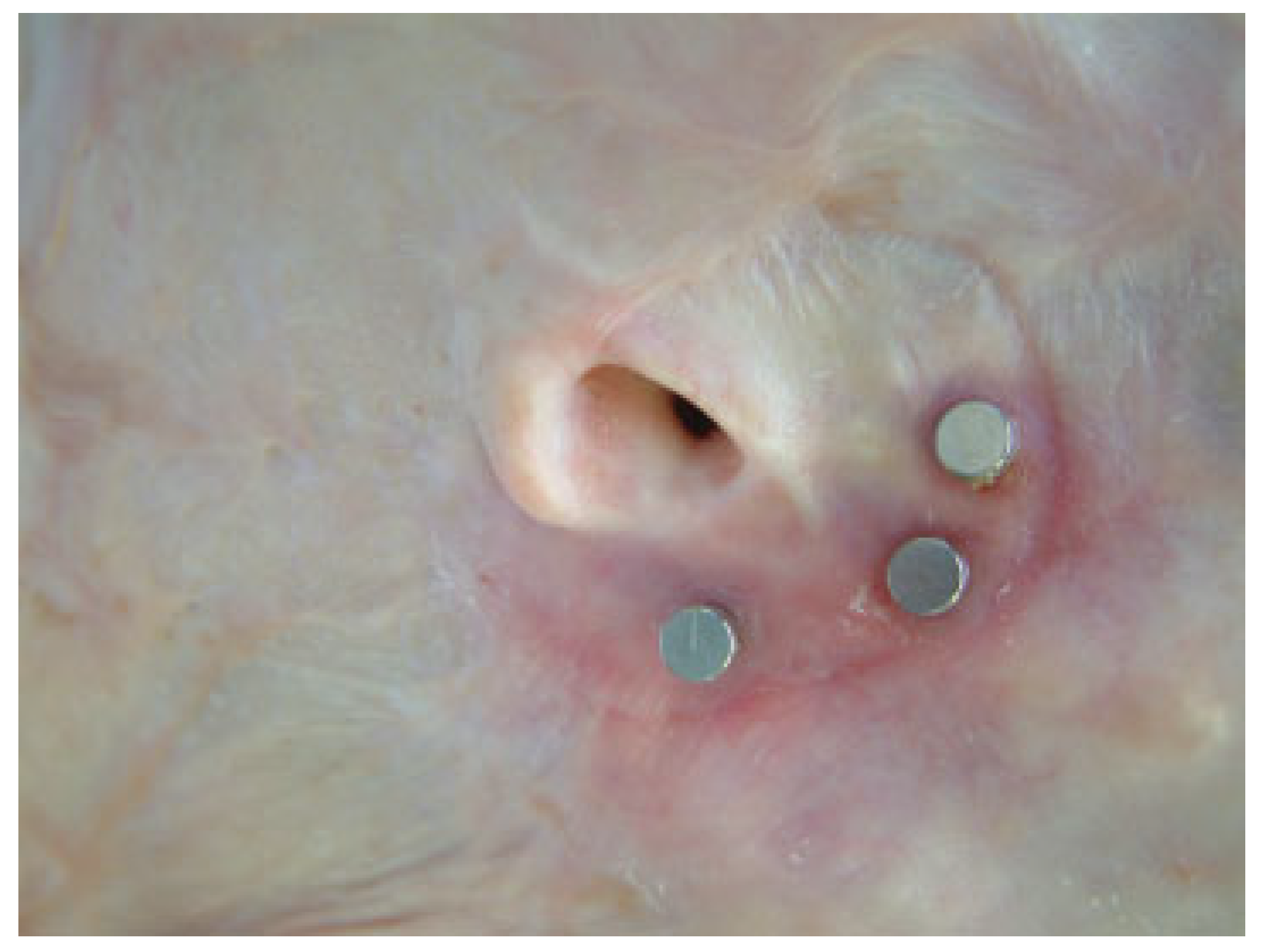
Figure 1. Patient following auriculectomy with placement of bone anchored posts to hold prosthesis.
Cmtr 11 00002 g001
Figure 2. Anterior view of auricular prosthesis.
Cmtr 11 00002 g002
Figure 3. Posterior view of auricular prosthesis showing mounting holes to fit onto osseous posts.
Cmtr 11 00002 g003
Figure 4. Previously shown patient in Figure 1 with placement of auricular prosthesis.
Cmtr 11 00002 g004
Figure 5. Patient with nasal defect following rhinectomy (eyes have been removed from image).
Cmtr 11 00002 g005
Figure 6. Nasal prosthesis to be used for reconstruction.
Cmtr 11 00002 g006
Figure 7. Previously shown patient in Figure 5 with placement of nasal prosthesis (eyes have been removed from image).
Cmtr 11 00002 g007
Figure 8. Patient with orbital defect following exoneration.
Cmtr 11 00002 g008
Figure 9. Orbital prosthesis custom designed for patient in Figure 8.
Cmtr 11 00002 g009
Figure 10. Patient with orbital prosthesis in place.
Cmtr 11 00002 g010