Next Article in Journal
A Hemangioma of the Zygomatic Bone: Management Ensuring Good Reconstructive and Aesthetic Results
Previous Article in Journal
New Management in Bilateral Masseter Muscle Hypertrophy
 
 
Craniomaxillofacial Trauma & Reconstruction is published by MDPI from Volume 18 Issue 1 (2025). Previous articles were published by another publisher in Open Access under a CC-BY (or CC-BY-NC-ND) licence, and they are hosted by MDPI on mdpi.com as a courtesy and upon agreement with Sage.
Technical Note

The Pull-Through Technique: A Viable Option for Preserving the Inferior Alveolar Nerve During Surgical Resection
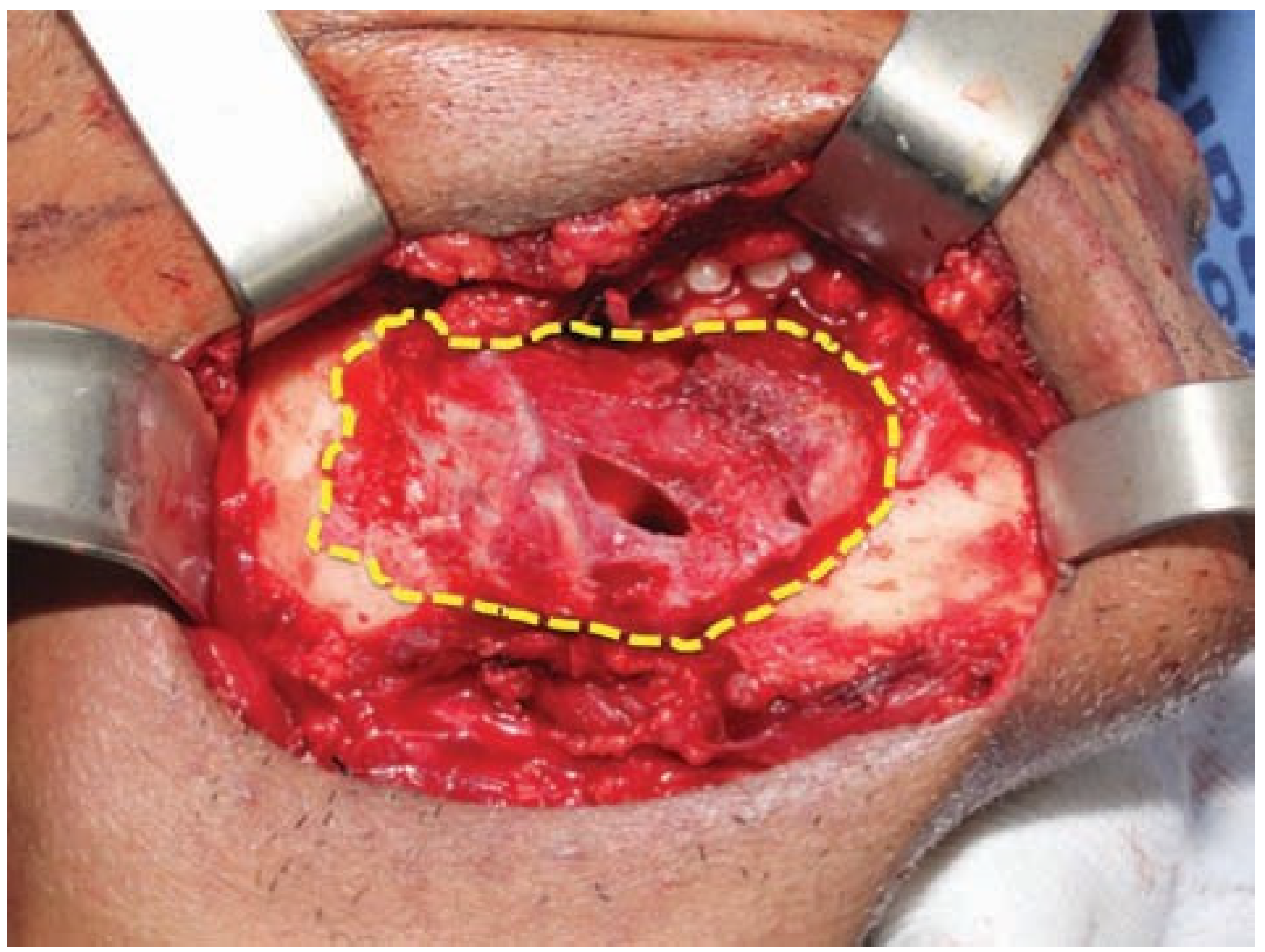
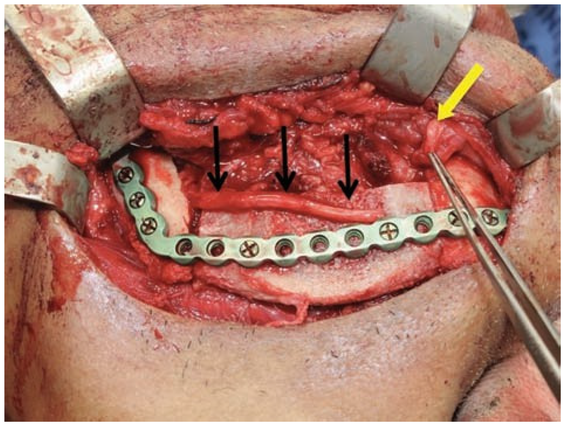
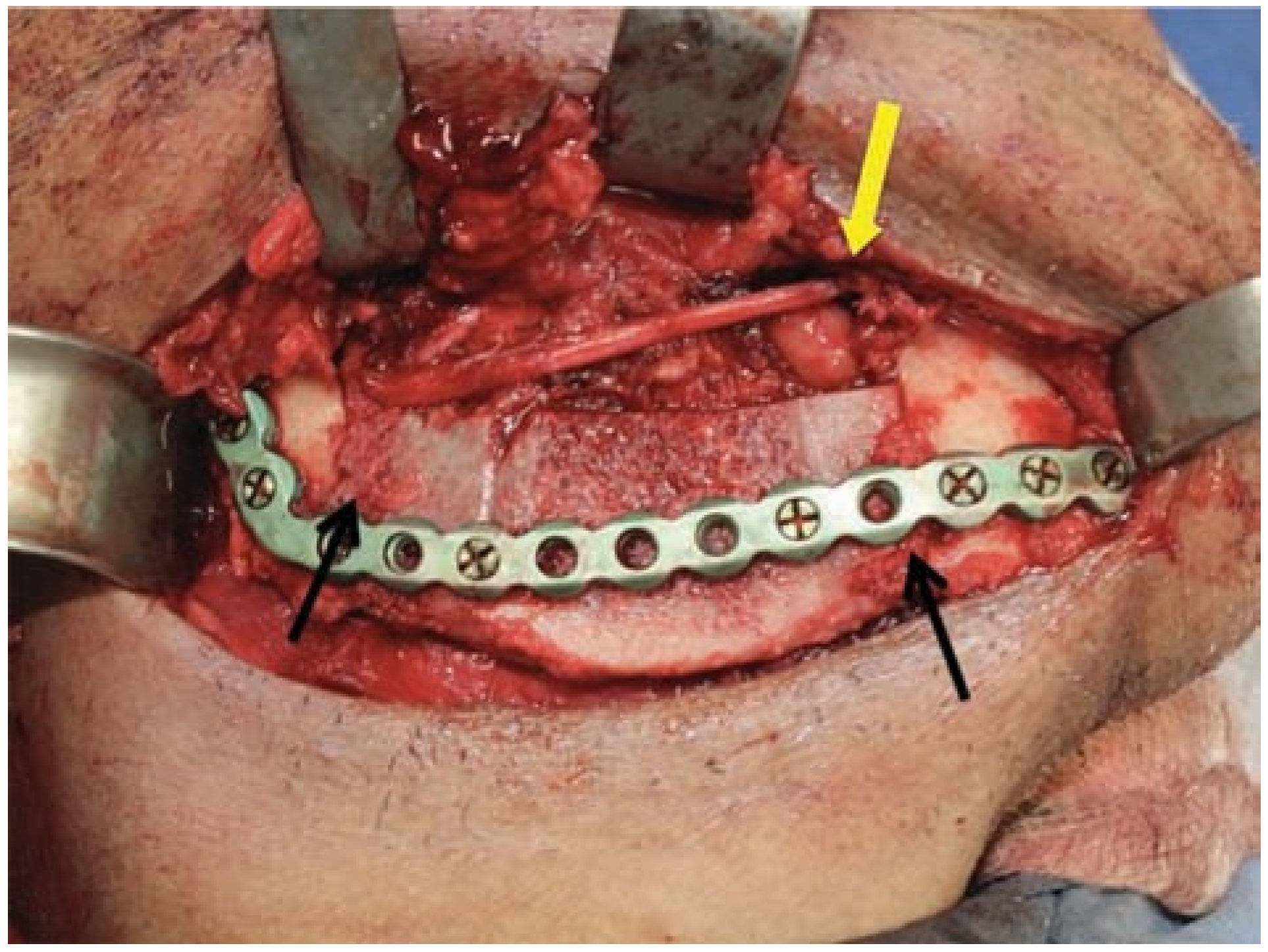
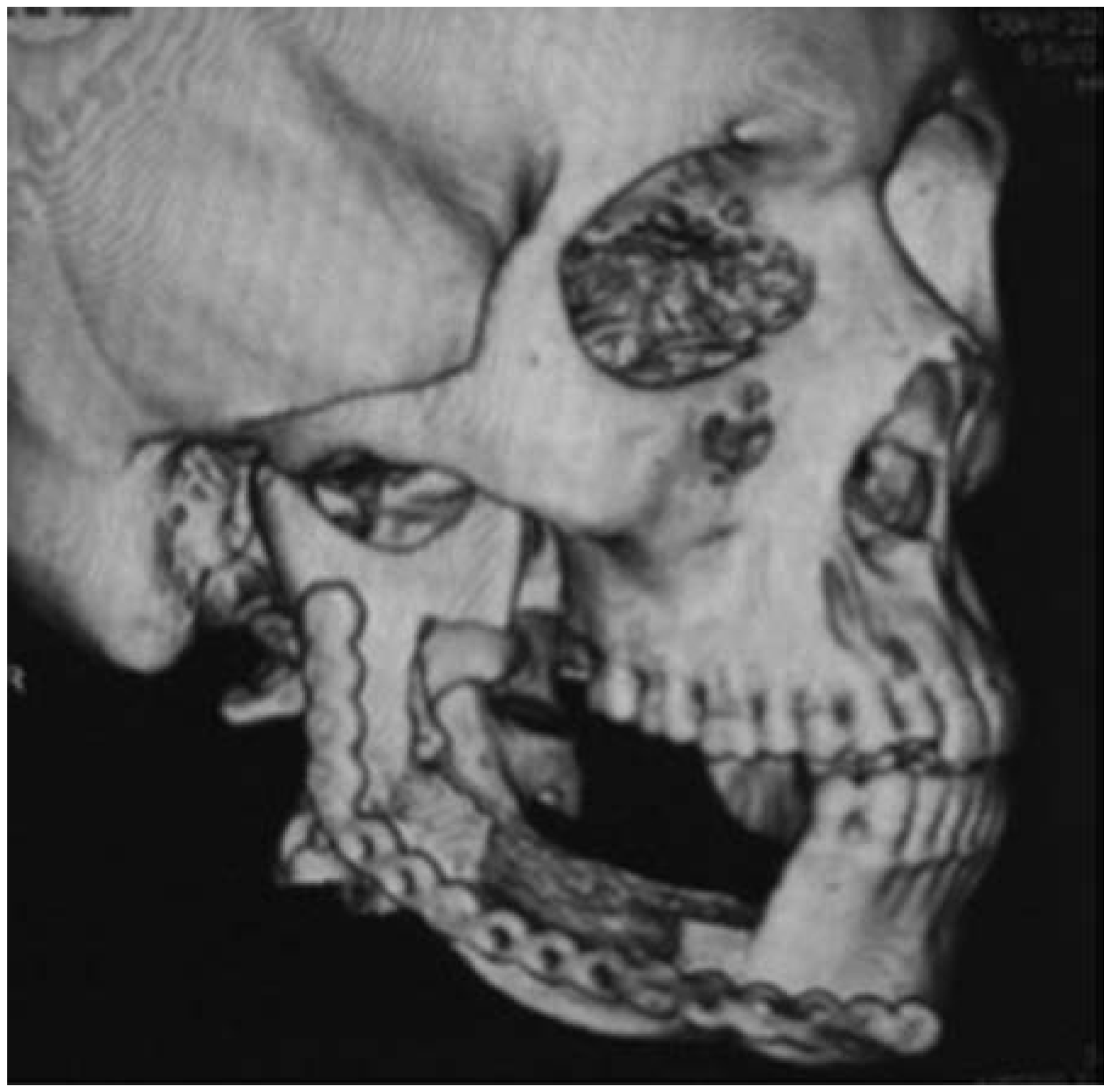
by
Gustavo Boehmer Leite
1,*,
Suelen Cristina Sartoretto
2,
Fernando César Amazonas Lima
3,
Mônica Calasans-Maia
4 and
Rafael Seabra Louro
4
1
Fluminense Federal University, Rio de Janeiro, Brazil
2
Graduate Program in Dentistry, Fluminense Federal University, Rio de Janeiro, Brazil
3
Oral and Maxillofacial Surgery Department of Federal Hospital of Employees of State, Rio de Janeiro, Brazil
4
Department of Oral Surgery, Dentistry School, Fluminense Federal University, Rio de Janeiro, Brazil
*
Author to whom correspondence should be addressed.
Craniomaxillofac. Trauma Reconstr. 2017, 10(4), 329-331; https://doi.org/10.1055/s-0036-1593893
Submission received: 13 July 2016 / Revised: 30 July 2016 / Accepted: 15 August 2016 / Published: 11 November 2016

Abstract

:
The aim of this study was to present a new surgical technique used to remove benign mandibular tumors with minimal damage to the inferior alveolar nerve. The pull-through technique was shown using an ameloblastoma surgical resection as an example. This technique consisted in the reconstruction of the lower jaw associating the resection of the lesion with nerve repair at the same surgical time. The resection was performed using the pull-through technique and the inferior alveolar nerve was preserved. After 6 months, the patient presented a recovery of approximately 80% of sensory function. The surgical technique presented should be considered an important method by which to produce a higher functional outcome to remove benign mandibular tumors with minimal damage to the inferior alveolar nerve and allows the maintenance of quality of life for the patient, as the consequences of this type of surgery are minimized.

Segmental surgical resection is a surgical procedure frequently indicated for the treatment of benign tumors of the lower jaw [1]. One of the most important complications stemming from this technique is permanent paresthesia of the inferior alveolar nerve. This complication can be avoided by preserving the inferior alveolar nerve in resections of benign mandibular pathologies. This technical note aims to present a surgical technique called the pull-through technique to preserve the inferior alveolar nerve during surgical mandibular resection procedures.
The pull-through technique is indicated in cases wherein the nerve cannot be directly dissected due to the extent of the tumor. This technique is not indicated for approaches to lesions with nerve infiltration or malignant character, which can affect neural tissue.
The patient presented a multicystic ameloblastoma, a lesion with locally aggressive behavior that did not infiltrate the neural tissue [1]. Surgical submandibular access was used to expose the tumor site (Figure 1), and the inferior alveolar and mental nerves were located. The nerve was cut with a no. 15 scalpel at the mental foramen region. After demarcating the margins of the tumor resection, the anterior portion of the tumor margin was osteotomized anterior to the mental foramen region. The posterior margin of the lesion was also osteotomized, but the inferior alveolar nerve was preserved within the resected area. The osteotomy technique was performed with a piezoelectric device to minimize injury to the nerve. The bone segment containing the lesion was pulled and “slid” forward, separating it from the nerve. The distal segment of the inferior alveolar nerve stayed intact and was uninjured. Thus, the resection was performed, and the nerve was maintained in position. The remaining bone was fixed with a 2.4 reconstruction plate (Figure 2), and the bone gap was grafted with autogenous iliac crest bone. The two extremities of the nerve were approximated, and to repair the mesial segment of the nerve, a tubing technique with a polytetrafluoroethylene (PTFE) membrane was used (GoreTex, Flagstaff, AZ) [2], thereby reattaching the nerve where it was first cut at the mental foramen (Figure 3)
In the immediate postoperative period, the patient reported paresthesia throughout the region innervated by the inferior alveolar nerve. At 6 months postoperation, the graft was within the normal limits (Figure 4).
To evaluate the recovery of sensitivity in the inferior alveolar region, the Semmes-Weinstein monofilament test was used as reported previously [3]. This test consists of pressing a monofilament on the skin and use the patient response to each spot to elaborate a score to evaluate the recovery. Three months after the surgical procedure, the patient presented an improvement of 40% local sensitivity. After 6 months, the patient presented a recovery of approximately 80% of sensory function.
Bone reconstruction that associates the resection of the lesion with nerve repair at the same surgical time allows the maintenance of quality of life for the patient because the consequences of this type of surgery are minimized. Thus, this surgical technique should be considered an important method by which to produce a higher functional outcome. It is also important to point out that when treating malignant lesions, this technique should not be used because the inferior alveolar nerve may be infiltrated by malignant cells.

Institutional Review Board Statement

This study was approved by the Ethics Committee of the Servidores do Estado Hospital, Rio de Janeiro, Brazil, and has been registered as # 32966014.9.0000.5252.

Conflicts of Interest

The authors have no conflict of interests.

References

  1. Hammarfjord, O.; Roslund, J.; Abrahamsson, P.; et al. Surgical treatment of recurring ameloblastoma, are there options? Br J Oral Maxillofac Surg 2013, 51, 762–766. [Google Scholar]
  2. Stanec, S.; Stanec, Z. Reconstruction of upper-extremity peripheral-nerve injuries with ePTFE conduits. J Reconstr Microsurg 1998, 14, 227–232. [Google Scholar] [CrossRef]
  3. Poort, L.J.; van Neck, J.W.; van der Wal, K.G. Sensory testing of inferior alveolar nerve injuries: A review of methods used in prospective studies. J Oral Maxillofac Surg 2009, 67, 292–300. [Google Scholar] [CrossRef] [PubMed]
Figure 1. Exposure of the tumor area. Intraoperative view of multicystic ameloblastoma area (border) exposed by submandibular access.
Cmtr 10 00053 g001
Figure 2. Fixation of graft. Using a 2.4 plate, the bone gap was grafted with autogenous iliac crest bone. Black arrows indicate the inferior alveolar nerve preserved within the resected area. The yellow arrow indicates the anterior extremity of the nerve.
Cmtr 10 00053 g002
Figure 3. Nerve repair. The two extremities of the nerve were approximated (yellow arrow). Observe the particulate bone following the gap between the mandibular bone and the graft (black arrows).
Cmtr 10 00053 g003
Figure 4. Aspect of region after 3 months. Postoperative 3D tomographic image showing the region of surgical procedure.
Cmtr 10 00053 g004

