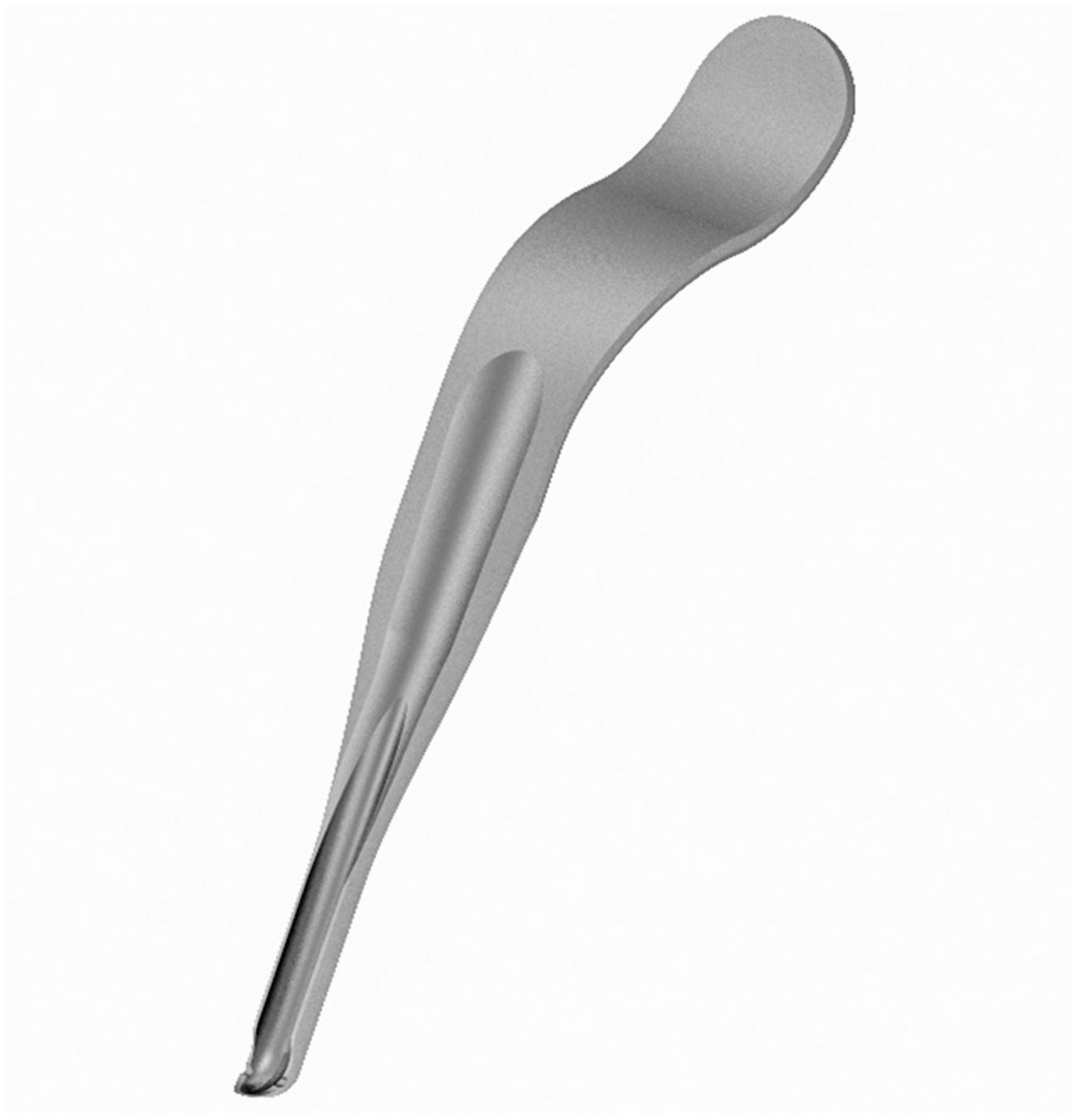
Next Article in Journal
In-Office Guided Implant Placement for Prosthetically Driven Implant Surgery
Previous Article in Journal
Orbital Compartment Syndrome despite Significant Traumatic Expansion of the Orbital Cavity
 
 
Craniomaxillofacial Trauma & Reconstruction is published by MDPI from Volume 18 Issue 1 (2025). Previous articles were published by another publisher in Open Access under a CC-BY (or CC-BY-NC-ND) licence, and they are hosted by MDPI on mdpi.com as a courtesy and upon agreement with Sage.
Technical Note

Narrow-Bladed “Endo” Sagittal Split Osteotomy Retractor

by
Maurice Yves Mommaerts
1,2
1
European Face Centre, Universitair Ziekenhuis Brussel, Brussel, Belgium
2
Orthoface Clinic, Oost-Vlaanderen, Belgium
Craniomaxillofac. Trauma Reconstr. 2017, 10(3), 244-245; https://doi.org/10.1055/s-0036-1584889
Submission received: 21 February 2016 / Revised: 31 March 2016 / Accepted: 3 April 2016 / Published: 30 June 2016

Abstract

:
A modification of the Obwegeser sagittal split retractor is presented. It is slender while still protecting the soft tissues and is particularly suitable for endoscopically assisted surgery.

The narrow- and wide-channeled retractors designed by Hugo L. Obwegeser used during his sagittal split osteotomy [1] have stood the test of half a century. These retractors have been designed to expose the surgical field and to protect the facial artery and mandibular branch of the facial nerve [2]. Modifications, apart from manufacturers’ deviations from the original, have rarely been described. The original Obwegeser retractors are distributed by KLS-Martin (Jacksonville, FL) and Medicon (Tuttlingen, Germany).
When coming across the article of Matsushita in the February 2015 issue of this journal [3], in which the author describes a wider spatula-like modification, I considered it interesting to publish on a narrower gutter-shaped modification [4,5]. This one was designed for the endoscopic approach.
However, many colleagues and myself find the instrument useful for “open-sky” sagittal split ramus osteotomies because it has a favorable angle of attack and a sharp tip that allows for easy subperiosteal dissection (both buccally and lingually; Figure 1 and Figure 2). It snugly fits the lower and posterior border (Figure 3), and it protects the soft tissues not by being wider, but by its “guarding” relationship when placed against the bony surface (Figure 3 and Figure 4). Since its design nearly 10 years ago, I have performed approximately 800 bilateral and unilateral sagittal split osteotomies with it and find it highly effective. Test models were made by DePuy Synthes (Solothurn, Switzerland). The instrument is available at Surgi-Tec NV, Sint-Denijs-Westrem, Belgium (www.surgi-tec.com).

References

  1. Trauner, R.; Obwegeser, H. The surgical correction of mandibular prognathism and retrognathia with consideration of genioplasty. I. Surgical procedures to correct mandibular prognathism and reshaping of the chin. Oral Surg Oral Med Oral Pathol 1957, 10, 677–689. [Google Scholar] [CrossRef] [PubMed]
  2. Obwegeser, H.L. Mandibular Growth Anomalies; Springer: Berlin/Heidelberg, Germany, 2011; p. 382. [Google Scholar]
  3. Matsushita, K. Wide-bladed mandibular channel retractor efficiently secures surgical manoeuvres during ramus osteotomy. Br J Oral Maxillofac Surg 2015, 53, 210–211. [Google Scholar] [CrossRef] [PubMed]
  4. Mommaerts, M.Y. Endoscopically assisted sagittal split osteotomy for mandibular lengthening: Technical note and initial experience. J Craniomaxillofac Surg 2010, 38, 108–112. [Google Scholar] [CrossRef] [PubMed]
  5. Mommaerts, M.Y. The surgical art of facial makeover. Planning and operative techniques. Chapter “Endoscopic bilateral sagittal split osteotomy (endo-BSSO)”. Sint Martens Latem 2013, 1, 189–196. [Google Scholar]
Figure 1. 3D graphical representation of the “endo BSSO retractor.” The lower shaft is 7 mm wide.
Cmtr 10 00038 g001
Figure 2. Angle of attack to the bony surface and the sharp tip.
Cmtr 10 00038 g002
Figure 3. The instrument fits well around the mandibular border and prevents soft-tissue prolapse into the danger area.
Cmtr 10 00038 g003
Figure 4. The “endo BSSO retractor” providing efficient access and effective soft-tissue protection from the Lindemann bur (endoscopic view in the lingual tunnel—arrow is pointing at the fat pad surrounding the mandibular foramen).
Cmtr 10 00038 g004

