Next Article in Journal
Penetrating Craniomaxillofacial Injury Caused by a Pneumatic Nail Gun
Previous Article in Journal
Treatment of a Large Traumatic Encephalocele with Titanium Mesh
 
 
Craniomaxillofacial Trauma & Reconstruction is published by MDPI from Volume 18 Issue 1 (2025). Previous articles were published by another publisher in Open Access under a CC-BY (or CC-BY-NC-ND) licence, and they are hosted by MDPI on mdpi.com as a courtesy and upon agreement with Sage.
Case Report

Immediate Surgical Management of Traumatic Dislocation of the Eye Globe into the Maxillary Sinus: Report of a Rare Case and Literature Review

by
Samer Abduljabar Noman
1,* and
Mostafa Ibrahim Shindy
2
1
Department of Oral and Maxillofacial Surgery, Faculty of Oral and Dental Medicine, Sana'a University, Algahm’ah Street, Sana'a, Yemen
2
Department of Oral and Maxillofacial Surgery, Faculty of Oral and Dental Medicine, Cairo University, Cairo, Egypt
*
Author to whom correspondence should be addressed.
Craniomaxillofac. Trauma Reconstr. 2017, 10(2), 151-158; https://doi.org/10.1055/s-0036-1584393
Submission received: 25 November 2015 / Revised: 31 December 2015 / Accepted: 28 February 2016 / Published: 24 June 2016

Abstract

:
We report a case of complete dislocation of the globe into the maxillary sinus, with immediate repositioning of the globe. This report highlights the importance of early surgical repair of orbital fracture and globe repositioning to regain the maximum amount of ocular functions. A review of literature found 19 cases of globe dislocation into the maxillary sinus: One case was enucleated 2 months after misdiagnosis as traumatic enucleation, six cases were documented no vision or no light perception, three cases did not have reported vision (patients did not survive), and nine cases with postoperative vision. We recommend early surgical intervention to restore the cosmetic and visual function of the dislocated eye.

Traumatic orbital floor fractures with complete globe dislocations into the maxillary sinus are rare presentation and often result in no light perception vision. We report a case with recovery of visual acuity after initial no light perception vision.
Displacement of the globe into the paranasal sinuses can be explained by mechanism of blowout fracture. Orbital blowout fracture will act similar to a pressure valve in that the force of the blow to the eye, in addition to being absorbed by the adnexal structures of the globe, will also be absorbed by the bony rupture of the medial orbital wall, the orbital floor, and compression of air within the paranasal sinuses. [1]
Two mechanisms are possible explanations for blowout fractures. The first is called the buckling effect: forces affecting the orbital rims generating transient deformations that are transmitted to the orbital floor and leading to the blow out. The second mechanism is called the hydraulic effect. In this theory, the traumatic force causes an increase in orbital pressure that results in a fracture of the orbital floor as one of the weak borders of the orbit [2].
The periorbital tissues act as soft tissue padding and are able to dissipate considerable energy. Thus, blunt trauma to the ocular region can produce significant contusions and fractures of the orbit (especially the orbital floor), while causing little or no damage to the globe itself [1,3].
According to Amaral et al., [4] traumatic globe luxation can be divided in two types: (1) when the globe is displaced forward the orbit anteriorly (anterior globe luxation) and (2) when the globe is displaced into the paranasal sinuses or anterior cranial fossa.
Hypothetical causes of anterior globe luxation were proposed by Morris et al., [5]: (1) an elongated object enters the medial orbit using the nasal sidewall as a fulcrum, propelling the globe forward and (2) a wedge-shaped object enters the orbit medially and displaces the globe anteriorly.
Displacement of the globe into the paranasal sinuses can be explained by mechanism of blowout fracture. When strong blunt forces are applied to the globe fracturing, the thin orbital walls displace the globe. Direct orbital trauma with fractures of medial and floor walls displacing the globe into the paranasal sinuses proved to be the most common cause of traumatic globe luxation [6].
The present report describes a rare case of complete dislocation of the globe into the maxillary sinus, with immediate repositioning of the globe. This report highlights the importance of early surgical repair of orbital fractures and globe repositioning to regain the maximum amount of ocular functions.
Case Report
A 24-year-old woman was referred to the emergency unit because of a blunt injury to the right orbital region when she was accidentally hit by blunt wooden object during her rehearsal session at the opera theater.
On examination, the patient had a right periorbital hematoma. The eyelids were closed and could not be opened by the patient. During palpation, no disruption of the orbital rims could be found. Skin laceration of the upper right eyelid was noticed. Manual opening of the eyelids were performed and the right globe could not be visualized (anophthalmic right orbital cavity) (Figure 1). Using a fiber optic bright light for transmaxillary illumination on the right maxillary region, the patient had no light perception.
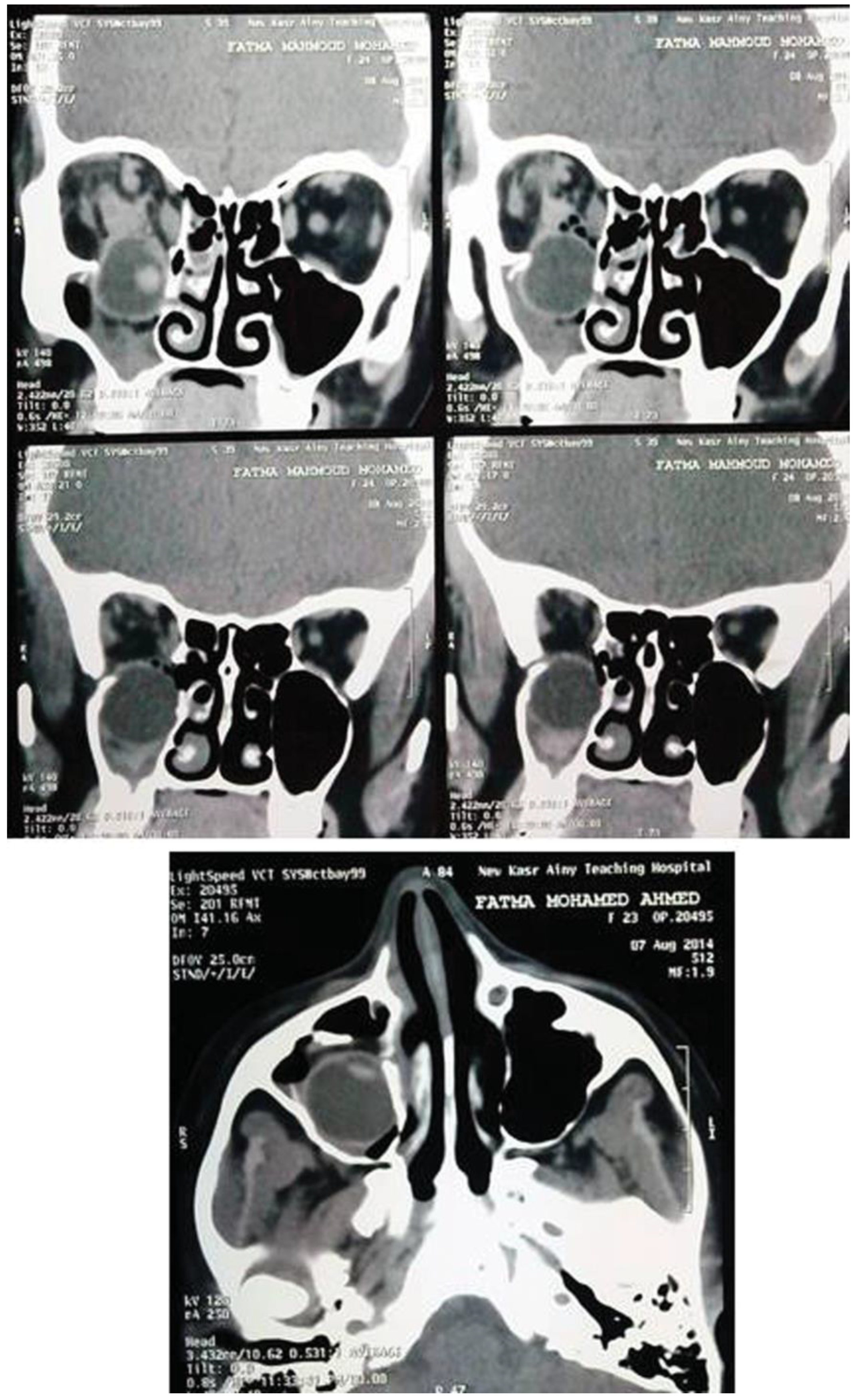
Computed tomography (CT) scan of the orbit and the midface revealed a blowout fracture of the posterior part of the right orbital floor with a complete dislocation of the intact globe into the right maxillary sinus (Figure 2).
The patient was taken immediately to the operating room and the dislocated globe was repositioned manually into the orbit via combined transmaxillary and transconjunctival approaches.
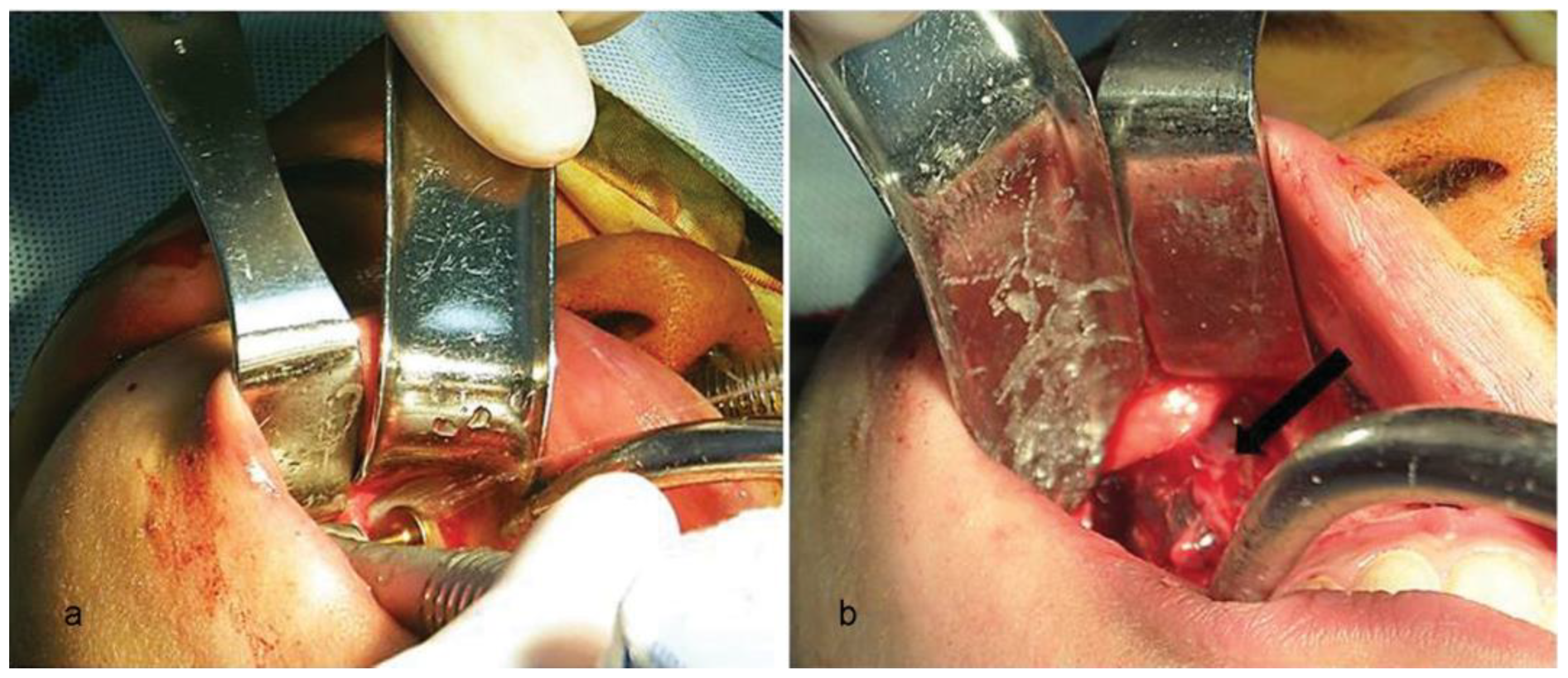
A right maxillary antrostomy was performed utilizing 8 mm diameter atraumatic dome-shaped sinus lateral window bur to avoid further injury to the displaced globe. After the antrostomy, the globe was noted to be completely contained within the right maxillary sinus (Figure 3). Gentle finger pressure was applied at the inferior aspect of the globe to guide the globe cephalically from the maxillary sinus into the orbital cavity. The tone and consistency of the globe was normal on palpation (Figure 4).
Titanium mesh carefully adapted to fit the bony anatomic contours of the remaining orbital walls. Stable bone around the defect was exposed to allow the mesh to rest on sound bone on all margins of the defect. Anteriorly, the mesh was secured to the inferior orbital rim with screw fixation. The proximal extent of the floor defect was observed extending far posteriorly without sound bony margin, thus a second mesh applied directly through maxillary sinus antrostomy inferiorly and fixed at the superior–posterior maxillary sinus wall to support the globe and orbital contents posteriorly. Maxillary antrostomy was repaired by titanium mesh and screw fixation (Figure 5) followed by intraoral and transconjunctival wounds closer.
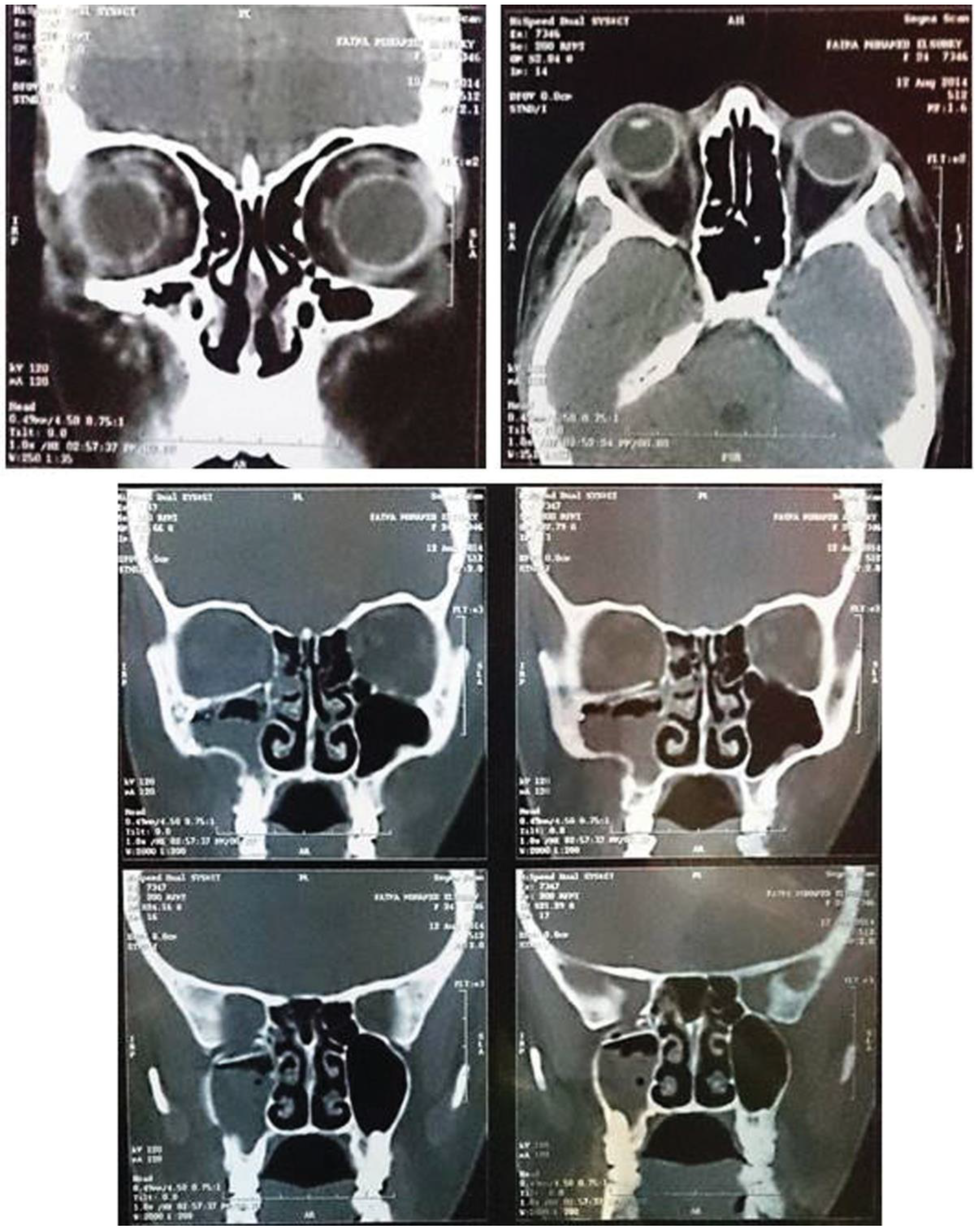
Postoperative, 250 mg of intravenous methylprednisolone was administered every 6 hours for 3 days. Postoperative CT showed good repositioning of the globe (Figure 6).
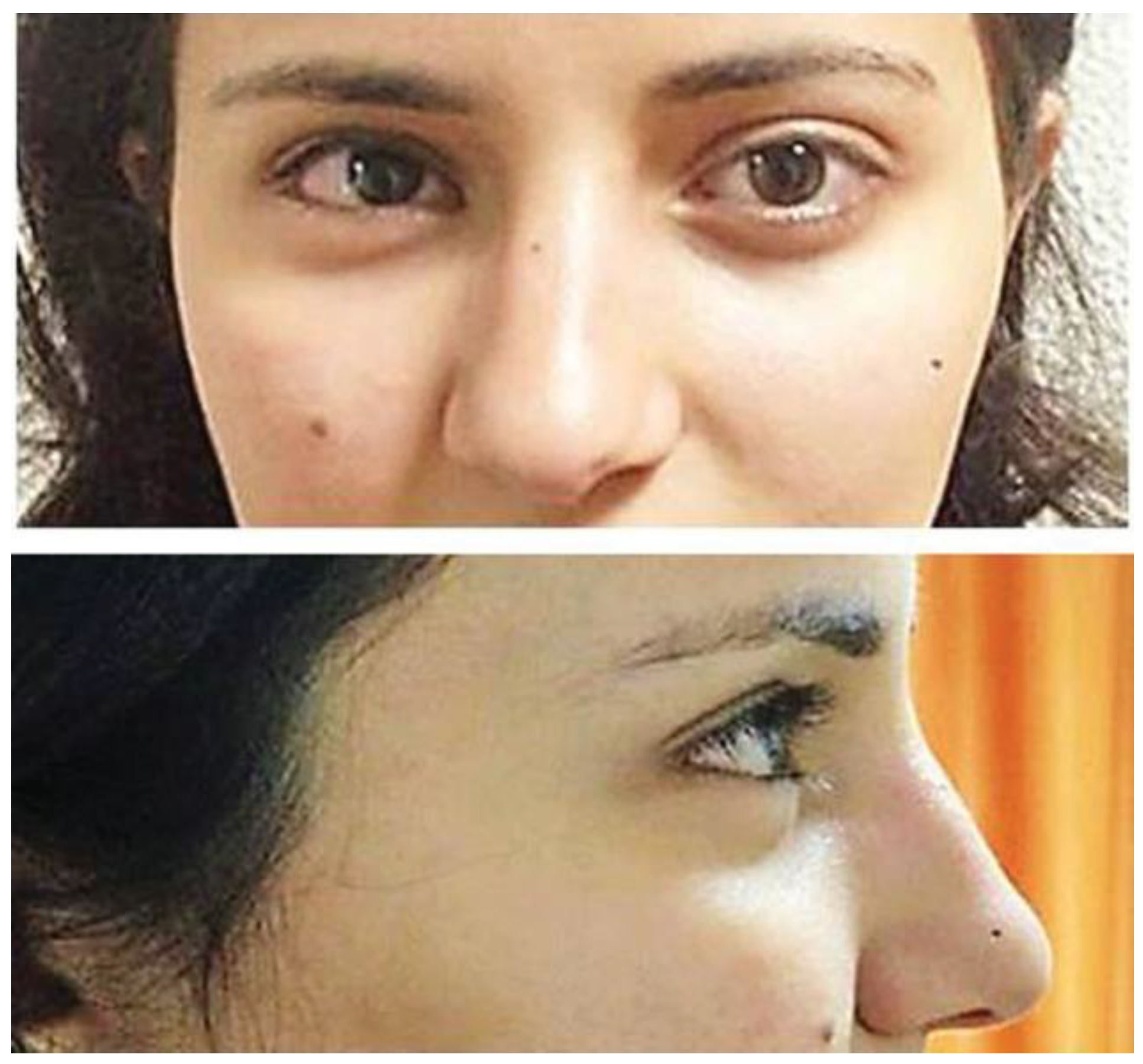
Immediate postoperative patient reported no light perception. Light perception vision was tested daily with multiple attempts. The patient was discharged to home with a rapid oral prednisone taper over 7 days. On postoperative day 8, the visual acuity improved to light perception. The patient reported marked improvement in vision at postoperative week 4. On postoperative week 7, her vision was noted to be 6/12 OD equivalent to United States (20/40 OD). Over the next several months, the visual acuity stabilized at 6/12 and she continued to have an afferent pupillary defect with reduced motility and diplopia in the lateral gaze, her visual function continues to remain stable 10 months after the initial trauma and the patient has a good cosmetic result (Figure 7).
Discussion
Traumatic displacement of the globe is a rare event that can be classified into three categories: luxation, dislocation, and avulsion. Dislocation can be defined as the migration of the globe into the paranasal sinuses, nasal cavity, or anterior cranial fossa [3].
Previous case reports were researched by detailed investigation of the English-language literature by searching for the following keywords: traumatic dislocation of the globe, traumatic luxation of the globe, traumatic prolapse of the globe, and traumatic displacement of the globe. In the English language literature, 45 cases (including anterior luxation, dislocation into anterior cranial fossa, and dislocation into paranasal sinuses) have been reported.
We found 19 cases of globe dislocation into the maxillary sinus summarized in Table 1 as follows:
  • One was enucleated 2 months after misdiagnosis as traumatic enucleation [32].
  • Seven were documented no vision or no light perception [6,29,31,34,35,42].
  • Three did not have reported vision [1,3,38].
  • Nine had postoperative vision [3,28,30,33,36,37,39,40,41].
Our case represents the 20th reported case ofdislocated globe into maxillary sinus, the 10th case with postoperative vision recovery, and the 2nd reported case with recovery of visual acuity after initial postoperative no light vision (Table 1).
The optic nerve has an excess reserve length of 8 mm inside the orbit, which saves it in the event of huge proptosis and globe displacements. The possible mechanism of the optic nerve injury was assumed to be stretching, rotation, and kinking of the nerve fibers causing mechanical damage to the nerve and a possible ischemia due to shearing of vessels and vascular compression. Being a sensory tract, it is not covered by the neurilemma; therefore, there is no regenerative potential when severed and any resulting visual loss will be permanent [27].
The pathogenesis outlined earlier may explain blindness as Roth et al., [43] concluded that traumatic optic nerve neuropathy should be treated as a cell death inducing increased intraneuronal pressure complicated by ischemia, axone rupture, and electrolyte imbalance of adjacent tissues just like in spinal paraplegia.
Salvaging the eye in this situation is a surgical challenge. Visual prognosis is generally poor in such cases since the retina and optic nerve are very sensitive to the injury and ischemia, although recovery of a normal vision has been reported in some cases [3,28,30,33,36,37,39,40,41].
The obtained outcome in the present case supports the opinion of Müller-Richter et al., [36] as they stated in their discussion that the very satisfying results were due to the fact that the orbital floor broke in the posterior part, reducing the strain on the muscles and optic nerve during the dislocation, and preventing their laceration.
Time is a major factor in the management of traumatic globe dislocations. An eye dislocated out of the orbital socket is under serious vascular compromise and sustains severe mechanical damage and early surgical intervention significantly reduces the ocular morbidity [27].
Although controversy exists regarding the specific surgical and medical management of grossly displaced globes, most investigators have agreed that every attempt should be made to anatomically reposition a displaced globe within the orbital pyramid as soon as possible [42]. Treatment delays increase the risk of complications as edema and strain on the optic nerve and central retinal artery increase with time. The period that the central retinal artery is twisted or stretched further compromises the blood supply to the optic nerve, increasing the probability of irreversible vision loss [6].
Early surgical intervention significantly reduces the ocular morbidity. Restoration of the eye movements and orbital volume could be aesthetically and emotionally acceptable to the patient as a minimum, if function could not be restored with the best possible efforts.
Conclusions
The present case shows that there is a possibility of regaining fair visual acuity, even though the initial visual acuity was extremely poor due to traumatic dislocation of the eyeball into the maxillary sinus. We recommend early treatment in cases such as our case. Early surgical interventions would help restore the cosmetic and visual function of the dislocated eye.

Acknowledgments

We express our thanks to the patient for her consent to being reported on in this article. Special thanks to Dr. Aya El-Hamalawy for photo documentation of this article.

References

  1. Pelton, R.W.; Rainey, A.M.; Lee, A.G. Traumatic subluxation of the globe into the maxillary sinus. AJNR Am. J. Neuroradiol. 1998, 19, 1450–1451. [Google Scholar] [PubMed]
  2. Ahmad, F.; Kirkpatrick, W.N.; Lyne, J.; Urdang, M.; Garey, L.J.; Waterhouse, N. Strain gauge biomechanical evaluation of forces in orbital floor fractures. Br. J. Plast. Surg. 2003, 56, 3–9. [Google Scholar] [CrossRef] [PubMed]
  3. Jellab, B.; Baha, A.T.; Moutaouakil, A.; et al. Management of a severe cranio-orbito-facial trauma with a dislocation of the globe into the maxillary sinus. Bull. Soc. Belge. Ophtalmol. 2008, 309–310, 37–41. [Google Scholar]
  4. Amaral, M.B.; Carvalho, M.F.; Ferreira, A.B.; Mesquita, R.A. Traumatic globe luxation associated with orbital fracture in a child: A case report and literature review. J. Maxillofac. Oral. Surg. 2015, 14 (Suppl. S1), 323–330. [Google Scholar] [CrossRef] [PubMed]
  5. Morris, W.R.; Osborn, F.D.; Fleming, J.C. Traumatic evulsion of the globe. Ophthal Plast. Reconstr. Surg. 2002, 18, 261–267. [Google Scholar] [CrossRef]
  6. Kreiner, B.; Amer, R.; Sharfi, G.; Solomon, A.; Ilsar, M. Traumatic displacement of the globe into the paranasal sinuses: Case report and guidelines for treatment. J. Oral. Maxillofac. Surg. 2008, 66, 826–830. [Google Scholar] [CrossRef]
  7. Reuling, F.H.; Hadlund, R.L. Traumatic luxation of the globe. Eye Ear Nose Throat Mon. 1970, 49, 129–130. [Google Scholar]
  8. Pillai, S.; Mahmood, M.A.; Limaye, S.R. Complete evulsion of the globe and optic nerve. Br. J. Ophthalmol. 1987, 71, 69–72. [Google Scholar] [CrossRef] [PubMed]
  9. van der Wal, K.G.; van der Pol, B.A. Traumatic luxation of the eyeball. J. Craniomaxillofac Surg. 1991, 19, 205–207. [Google Scholar] [CrossRef]
  10. Zengin, N.; Karakurt, A.; Gültan, E.; Kural, G. Traumatic globe luxation. Acta Ophthalmol. 1992, 70, 844–846. [Google Scholar] [CrossRef]
  11. Arkin, M.S.; Rubin, P.A.; Bilyk, J.R.; Buchbinder, B. Anterior chiasmal optic nerve avulsion. AJNR Am. J. Neuroradiol. 1996, 17, 1777–1781. [Google Scholar] [PubMed]
  12. Kiratli, H.; Tümer, B.; Bilgiç, S. Management of traumatic luxation of the globe: A case report. Acta Ophthalmol. Scand. 1999, 77, 340–342. [Google Scholar] [CrossRef]
  13. Bajaj, M.S.; Kedar, S.; Sethi, A.; Gupta, V. Traumatic globe luxation with optic nerve transection. Orbit 2000, 19, 165–170. [Google Scholar] [CrossRef]
  14. Alp, B.; Yanyali, A.; Elibol, O.; Talu, H.; Karabas, L. A case of traumatic globe luxation. Eur. J. Emerg. Med. 2001, 8, 331–332. [Google Scholar] [CrossRef] [PubMed]
  15. Bajaj, M.S.; Pushker, N.; Nainiwal, S.K.; Balasubramanya, R. Traumatic luxation of the globe with optic nerve avulsion. Clin. Experiment Ophthalmol. 2003, 31, 362–363. [Google Scholar] [CrossRef]
  16. Song, A.; Carter, K.D. Bilateral traumatic globe subluxation. Ophthal Plast. Reconstr. Surg. 2006, 22, 136–137. [Google Scholar] [CrossRef]
  17. Lelli, G.J., Jr.; Demirci, H.; Frueh, B.R. Avulsion of the optic nerve with luxation of the eye after motor vehicle accident. Ophthal Plast. Reconstr. Surg. 2007, 23, 158–160. [Google Scholar] [CrossRef]
  18. de Saint Sardos, A.; Hamel, P. Traumatic globe luxation in a 6-year-old girl playing with a tube of wrapping paper. J. AAPOS 2007, 11, 406–407. [Google Scholar] [CrossRef] [PubMed]
  19. Roldan-Valadez, E.; Corona-Cedillo, R.; Rojas-Marin, C.; Valdivieso Cardenas, G.; Sanchez-Sanchez, J.M.; Quiroz-Mercado, H. Tomographic findings in traumatic globe evulsion caused by blunt head trauma. Br. J. Radiol. 2007, 80, e247–e249. [Google Scholar] [CrossRef]
  20. Shimia, M.; Sayyahmelli, S. Traumatic Displacement of the Globe into Brain. RMJ 2009, 34, 234–235. [Google Scholar]
  21. Vahdati, S.S.; Sadeghi, H. Orbital roof fracture: Dislocation of globe into the anterior cranial fossa. JAEMCR 2011, 2, 47–49. [Google Scholar] [CrossRef]
  22. Gollapudi, P.R.; Nandigama, P.K.; Maila, S.K.; Karla, R.; Kotakadira, S. Traumatic intracranial prolapse of eye ball—A case report. Br. J. Neurosurg. 2013, 27, 114–116. [Google Scholar] [CrossRef] [PubMed]
  23. Risco, J.M.; Stratas, B.A.; Knott, R.H. Prolapse of the globe into the ethmoid sinus. Am. J. Ophthalmol. 1984, 97, 659–660. [Google Scholar] [CrossRef] [PubMed]
  24. Moon, M.; Pietris, G.; Shapter, M. Dislocation of the globe into the ethmoid sinuses. Aust. N. Z. J. Ophthalmol. 1997, 25, 175–176. [Google Scholar] [CrossRef]
  25. Kang, B.D.; Jang, M.H. A case of blowout fracture of the orbital wall with eyeball entrapped within the ethmoid sinus. Korean J. Ophthalmol. 2003, 17, 149–153. [Google Scholar] [CrossRef]
  26. Okabe, H.; Kimura, K.; Sonoda, S.; Sakamoto, T. Displacement of globe into ethmoid sinus by orbital medial wall fracture with good recovery of vision. Jpn. J. Ophthalmol. 2005, 49, 426–428. [Google Scholar] [CrossRef]
  27. Tranfa, F.; Di Matteo, G.; Di Salle, F.; Strianese, D.; Bonavolontà, G. Traumatic displacement of the globe into the ethmoid sinus. Am. J. Ophthalmol. 2000, 130, 253–254. [Google Scholar] [CrossRef]
  28. Berkowitz, R.A.; Putterman, A.M.; Patel, D.B. Prolapse of the globe into the maxillary sinus after orbital floor fracture. Am. J. Ophthalmol. 1981, 91, 253–257. [Google Scholar] [CrossRef]
  29. Smit, T.J.; Koornneef, L.; Zonneveld, F.W. A total orbital floor fracture with prolapse of the globe into the maxillary sinus manifesting as postenucleation socket syndrome. Am. J. Ophthalmol. 1990, 110, 569–570. [Google Scholar] [CrossRef]
  30. Ziccardi, V.B.; Patterson, G.T.; Ramasastry, S.; Sotereanos, G.C. Management of a severe zygomatico-orbital fracture with dislocation of the globe into the antrum. J. Craniofac Surg. 1993, 4, 95–101. [Google Scholar] [CrossRef]
  31. Tung, T.C.; Chen, Y.R.; Santamaria, E.; Chen, C.T.; Lin, C.J.; Tsai, T.R. Dislocation of anatomic structures into the maxillary sinus after cranio-facial trauma. Plast. Reconstr. Surg. 1998, 101, 1904–1908. [Google Scholar] [CrossRef]
  32. Saleh, T.; Leatherbarrow, B. Traumatic prolapse of the globe into the maxillary sinus diagnosed as traumatic enucleation of the globe. Eye 1999, 13 Pt 5, 678–680. [Google Scholar] [CrossRef] [PubMed]
  33. Avadhanam, V.S.; Sreedhara, S.P.; Ameerally, P.; El-Ghazali, K.M.S. Traumatic globe dislocations-Significance of early intervention. Inj. Extra 2008, 39, 356–358. [Google Scholar] [CrossRef]
  34. Kim, S.; Baek, S. Traumatic dislocation of the globe into the maxillary sinus associated with extraocular muscle injury. Graefes Arch. Clin. Exp. Ophthalmol. 2005, 243, 1280–1283. [Google Scholar] [CrossRef] [PubMed]
  35. Abrishami, M.; Aletaha, M.; Bagheri, A.; Salour, S.H.; Yazdani, S. Traumatic subluxation of the globe into the maxillary sinus. Ophthal Plast. Reconstr. Surg. 2007, 23, 156–158. [Google Scholar] [CrossRef]
  36. Müller-Richter, U.D.; Kohlhof, J.K.; Driemel, O.; Wagener, H.; Reichert, T.E. Traumatic dislocation of the globe into the maxillary sinus. Int. J. Oral. Maxillofac. Surg. 2007, 36, 1207–1210. [Google Scholar] [CrossRef]
  37. Ramstead, C.; McCabe, J.; Alkahtani, M.; Leong-Sit, J.; Morhart, M. Traumatic dislocation of the globe into the maxillary sinus. Can. J. Ophthalmol. 2008, 43, 364–366. [Google Scholar] [CrossRef]
  38. Akhaddar, A.; Elmostarchid, B.; Boucetta, M. Images in emergency medicine. Traumatic intraorbital stone with globe displacement into the maxillary sinus. Emerg. Med. J. 2010, 27, 828. [Google Scholar] [CrossRef]
  39. Damasceno, N.A.P.; Damasceno, E.F. Traumatic orbital fracture with intact ocular globe displacement into the maxillary sinus. Rev. Bras. Oftalmol. 2010, 69, 52–54. [Google Scholar] [CrossRef]
  40. Zhang-Nunes, S.X.; Jarullazada, I.; Mancini, R. Late central visual recovery after traumatic globe displacement into the maxillary sinus. Ophthal Plast. Reconstr. Surg. 2012, 28, e17–e19. [Google Scholar] [CrossRef]
  41. Xu, B.; Xu, X.L.; Yan, J. Treatment of traumatic globe dislocated completely into the maxillary sinus. Int. J. Ophthalmol. 2013, 6, 106–107. [Google Scholar] [PubMed]
  42. Haggerty, C.J.; Roman, P. Repositioning of a traumatically displaced globe with maxillary antrostomy: Review of the literature and treatment recommendations. J. Oral. Maxillofac. Surg. 2013, 71, 1915–1922. [Google Scholar] [CrossRef] [PubMed]
  43. Roth, J.; Hingst, V.; Lenz, J.H. Blindness following severe midfacial trauma—Case report and review. J. Craniomaxillofac Surg. 2012, 40, 608–613. [Google Scholar] [CrossRef] [PubMed]
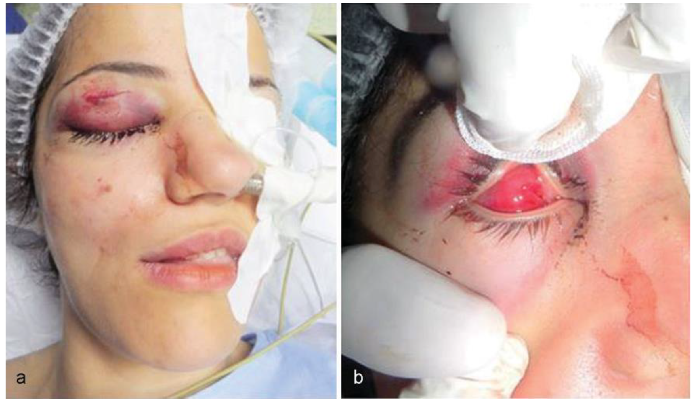
Figure 1. (a) Periorbital hematoma and skin laceration of the upper right eyelid. (b) Manual opening of the eyelid without any evidence of the eyeball (anophthalmic right orbital cavity).
Cmtr 10 00023 g001
Figure 2. Preoperative coronal and axial computed tomography scan images showing the right globe completely dislocated into the maxillary sinus.
Cmtr 10 00023 g002
Figure 3. (a) Antrostomy by 8 mm diameter atraumatic dome-shaped sinus lateral window bur. (b) The globe (arrow) completely dislocated and contained within the right maxillary sinus.
Cmtr 10 00023 g003
Figure 4. (a) Gloved finger (arrow) introduced inferiorly from the maxillary sinus protruding through orbital floor defect. (b) Right globe repositioned within the orbital cavity.
Cmtr 10 00023 g004
Figure 5. (a) Titanium mesh reconstruct orbital floor defect. (b) Second mesh applied through antrostomy to support the globe posteriorly. (c) Maxillary antrostomy repaired by titanium mesh.
Cmtr 10 00023 g005
Figure 6. Postoperative coronal and axial CT scan images showing good repositioning of the globe. Coronal CT scan images showing the posterior extent of the reconstructed defect. CT, computed tomography.
Cmtr 10 00023 g006
Figure 7. Postoperative appearance after repositioning of the right globe with good cosmetic outcome.
Cmtr 10 00023 g007
Table 1. Reported cases of globe dislocation into the maxillary sinus.
AgeSexVisual AcuityMotilityPupillary DefectDiplopia
Berkowitz et al (1981)12FRecovery 20/20NANANA
Smit et al (1990)28MNoNANANA
Ziccardi et al (1993)35MRecovery 20/20Restricted motilityNAYes (primary gaze)
Pelton et al (1998)19MNANANANA
Tung et al (1998)17MNoMinimal horizontal gaze and no vertical gazeNANA
Saleh and Leatherbarrow (1999)29MNoNANANA
Kim and Baek (2005)68MNoRestricted motilityNonreactive mydriatic pupilNA
Müller-Richter et al (2007)62MRecovery 20/20Reduced motilityAnisocoria with dilation of the pupilYes (lateral gaze and upward)
Abrishami et al (2007)18MNoLimited in all gazesNonreactive mydriatic pupilNA
Avadhanam et al (2008)30FNoGood motility in all gazesNonreactive mydriatic pupilNA
Kreiner et al (2008)50MNoloss of ocular motilityNonreactive mydriatic pupilNA
Ramstead et al (2008)32MRecovery 20/ 200Minimal horizontal gaze and no vertical gazeAfferent pupillary defectNA
Jellab et al (2008)24MLight perceptionReduced motilityNANA
Jellab et al (2008)50FNANANANA
Akhaddar et al (2010)62MNANANANA
Damasceno and Damasceno (2010)42MRecovery 20/20Loss of ocular motility (except for abduction)NoYes
Zhang-Nunes et al (2012)20MRecovery 20/25NAAfferent pupillary defectNA
Xu et al (2013)46FLight perceptionRestricted motilityNANA
Haggerty and Roman (2013)35MLight perceptionRestricted motilityNonreactive mydriatic pupilNA
Noman and Shindy (2015)24FRecovery 20/40Reduced motility in lateral gazeAfferent pupillary defectYes (lateral gaze)
Abbreviation: NA, not available.