


Abstract
Background: Isolated medial cuneiform fracture is a rare but diagnostically challenging condition. Diagnostic delay in these cases may lead to delays in ideal treatment approaches and prolonged symptoms. An understanding of clinical presentation is needed to expedite diagnosis, facilitate decision making, and guide treatment approach. Methods: Case studies/series were searched in four databases until September 2019. Included studies had participants with a history of traumatic closed medial cuneiform fracture. Studies were excluded if the medial cuneiform fractures were open fractures, associated with multitrauma, or associated with dislocation/Lisfranc injury. Three blinded reviewers assessed the methodological quality of the studies, and a qualitative synthesis was performed. Results: Ten studies comprising 15 patients were identified. Mean ± SD patient age was 38.0 ± 12.8 years, with 86.7% of reported participants being men. The overall methodological quality was moderate to high, and reporting of the patient selection criteria was poor overall. The most commonly reported clinical symptoms were localized tenderness (60.0%) and edema (53.3%). Direct blow was the most common inciting trauma (46.2%), followed by axial load (30.8%) and avulsion injuries (23.1%). Baseline radiographs were occult in 72.7% of patients; magnetic resonance imaging and computed tomography were the most common diagnostic modalities. Mean ± SD diagnostic delay was 64.7 ± 89.6 days. Conservative management was pursued in 54.5% of patients, with reported resolution of symptoms in 3 to 6 months. Surgical intervention occurred in 45.5% of patients and resulted in functional restoration in 3 to 6 months in all but one patient. Conclusions: Initial radiographs for isolated medial cuneiform fractures are frequently occult. Due to expedience and relatively low cost, radiographs are still a viable first-line imaging modality. If clinical concern remains, magnetic resonance imaging may be pursued to minimize diagnostic delay. Conservative management is a viable treatment method, with expected return to full function in 3 to 6 months.
Traumatic foot injuries are a common presentation in emergency departments [1,2,3]. These injuries can cost up to $21,801 to manage and are compounded by loss of productivity and salary by the employer and employee, respectively [4,5]. The three cuneiform bones of the foot (medial, intermediate, and lateral) and their articulations are relatively small and well protected from injury, primarily due to the extensive ligamentous structures around these articulations [6]. Although there is motion between the cuneiform bones, it is slight and limited to gliding motions [7]. Due to the inherent stability of these joints, isolated fracture in the absence of ligamentous disruption or dislocation is rare, accounting for approximately 2% of all tarsal fractures [8].
Evidence-based guidelines such as the Ottawa Ankle Rules and the American College of Radiology Appropriateness Criteria are effective in aiding clinicians to rule out fracture in patients with foot trauma [9,10,11,12]. Although these guidelines currently constitute best practice, they do not achieve 100% sensitivity to rule out fracture and may lead to a lack of baseline imaging in isolated cuneiform fractures [13,14,15]. When radiographs are ordered, midfoot fractures are often occult, which can lead to delayed diagnosis and prolonged patient disability [16,17,18,19,20,21]. Olson et al [22] previously reported that the diagnosis of nondisplaced medial cuneiform fractures may be delayed or even missed completely because these fractures are subtle or occult on plain film radiographs, especially when taken in a nonweightbearing position. Furthermore, in this region of the foot, radiographs can be particularly difficult to interpret because of multiple overlapping shadows of the cuneiform bones [7]. Therefore, advanced imaging, such as skeletal scintigraphy, computed tomography (CT), or magnetic resonance imaging (MRI), may be necessary during patient evaluation to make the correct diagnosis in a timely manner [7,22].
Although individual case studies detail the diagnosis and management of isolated medial cuneiform injuries, we were unable to locate any research publications that comprehensively describe the evaluation and management of isolated fracture of the medial cuneiform. No known composite patient presentation has been established to supplement clinical decision making where imaging guidelines may fail. Although the American College of Radiology Appropriateness Criteria support advanced imaging in patients with suspected occult foot fractures, no systematic review has reported which modality may be ideal for isolated medial cuneiform fractures. Although individual case reports have documented success with both conservative and surgical interventions in this patient population, no comprehensive study exists to direct which treatment approach may be best pursued [23,24].
The main purpose of this study was to conduct a systematic search, critical appraisal, and qualitative synthesis of the evidence on the clinical presentation of patients with medial cuneiform fractures. A secondary aim of this study was to identify a potential diagnostic imaging pathway for these injuries. Finally, the study aimed to establish an understanding of conservative management and surgical intervention treatment outcomes.
Materials and Methods
Search Strategy
A comprehensive literature search was completed using the following databases: CINAHL, SPORTdiscus, PubMed (Medline), and Scopus. Limits applied included English language and studies on human subjects, and it was conducted from inception until September 2019. Search terms used were (fracture) AND (medial cuneiform OR first cuneiform OR 1st cuneiform).
Inclusion and Exclusion Criteria
Studies were considered eligible for inclusion if they were case vignettes, case reports, or case series with participants who had a history of traumatic closed medial cuneiform fracture.
Studies were not considered eligible if any of the following were found: 1) studies in which the medial cuneiform fracture was considered to be a stress (fatigue or insufficiency) fracture; 2) studies in which the medial cuneiform fracture was an open fracture, was associated with concurrent dislocation or Lisfranc injury, or was associated with multitrauma (such as multiple fractures); 3) studies that did not report clinical presentation or clinical course of care; and 4) cadaveric studies.
Study Selection
After articles were identified using the previously defined search strategy, duplicates were removed. Two independent reviewers, L.M.M. and T.N.P., screened titles and abstracts to determine eligibility for full-text consideration. Reviewers were blinded to one another’s results during the screening process. After screening titles and abstracts, fulltext articles were reviewed for final inclusion consideration. When a consensus could not be reached between reviewers, a third reviewer (A.S.G.) was consulted.
Data Extraction
Data were extracted independently by L.M.M. and A.S.G. using a standardized spreadsheet to record author(s), year of publication, and study population (sex, age); mechanism of injury; trauma type; initial/ managing provider; ability to walk immediately after injury; baseline and secondary imaging types; presence of antalgic gait pattern; presence of ecchymosis, edema, or erythema; palpatory tenderness; diagnostic delay period; treatment approach; and outcome. Consensus was reached by discussion.
Assessment of Methodological Quality
Both L.M.M. and T.N.P. completed a bias assessment of the final ten articles using the Murad criteria [25]. Two of the eight bias assessment criteria were excluded because they pertained specifically to pharmaceutical studies: was there a challenge/ rechallenge phenomenon and was there a doseresponse effect. Therefore, six of the eight criteria were used for the bias assessment. Both L.M.M. and T.N.P. were blinded to one another’s quality scores during the scoring process, and A.S.G. resolved any discrepancies between the two reviewers on the quality score of each article.
Synthesis of Results
Qualitative synthesis of the articles was performed with Microsoft Excel 2016 (Microsoft Corp, Redmond, Washington). Descriptive statistics, including ranges, counts, and averages, were calculated with either Microsoft Excel 2016 or the Microsoft calculator application 2018 version 10.1804.11545.0 (Microsoft Corp).
Results
Study Selection
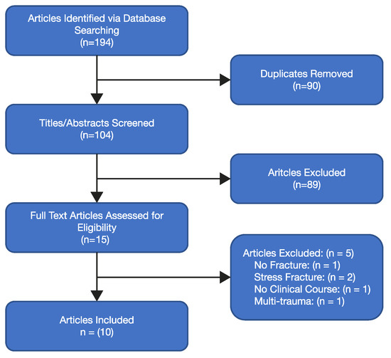
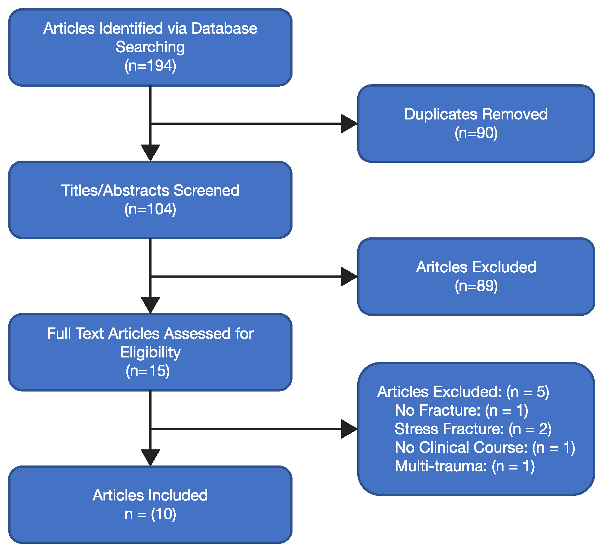
In total, 194 studies were identified from the database searches (Fig. 1). After removal of duplicates and screening of titles and abstracts, 15 studies remained for full-text analysis. Of those 15 studies, five were eliminated for the following reasons: no defined medial cuneiform fracture [26], atraumatic medial cuneiform fracture [27,28], no clinical course reported [29], and multitrauma [30]. Ten studies were, therefore, included in the qualitative analysis. Fifteen patient cases from these ten peer-reviewed publications were found [22,23,24,31,32,33,34,35,36,37].
Figure 1.
Study selection flow diagram.
Study Characteristics
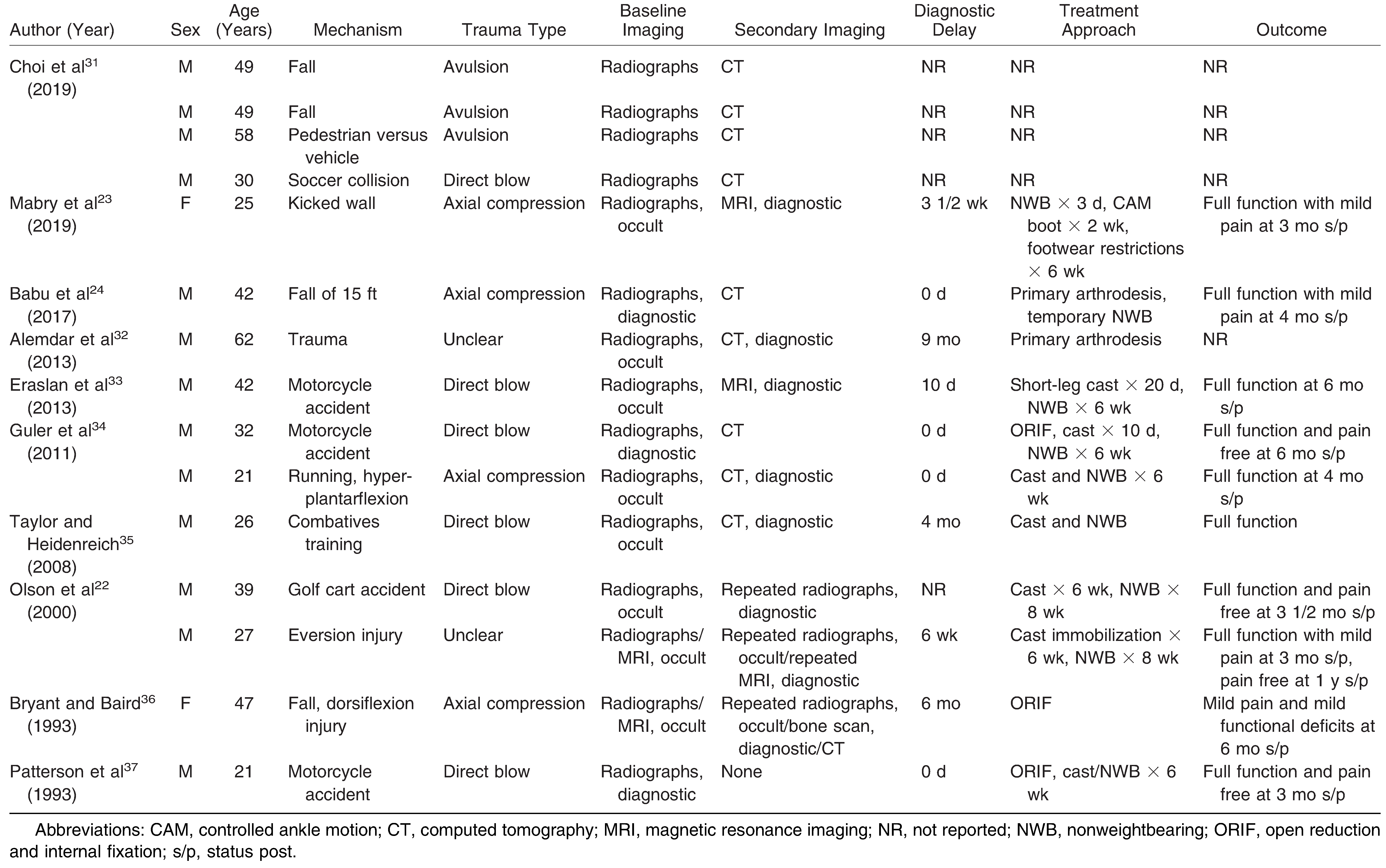
Table 1 reports the main characteristics of the included studies and patients. Patient ages ranged from 21 to 62 years (mean ± SD, 38.0 ± 12.8 years), with 13 men (86.7%) and two women (13.3%). Thirteen patients reported a type of trauma, of which a direct blow was the most common etiology (46.2%), followed by axial compression (30.8%), with avulsion fractures having the lowest etiologic frequency (23.1%). Ten patients detailed first-line providers, the most common of which was the emergency department (60.0%), although a portion of initial presentations occurred at the primary care provider (20.0%) or orthopedics (20.0%). Seven of 11 patients were finally managed by orthopedics (63.6%), with podiatry treating three patients (27.3%) and physical therapy treating one (9.1%).
Table 1.
Demographic, Clinical, Diagnostic, and Treatment Presentations.
Localized palpatory tenderness was the most commonly reported clinical finding, occurring in nine patients (60.0%). An antalgic gait pattern was present in four patients (26.7%). Localized edema was present in eight patients (53.3%), ecchymosis in three (20.0%), and erythema in one (6.7%). Of note, many case reports failed to mention either the presence or absence of these findings, which may suggest that these percentages are lower than what were clinically present.
Baseline radiographs were reported in 11 of the 15 patients, and of these, eight were occult (72.7%). Of the eight patients with occult imaging, three with MRI [22,23,33], four with CT [32,34,35,36], one with bone scan, and one of three with repeated radiographs (33.3%)[22,36] were diagnostic. In the patient with the bone scan, CT was also ordered due to the inherent low specificity of bone scan [22].
Eleven cases reported treatment approaches with clinical outcomes. Six patients were treated conservatively (54.5%) and five were treated surgically (45.5%). Those treated conservatively were commonly prescribed 6 to 8 weeks of nonweightbearing, with return to full function in 3 to 6 months [22,33,34]. Note, however, that in one case the patient advanced herself to full weightbearing 3 days after the injury and still achieved full function with conservative care at the 3-month mark [23]. Although most surgical interventions reported a return to full function in 3 to 6 months [24,34,37], one patient reported ongoing functional deficits at the 6-month mark [36].
Diagnostic delay was reported in ten patients and ranged from 0 days to 9 months. The mean ± SD diagnostic delay was 64.7 ± 89.6 days and was primarily composed of provider delay. One case did, however, report a 2-month patient delay before first seeking treatment [35].
Methodological Quality Assessment of Studies
Bias for all ten studies was evaluated on a 6-point scale where higher percentages suggest lower levels of bias and lower percentages suggest higher levels of bias (Table 2). Bias scores ranged from 2 to 5 points on the 6-point scale (33.3%–100.0%). The average bias score across all studies was 4.5 points (75.0%), suggesting a low-to-moderate level of bias [25]. A common weakness of the studies was a failure to report the patient selection method. The following metrics were reasonably well represented by the included studies: adequate exposure; adequate outcome; alternative causes of symptoms; sufficient follow-up; and reproducibility.
Table 2.
Methodological Quality Analysis.
Discussion
Isolated medial cuneiform fractures are rare and diagnostically challenging; they are typically occult on baseline radiographs [23,32,33]. Noncontributory imaging may lead to diagnostic delay and may unnecessarily prolong patient suffering. Because this condition is exceedingly rare, to our knowledge, no studies have been published with greater methodological rigor than the case studies/series presented in these findings. This is the first article to present a systematic search and comprehensive synthesis of presentation, diagnosis, and treatment of isolated medial cuneiform fractures.
Only one patient reported an inability to walk at the time of injury, indicating a positive finding on the Ottawa Ankle Rules [38,39,40]. This would suggest a risk of a patient failing to receive baseline imaging if evidence-based guidelines for imaging are followed. Because the Ottawa Ankle Rules do not always achieve 100% sensitivity [41,42,43], it may be of value to add medial cuneiform tenderness to the rules to minimize the chance of a missed fracture. Other authors have similarly suggested updates to the Ottawa Ankle Rules to improve overall yield [44]. Although this addition may improve the sensitivity of the rules, it may also have a negative effect on specificity, which warrants further investigation.
Localized palpatory tenderness, edema, and antalgic gait pattern were present in all of the reported patients. Although this may aid in diagnosis, these symptoms would be similarly experienced in patients with a diagnosis of a Lisfranc injury [45]. Mechanisms for isolated medial cuneiform fractures are also consistent with those for Lisfranc injuries [46]. Similar to medial cuneiform fractures, Lisfranc injuries are frequently misdiagnosed [47,48,49] and may present as occult on baseline radiographs [45,50]. Distinguishing between these diagnoses is of utmost importance because isolated medial cuneiform fractures may be treated conservatively [23,33,35] whereas Lisfranc injuries may result in poor outcomes unless treated surgically [51,52,53]. Note that these articles were largely silent on the presence or absence of clinical findings typically associated with Lisfranc injuries, such as loss of arch height, the plantar ecchymosis sign, or toe gapping [54,55,56].
Although baseline radiographs were commonly occult, they were diagnostic in three of 11 patients. This finding is consistent with previous evidence suggesting that midfoot fractures are typically occult on baseline radiographs [57,58]. However, given the low radiation exposure, expedience, and costeffectiveness of plain film radiographs, these may still offer value as a primary imaging modality [59,60]. Lisfranc injury would be a typical differential diagnosis of isolated medial cuneiform fractures before imaging results. Given the ability for weightbearing foot radiographs to aid in ligamentous disruption via expansion of joint spaces, we suggest weightbearing foot radiographs as tolerated to aid in this differential diagnosis [61,62,63]. This recommendation is consistent with the American College of Radiology Appropriateness Criteria for traumatic midfoot injuries [9].
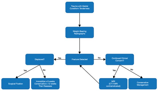
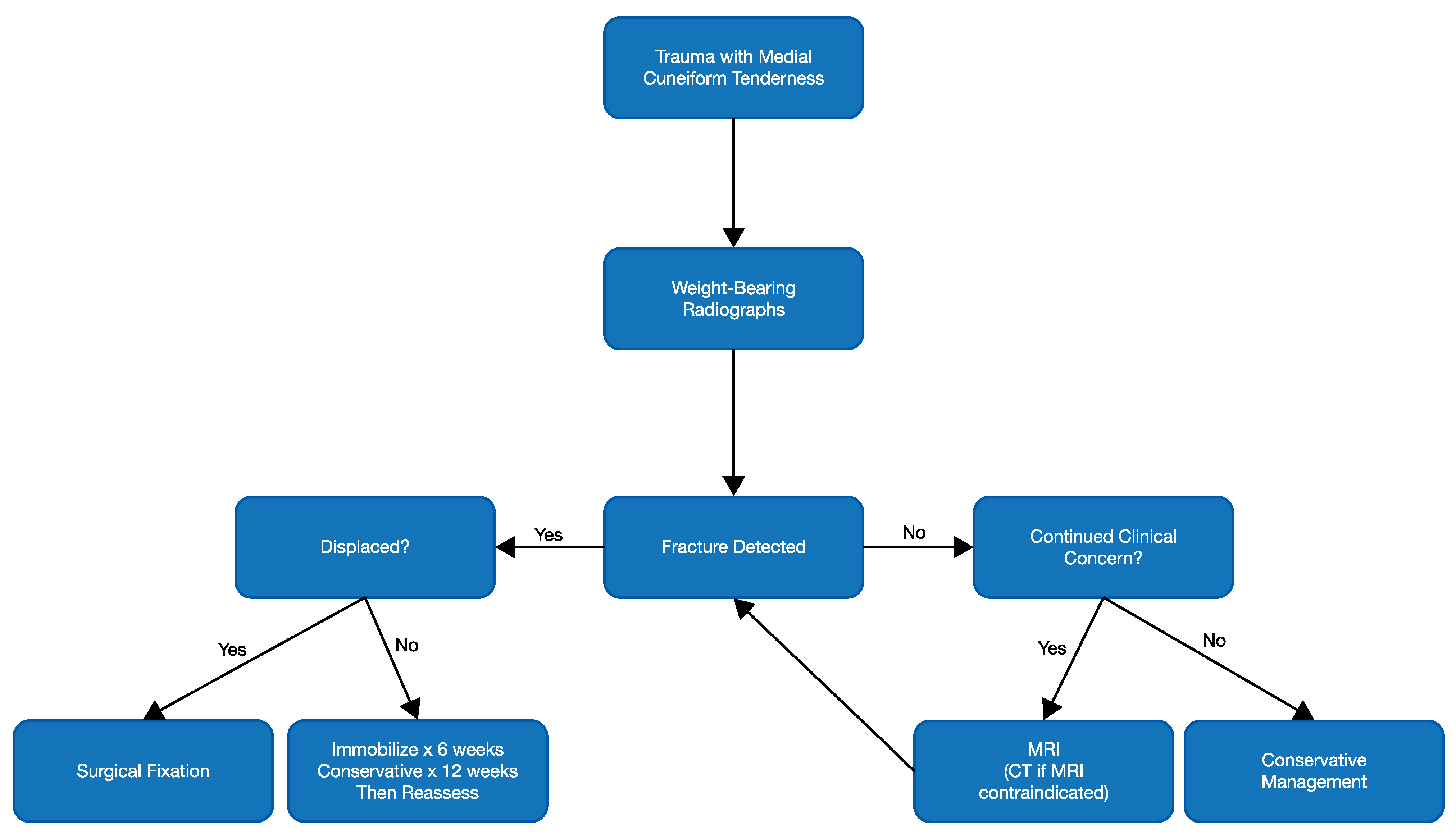
The present study found that neither repeated radiographs nor bone scans were definitively diagnostic in patients with isolated medial cuneiform fractures [22,36]. These findings suggest that repeated radiographs or bone scans are not the optimal secondary imaging choice. Conversely, MRI or CT may potentially be of greatest diagnostic value because both modalities led to a definitive diagnosis as a secondary imaging modality [22,23,32,33,34,35,36]. Given the ability of MRI to rule out a concomitant Lisfranc injury, it may hold superior diagnostic capability over CT [64,65,66]. With that in mind, we propose a diagnostic flow diagram to aid in identifying isolated medial cuneiform fractures (Fig. 2).
Figure 2.
Isolated medial cuneiform fracture diagnostic and treatment flow diagram. CT, computed tomography; MRI, magnetic resonance imaging.
Diagnostic delay in these patients was primarily composed of provider delay. Patient delay was minimized because many patients reported immediately to the emergency department after the injury. This finding is consistent with delays reported in other traumatic isolated midfoot frac-tures [7,67]. More insidious sources of foot pain tend to lead to greater duration of patient delays compared with provider delays [68,69,70]. Given the qualitative analysis in this study, progressing to MRI in patients with traumatic foot injuries with tenderness at the medial cuneiform may minimize provider delay while expediting the requisite rehabilitative treatments.
This article is limited by the methodological rigor of its included studies, as the highest level of evidence for isolated medial cuneiform fractures in this review is case studies/series. Although studies overall had low-to-moderate bias, selection bias was common among the studies. Future studies should explore changes in the diagnostic properties of the Ottawa Ankle Rules if medial cuneiform tenderness is added to the foot protocol.
Conclusions
An isolated medial cuneiform fracture is a rare but diagnostically challenging injury that leads to average diagnostic delays of 2 months. Patients commonly present with localized tenderness and edema. Initial radiographs are frequently occult; however, due to expedience and relatively low cost, radiography is still a viable first-line imaging modality. If clinical concern remains, findings from this review suggest that MRI may be pursued to minimize diagnostic delay. Conservative management is a viable treatment method, with expected return to full function in 3 to 6 months.
Financial Disclosure
None reported.
Conflict of Interest
None reported.
References
- STRUDWICK K, MCPHEE M, BELL A, ET AL: Review article: best practice management of common ankle and foot injuries in the emergency department (part 2 of the musculoskeletal injuries rapid review series). Emerg Med Australas 30: 152, 2018. [CrossRef]
- DISCHINGER PC, READ KM, KUFERA JA, ET AL: Consequences and costs of lower extremity injuries. Annu Proc Assoc Adv Automot Med 48: 339, 2004.
- DE BOER AS, SCHEPERS T, PANNEMAN MJ, ET AL: Health care consumption and costs due to foot and ankle injuries in the Netherlands, 1986-2010. BMC Musculoskelet Disord 15: 128, 2014. [CrossRef]
- GRIMM DJ, FALLAT L: Injuries of the foot and ankle in occupational medicine: a 1-year study. J Foot Ankle Surg 38: 102, 1999. [CrossRef]
- BIELSKA IA, WANG X, LEE R, ET AL: The health economics of ankle and foot sprains and fractures: a systematic review of English-language published papers: part 2. The direct and indirect costs of injury. Foot (Edinburgh, Scotland) 39: 115, 2019.
- McGlamry ED, Banks AS, Downey MS: Comprehensive Textbook of Foot Surgery, 2nd Ed, Vol 2, Williams & Wilkins, Baltimore, 1992.
- SHAH K, ODGAARD A: Fracture of the lateral cuneiform only: a rare foot injury. JAPMA 97: 483, 2007. [CrossRef]
- WILSON PD: Fractures and dislocations of the tarsal bones. South Med J 26: 833, 1933. [CrossRef]
- Bancroft LW, Kransdorf MJ, Adler R, et al: ACR Appropriateness Criteria acute trauma to the foot. J Am Coll Radiol 12: 575, 2015. [CrossRef]
- MACLELLAN J, SMITH T, BASERMAN J, ET AL: Accuracy of the Ottawa Ankle Rules applied by non-physician providers in a pediatric emergency department. CJEM 20: 746, 2018. [CrossRef]
- LI N, LIU YS, XIN JY, ET AL: Efficiency of the Modified Ottawa Ankle Rules for the differential diagnosis of fracture in acute foot and ankle injury [in Chinese]. Zhonghua Yi Xue Za Zhi 97: 2742, 2017.
- BACHMANN LM, KOLB E, KOLLER MT, ET AL: Accuracy of Ottawa ankle rules to exclude fractures of the ankle and mid-foot: systematic review. BMJ 326: 417, 2003. [CrossRef]
- PIRES R, PEREIRA A, ABREU ESG, ET AL: Ottawa ankle rules and subjective surgeon perception to evaluate radiograph necessity following foot and ankle sprain. Ann Med Health Sci Res 4: 432, 2014. [CrossRef]
- MARKERT RJ, WALLEY ME, GUTTMAN TG, ET AL: A pooled analysis of the Ottawa ankle rules used on adults in the ED. Am J Emerg Med 16: 564, 1998. [CrossRef]
- JONCKHEER P, WILLEMS T, DE RIDDER R, ET AL: Evaluating fracture risk in acute ankle sprains: any news since the Ottawa Ankle Rules? a systematic review. Eur J Gen Pract 22: 31, 2016. [CrossRef]
- ANGOULES AG, ANGOULES NA, GEORGOUDIS M, ET AL: Update on diagnosis and management of cuboid fractures. World J Orthop 10: 71, 2019. [CrossRef]
- ALMEIDA RR, MANSOURI M, TSO DK, ET AL: The added value of cross-sectional imaging in the detection of additional radiographically occult fractures in the setting of a Chopart fracture. Emerg Radiol 25: 513, 2018. [CrossRef]
- AITKEN SA, SHORTT N: Dorsomedial fracture dislocation of the first ray and medial cuneiform: a case report. J Foot Ankle Surg 51: 795, 2012. [CrossRef]
- BURGE AJ, GOLD SL, POTTER HG: Imaging of sports-related midfoot and forefoot injuries. Sports Health 4: 518, 2012. [CrossRef]
- SIDDIQUI NA, GALIZIA MS, ALMUSA E, ET AL: Evaluation of the tarsometatarsal joint using conventional radiography, CT, and MR imaging. Radiographics 34: 514, 2014. [CrossRef]
- BAQUIE P, FOOKS L, POPE J, ET AL: The challenge of managing mid-foot pain. Aust Fam Physician 44: 106, 2015.
- OLSON RC, MENDICINO SS, ROCKETT MS: Isolated medial cuneiform fracture: review of the literature and report of two cases. Foot Ankle Int 21: 150, 2000. [CrossRef] [PubMed]
- MABRY LM, PATTI TN, BLEAKLEY CM: Radiographically occult medial cuneiform impaction fracture. J Orthop Sports Phys Ther 49: 675, 2019. [CrossRef]
- BABU NS, GAMBARDELLA GV, BOWLBY MA: Isolated fracture of the medial cuneiform: a case report. JAPMA 107: 436, 2017. [CrossRef]
- MURAD MH, SULTAN S, HAFFAR S, ET AL: Methodological quality and synthesis of case series and case reports. BMJ Evid Based Med 23: 60, 2018. [CrossRef]
- AZURZA K, SAKELLARIOU A: ’Ostoesynthesis’ of a symptomatic bipartite medial cuneiform. Foot Ankle Int 22: 499, 2001. [CrossRef]
- PAISAN G, MAGISTER S, BRIDGFORTH A, ET AL: Non-traumatic isolated medial cuneiform fracture: a unique mechanism of a rare injury. SAGE Open Med Case Rep 5: 2050313x17744483, 2017. [CrossRef] [PubMed]
- WARREN ET, ARMEN JH, BOOHER MA: Unusual cause of midfoot pain in a pole vaulter. Clin J Sport Med 14: 360, 2004. [CrossRef]
- PREIDLER KW, BROSSMANN J, DAENEN B, ET AL: MR imaging of the tarsometatarsal joint: analysis of injuries in 11 patients. AJR Am J Roentgenol 167: 1217, 1996.
- O’NEAL ML, GANEY TM, OGDEN JA: Fracture of a bipartite medial cuneiform synchondrosis. Foot Ankle Int 16: 37, 1995. [CrossRef]
- CHOI JY, LEE DJ, NGISSAH R, ET AL: Categorization of single cuneiform fractures and investigation of related injuries: a 10-year retrospective study. J Orthop Surg (Hong Kong) 27: 2309499019866394, 2019. [CrossRef] [PubMed]
- ALEMDAR C, UCAR BY, YILDIRIM A, ET AL: Nonunion of the medial cuneiform: a rare case. Case Rep Med 2013: 215756, 2013.
- ERASLAN A, OZYUREK S, EROL B, ET AL: Isolated medial cuneiform fracture: a commonly missed fracture. BMJ Case Rep 2013: bcr2013010093, 2013.
- GULER F, BAZ AB, TURAN A, ET AL: Isolated medial cuneiform fractures: report of two cases and review of the literature. Foot Ankle Spec 4: 306, 2011.
- TAYLOR SF, HEIDENREICH D: Isolated medial cuneiform fracture: a special forces soldier with a rare injury. South Med J 101: 848, 2008. [CrossRef] [PubMed]
- BRYANT MJ, BAIRD DS: A case of non-union of the medial cuneiform. Injury 24: 207, 1993. [CrossRef]
- PATTERSON RH, PETERSEN D, CUNNINGHAM R: Isolated fracture of the medial cuneiform. J Orthop Trauma 7: 94, 1993. [CrossRef]
- BARELDS I, KRIJNEN WP, VAN DE LEUR JP, ET AL: Diagnostic accuracy of clinical decision rules to exclude fractures in acute ankle injuries: systematic review and meta-analysis. J Emerg Med 53: 353, 2017. [CrossRef] [PubMed]
- DAVID S, GRAY K, RUSSELL JA, ET AL: Validation of the Ottawa Ankle Rules for acute foot and ankle injuries. J Sport Rehabil 25: 48, 2016. [CrossRef]
- ELLENBOGEN AL, RICE AL, VYAS P: Retrospective comparison of the low risk ankle rules and the Ottawa Ankle Rules in a pediatric population. Am J Emerg Med 35: 1262, 2017. [CrossRef]
- DERKSEN RJ, KNIJNENBERG LM, FRANSEN G, ET AL: Diagnostic performance of the Bernese versus Ottawa ankle rules: results of a randomised controlled trial. Injury 46: 1645, 2015. [CrossRef] [PubMed]
- DAS M, TEMIZ A, CEVIK Y: Implementation of the Ottawa ankle rules by general practitioners in the emergency department of a Turkish district hospital. Ulus Travma Acil Cerrahi Derg 22: 361, 2016. [CrossRef][Green Version]
- BECKENKAMP PR, LIN CC, MACASKILL P, ET AL: Diagnostic accuracy of the Ottawa Ankle and Midfoot Rules: a systematic review with meta-analysis. Br J Sports Med 51: 504, 2017. [CrossRef] [PubMed]
- CROSSWELL S, LEAMAN A, PHUNG W: Minimising negative ankle and foot X-rays in the Emergency Department: are the Ottawa ankle rules good enough? Injury 45: 2002, 2014. [CrossRef] [PubMed]
- GREENBERG ET, QUELLER HR: Tarsometatarsal (Lisfranc) joint injury in an athlete with persistent foot pain. J Orthop Sports Phys Ther 46: 494, 2016. [CrossRef] [PubMed]
- PATILLO D, RUDZKI JR, JOHNSON JE, ET AL: Lisfranc injury in a national hockey league player: a case report. Int J Sports Med 28: 980, 2007. [CrossRef]
- WRIGHT MP, MICHELSON JD: Lisfranc injuries. BMJ 347: f4561, 2013. [CrossRef]
- TZATZAIRIS T, FIRTH G, PARKER L: Adolescent Lisfranc injury treated with TightRope(TM): a case report and review of literature. World J Orthop 10: 115, 2019. [CrossRef]
- KAPLAN JD, KARLIN JM, SCURRAN BL, ET AL: Lisfranc’s fracture-dislocation: a review of the literature and case reports. JAPMA 81: 531, 1991. [CrossRef]
- RANKINE JJ, NICHOLAS CM, WELLS G, ET AL: The diagnostic accuracy of radiographs in Lisfranc injury and the potential value of a craniocaudal projection. AJR Am J Roentgenol 198: W365, 2012. [CrossRef]
- UPPAL HS: Open reduction internal fixation of the Lisfranc complex. J Orthop Trauma 32 (suppl 1): S42, 2018. [CrossRef]
- LESKO G, ALTMAN K, HOGUE G: Midfoot degenerative arthritis and partial fusion after pediatric lisfranc fracture-dislocation. J Am Acad Orthop Surg Glob Res Rev 2: e004, 2018. [CrossRef] [PubMed]
- DALAL D, CURRY C, CARTER R, ET AL: Surgical treatment of Lisfranc injury with plantar plate approach. J Foot Ankle Surg 57: 794, 2018. [CrossRef] [PubMed]
- PODOLNICK JD, DONOVAN DS, DEBELLIS N, ET AL: Is pes cavus alignment associated with Lisfranc injuries of the foot? Clin Orthop Relat Res 475: 1463, 2017. [CrossRef]
- ROSS G, CRONIN R, HAUZENBLAS J, ET AL: Plantar ecchymosis sign: a clinical aid to diagnosis of occult Lisfranc tarsometatarsal injuries. J Orthop Trauma 10: 119, 1996. [CrossRef]
- DAVIES MS, SAXBY TS: Intercuneiform instability and the ‘‘gap’’ sign. Foot Ankle Int 20: 606, 1999. [CrossRef]
- BAKER JC, HOOVER EG, HILLEN TJ, ET AL: Subradiographic foot and ankle fractures and bone contusions detected by MRI in elite ice hockey players. Am J Sports Med 44: 1317, 2016. [CrossRef]
- PREIDLER KW, PEICHA G, LAJTAI G, ET AL: Conventional radiography, CT, and MR imaging in patients with hyperflexion injuries of the foot: diagnostic accuracy in the detection of bony and ligamentous changes. AJR Am J Roentgenol 173: 1673, 1999. [CrossRef]
- LEAVER T, JOHNSON B, LAMPARD J, ET AL: The risks following the exposure to radiation associated with the surgical correction of limb deformities in children are minimal. Bone Joint J 101-B: 241, 2019. [CrossRef]
- MANNING BT, BOHL DD, IDARRAGA AJP, ET AL: Patient knowledge regarding radiation exposure from foot and ankle imaging. Foot Ankle Spec 13: 324, 2020. [CrossRef]
- KENNELLY H, KLAASSEN K, HEITMAN D, ET AL: Utility of weight-bearing radiographs compared to computed tomography scan for the diagnosis of subtle Lisfranc injuries in the emergency setting. Emerg Med Australas 31: 741, 2019. [CrossRef] [PubMed]
- MULCAHY H: Lisfranc injury: current concepts. Radiol Clin North Am 56: 859, 2018. [CrossRef]
- WYNTER S, GRIGG C: Lisfranc injuries. Aust Fam Physician 46: 116, 2017.
- RAIKIN SM, ELIAS I, DHEER S, ET AL: Prediction of midfoot instability in the subtle Lisfranc injury: comparison of magnetic resonance imaging with intraoperative findings. J Bone Joint Surg Ser Am 91: 892, 2009. [CrossRef] [PubMed]
- ABLIMIT A, DING HY, LIU LG: Magnetic resonance imaging of the Lisfranc ligament. J Orthop Surg Res 13: 282, 2018. [CrossRef]
- KITSUKAWA K, HIRANO T, NIKI H, ET AL: MR imaging evaluation of the Lisfranc ligament in cadaveric feet and patients with acute to chronic Lisfranc injury. Foot Ankle Int 36: 1483, 2015.
- HENSLEY CP, DIRSCHL DR: Diagnosis and rehabilitation of a middle cuneiform fracture in a hockey player. Am J Phys Med Rehabil 95: e98, 2016. [CrossRef] [PubMed]
- PEREDA C, TRAVES V, REQUENA C, ET AL: Clinical presentation of acral lentiginous melanoma: a descriptive study. Actas Dermosifiliogr 104: 220, 2013. [CrossRef]
- PAKARINEN TK, LAINE HJ, HONKONEN SE, ET AL: Charcot arthropathy of the diabetic foot: current concepts and review of 36 cases. Scand J Surg 91: 195, 2002. [CrossRef]
- HAROON M, GALLAGHER P, FITZGERALD O: Diagnostic delay of more than 6 months contributes to poor radiographic and functional outcome in psoriatic arthritis. Ann Rheum Dis 74: 1045, 2015. [CrossRef]

© 2021 American Podiatric Medical Association