Review

Surgical Management of Traumatic Meniscus Injuries

by
Hannah R. Popper
1,
Brian E. Fliegel
1,
Dawn M. Elliott
2 and
Alvin W. Su
2,3,*
1
Jefferson Health New Jersey, Stratford, NJ 08084, USA
2
Biomedical Engineering Department, University of Delaware, Newark, DE 19716, USA
3
Department of Orthopedics, Nemours (duPont) Children’s Hospital, Delaware, Wilmington, DE 19803, USA
*
Author to whom correspondence should be addressed.
Pathophysiology 2023, 30(4), 618-629; https://doi.org/10.3390/pathophysiology30040044
Submission received: 6 November 2023 / Revised: 28 November 2023 / Accepted: 30 November 2023 / Published: 4 December 2023

Abstract

:
The menisci increase the contact area of load bearing in the knee and thus disperse the mechanical stress via their circumferential tensile fibers. Traumatic meniscus injuries cause mechanical symptoms in the knee, and are more prevalent amongst younger, more active patients, compared to degenerative tears amongst the elderly population. Traumatic meniscus tears typically result from the load-and-shear mechanism in the knee joint. The treatment depends on the size, location, and pattern of the tear. For non-repairable tears, partial or total meniscal resection decreases its tensile stress and increases joint contact stress, thus potentiating the risk of arthritis. A longitudinal vertical tear pattern at the peripheral third red-red zone leads to higher healing potential after repair. The postoperative rehabilitation protocols after repair range from immediate weight-bearing with no range of motion restrictions to non-weight bearing and delayed mobilization for weeks. Pediatric and adolescent patients may require special considerations due to their activity levels, or distinct pathologies such as a discoid meniscus. Further biomechanical and biologic evidence is needed to guide surgical management, postoperative rehabilitation protocols, and future technology applications for traumatic meniscus injuries.

1. Introduction

The medial and lateral menisci are fibrous cartilaginous structures that serve to optimize transmission of mechanical loading across the tibiofemoral joint [1,2,3,4]. The menisci structures include an organized network of collagen, proteoglycan, glycoproteins, and other cellular elements and are crucial to protect the integrity of the articular surfaces of the knee during weight-bearing activities [2,5]. Meniscus injuries can result in mechanical symptoms in the knee such as pain, joint locking, and swelling. Loss of meniscus function can lead to accelerated development of osteoarthritis, which is particularly of concern in younger, active patients [6,7]. The present review focuses on the surgical perspective of treating traumatic meniscus injuries amongst such patients.

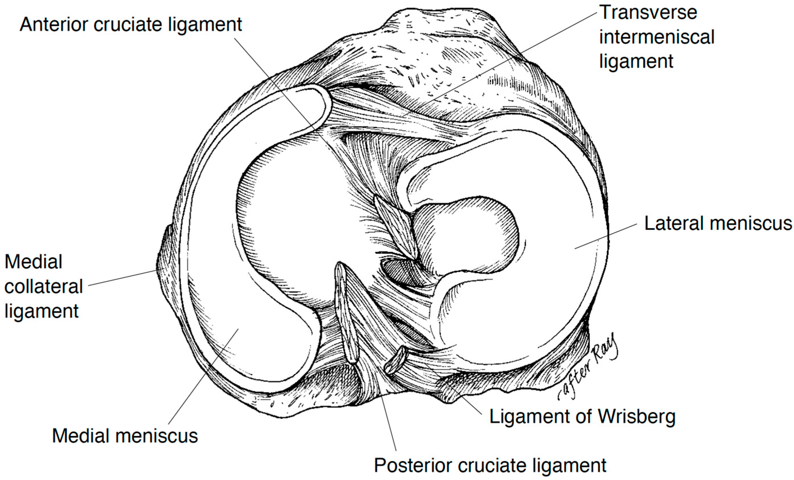
1.1. Anatomy of the Menisci

While the medial meniscus is C-shaped in appearance, the lateral meniscus is closer to a circular shape, both of which help with their function. Although they are independent structures, the medial and lateral menisci are connected by the transverse, or inter-meniscal, ligament at the anterior aspect of the tibia. They are stabilized to the tibia via the coronary ligaments with more rigid fixation focused on the medial meniscus. The meniscofemoral ligaments, also known as the Ligament of Humphrey, and the Ligament of Wrisberg, connect the menisci into the posterior cruciate ligament anteriorly and posteriorly, respectively [3,8]. (Figure 1) One or more of these ligamentous attachments may be absent in certain individuals who have anatomical variants.
It is necessary to understand the blood supply of the meniscus when addressing meniscal pathology. The middle, medial inferior, and lateral inferior genicular arteries supply blood to the periphery of the meniscus. Based on the perfusion of blood that flows to the area, the meniscus width, from the periphery toward the center, are described as the red-red zone, red-white zone, and white-white zone. This correlates with the ability to receive sufficient blood flow, with the red-red zone having the highest concentration of blood supply and the most likelihood of healing when injured. The white-white zone receives its nutrients primarily through diffusion via the synovial fluid of the knee joint, and having minimal blood supply heals very poorly [9,10]. Mechanical receptors are located in the anterior and posterior horns of the menisci contributing to proprioception, while the peripheral two-thirds contain type I and type II nociceptive free nerve endings [9,11].

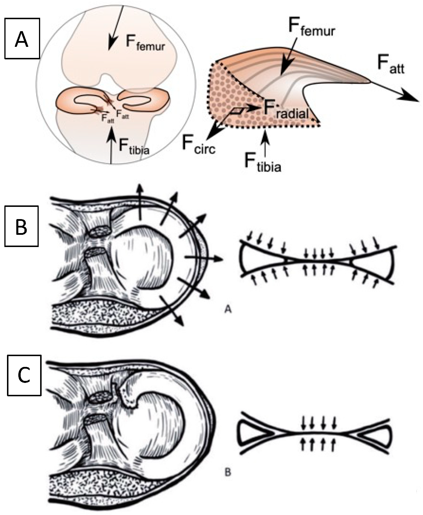
1.2. Clinical Biomechanics of the Menisci

The composition and structure of the menisci allows the transmission of joint loading by expanding under compressive forces and increasing the total contact area of the joint. The circumferential fibers provide hoop stress by converting axial compressive loading to tensile stress. (Figure 2A) While the medial meniscus is more rigid, the lateral meniscus has double the amount of excursion when under stress [2,12,13,14,15]. In the lateral compartment of the knee, approximately 70% of the load is sustained by the lateral meniscus while the cartilage directly sustains the remaining 30%; in the medial compartment, the medial meniscus shares about 50% of the load [16]. The ligamentous attachments on the meniscus to the tibia prevent peripheral extrusion under loading. The high frictional drag caused by the low permeability of the meniscal extracellular matrix protects the cartilage from high energies across the joint, as the articular cartilage is about six times as permeable [2] and thus more susceptible to strain and injury under excessive loading. Without menisci, the cartilage of the knee would undergo tremendous stresses leading to the accelerated development of osteoarthritis [17] (Figure 2B).
In addition to transmitting forces across the tibiofemoral joint, the menisci serve as secondary stabilizers. Particularly, the posterior horn of the medial meniscus is the main secondary stabilizer after the anterior cruciate ligament (ACL) to anterior translation of the tibia in relation to the femur [12,13,14,16,19]. ACL sustains an increased in situ force of 33% to 55% in the knee after medial meniscectomy [19]. When the meniscus is intact, they limit excess motion of the knee in all directions.
The integrity of the meniscus is essential to its biomechanical function. While pathology to the meniscus can occur to patients of all ages, the scope of this review focuses on the traumatic meniscus injury and its management in the younger patient population.

2. Injury Mechanism and Classification of Traumatic Meniscus Tears

2.1. Epidemiology of Traumatic Meniscus Tears

Meniscus injuries are seen in a bimodal distribution between traumatic injuries in younger patients, and oftentimes sports injuries in athletes, versus degenerative tears in the older population [20,21]. While meniscal injuries in children under 10 years old are rare, the incidence of meniscal tears in the adolescent population has been reported as 40–60 per 100,000 patients [6,22]. Recently, the incidence has risen significantly due to increased sports participation and early specialization [6,23].

2.2. Injury Mechanism of Traumatic Meniscus Tears

Most isolated meniscus injuries occur as a result of rotational or shearing forces placed across the knee joint during an increased axial load, namely the “load-and-shear” mechanism. These commonly occur during twisting or pivoting moments to the knee with the ipsilateral foot planted on the ground. They may also occur during an increased degree of knee flexion, during kneeling or squatting, while rapidly accelerating or decelerating, or while jumping [24,25], as well as through traumatic events such as motor vehicle accidents, falling from a height, or a trampoline injury. Concomitant ligamentous injuries are not uncommon. Although there is a higher incidence of medial meniscus tears overall, lateral meniscus tears are more prevalent in the setting of an acute ACL tear due to its commonly associated pivot-shift injury mechanism where the lateral femoral condyle loads and shears on the posterior tibial plateau, with the lateral meniscus in between the two [26].

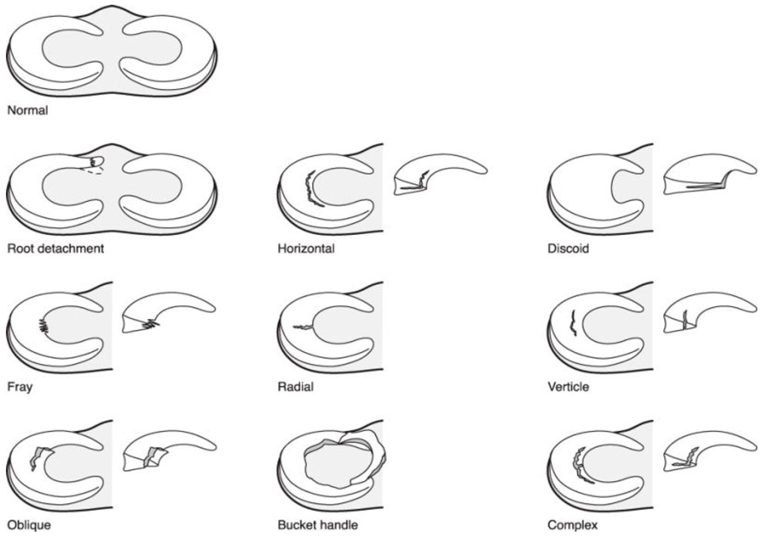
2.3. Classification of Meniscus Tears

Meniscus tears are often classified by their location, position, size, and pattern, and are treated differently based on these orientations. The location of the tear in the red-red, red-white, or white-white zone can help determine if the tear has reasonable healing potential for surgical intervention [27]. The position and size, whether it be at the anterior, middle, posterior, or at the root, plays a role in the integrity of the stability that the meniscus contributes to the knee joint itself [21].
Meniscus tear patterns are illustrated in Figure 3. Vertical, or longitudinal vertical, tears occur in-line with the circumferential collagen fibers, thus leading to less deterioration of the hoop stress mechanism. Smaller vertical tears may remain asymptomatic as the biomechanics are not significantly disrupted; however, complete vertical tears have the tendency to flip within the joint causing what is commonly known as a “bucket handle” tear [28]. These tears may cause mechanical symptoms such as clicking, popping, or even locking of the knee in flexion or extension resulting in limited knee range of motion (ROM). Complete vertical tears may also compromise the ability to distribute axial loading properly. Vertical tears are very common in the adolescent population, especially with a concomitant ACL tear [12,19,25,29]. Horizontal tears split the meniscal body into an upper and lower part. If a flap from one of the parts forms, also known as “leaflets”, the patient may present with mechanical symptoms as well. Otherwise, they may remain almost asymptomatic with low impact daily activities.
Radial tears occur in a perpendicular plane to the collagen fibers of the meniscus, thus disrupting the hoop stress mechanism. Complete radial tears that extend to the meniscocapsular junction, or greater than 90% of the width of the meniscus, lead to instability and loss of mechanical functionality of the meniscus, and are thus biomechanically similar to posterior root tears. Of note, radial meniscus tears up to 60% of the width may have less detrimental effect on the load distribution in the compartment of the knee [30]. Oblique, sometimes called parrot beak, tears give rise to large flaps of the meniscus. These flaps are mechanically unstable and may also become caught within the joint during knee ROM. Unfortunately, oblique tears have a tendency to propagate if not addressed surgically [31]. Complex meniscus tears are a combination of the above tear patterns.
Meniscal root tears can be defined as either a radial tear within 1 cm of the root attachment, or a complete avulsion of the meniscal root from the tibia. If the meniscal roots are torn, the stability of the entire meniscus is compromised and it is equivalent to a total meniscectomy [14,18,32], which indicates the total loss of the biomechanical function of the meniscus. (Figure 2C) For this reason, meniscal root tears in younger patients often benefit from surgical repair, which mitigates the risk of osteoarthritis progression and restores proper biomechanics to the knee joint.
Complex meniscus tears are any combination of the previously discussed meniscal tear patterns and usually involve the majority of the width of the meniscus. Although commonly seen in degenerative arthritic knees, these complex tears can result from propagation of smaller, simpler traumatic tears in the younger population, and they are frequently symptomatic [33,34].

2.4. The Discoid Meniscus

A discoid meniscus is an anatomical variant that presents in approximately 2–3% of the general population in the US, albeit higher prevalence has been reported in other countries [35]. The majority of these present in the lateral meniscus while the medial meniscus can have a discoid variant as well. The Watanabe classification system is commonly used to describe three types of the discoid meniscus: stable, complete discoid meniscus that covers the entire tibial plateau (type I), stable, partial discoid meniscus (type II), and unstable discoid meniscus (type III) that typically lacks menisco-tibial attachment [36]. While a discoid meniscus can remain asymptomatic, these typically become symptomatic before adulthood and are thus commonly discovered and treated in pediatric facilities. Patients more than 10 years old may present symptoms of knee locking, catching, swelling, limited ROM, pain, and clunking sound from time to time more commonly with an unstable or torn discoid meniscus. Patients younger than 10 years old may present with occasional knee clunking and discomfort due to a relatively stable but complete discoid meniscus. A stable discoid meniscus may be asymptomatic initially, with gradual development of symptoms as the patient grows in age and size, and may eventually progress toward meniscus tears. The tissue can be dysplastic across the whole discoid meniscus and not only at the portion where the excessive tissue covers the tibial plateau [37,38].
Figure 3. Patterns of meniscus tears; note that vertical tears are also known as longitudinal vertical tears; bucket handle tears result from an extensive vertical tear with or without other associated tear patterns. (Courtesy from Shieh et al. [34]).
Pathophysiology 30 00044 g003

3. Surgical Management of Meniscus Tears

Treatment of traumatic meniscus tears should preserve the meniscus when possible, especially in the young population. It is important to preserve the meniscus to maintain joint congruency and load transfer, thus mitigating the risk of post-traumatic osteoarthritis. Special considerations are taken depending on the location, tear pattern (morphology), and chronicity of the meniscus tear [22,39]. Meniscal repairs have positive clinical outcomes in the general population when performed for suitable tear patterns. Although there is limited literature focusing on the pediatric population and current preference leans towards repair versus meniscectomy when possible, in light of longer lifespans and more active lifestyles for the younger population [22].

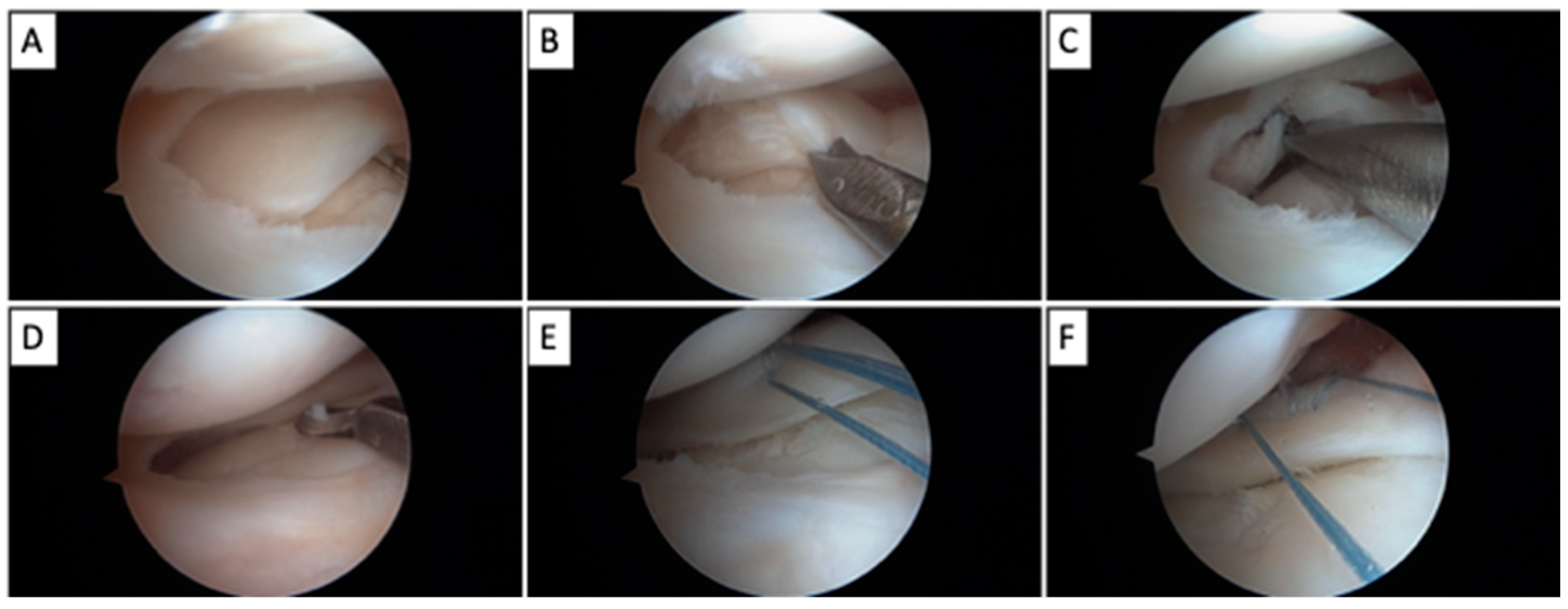
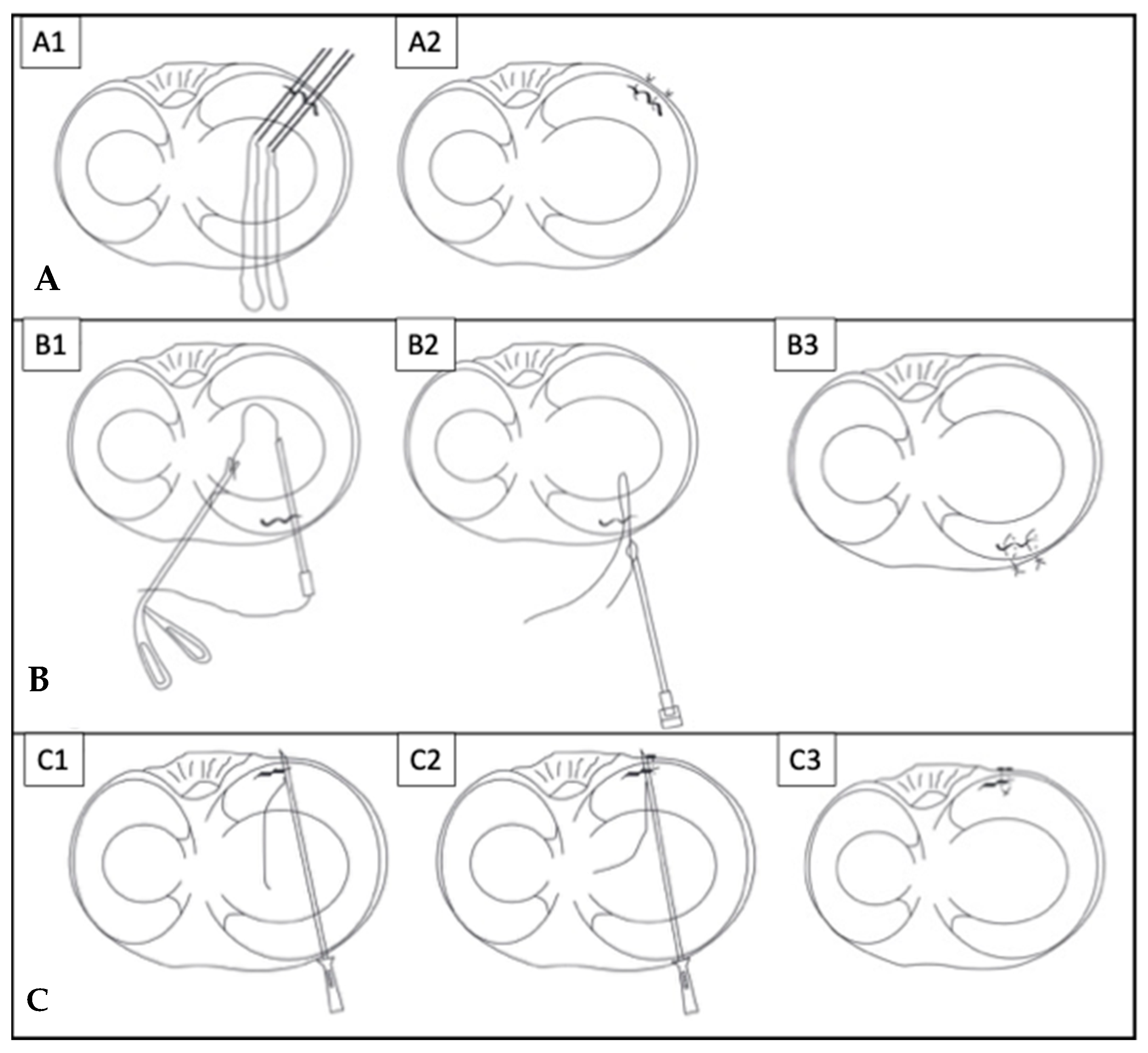
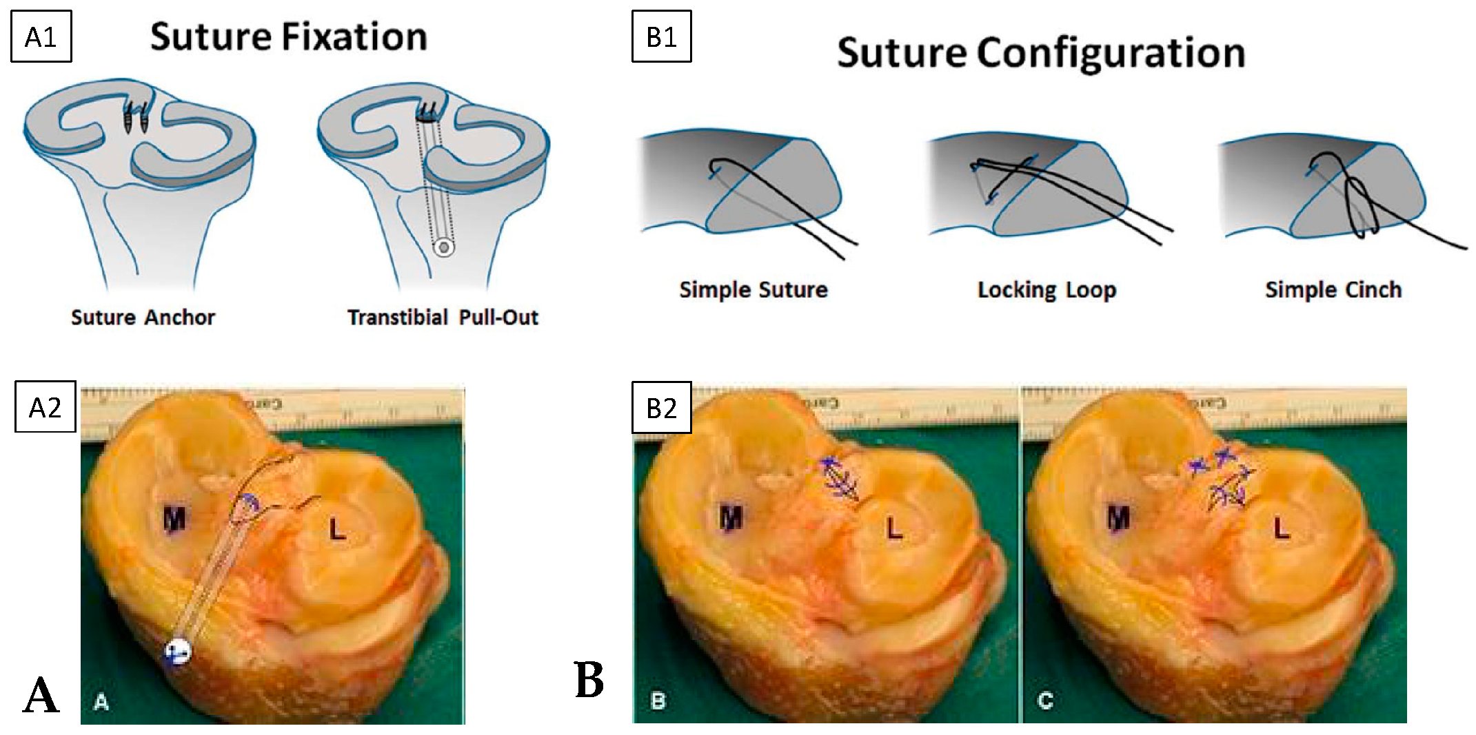
3.1. Surgical Techniques for Meniscus Repair

A number of repair techniques have been developed that can be categorized as in-side-out, outside-in, and all-inside (Figure 4). The inside-out technique combines an arthroscopic and an open technique. This involves passing sutures arthroscopically which are then retrieved through the meniscus from an incision outside the capsule. These inside-out techniques use double-armed sutures with mostly vertical mattress sutures due to their higher strength, while needing horizontal mattress sutures periodically for anatomic reduction. An incision in the posteromedial or posterolateral aspect of the knee is then used to retrieve the sutures and tie them over the joint capsule avoiding neurovascular structures [6]. The outside-in technique, on the other hand, involves passing sutures through the meniscus which are then tied over the capsule through a small incision where the suture exits the skin. Lastly, the all-inside technique involves the use of suture anchors through the arthroscopic portals without the need for an additional open approach to the knee. In this case, the repair sutures are anchored and cinched toward a synthetic anchor on the joint capsule or the edge of the meniscus. Alternatively, all-inside repairs can be performed using various suture-passing devices through the arthroscopic portals [40].
All-inside and inside-out techniques are the most widely used and they have similar repair strength biomechanically, as well as comparable clinical outcomes [21,41,42]. For anterior horn tears, the outside-in technique may provide better access to place the sutures [27]; however, recent advances of meniscus repair implant design, or creative use of suture-passing techniques, extends the application of the all-inside technique to a wider range of tear patterns, including anterior horn tears while minimizing the risk of further injury to the articular cartilage or the meniscus when passing the sutures [43].
Figure 4. Meniscus repair techniques of (A) inside-out repairs, classic technique for posterior horn to mid-body tears: (A1) the sutures are passed from inside of the joint under knee arthroscopy, and (A2) retrieved posterior to the joint capsule accessed via open incision; (B) outside-in repairs, suitable for tears in the anterior horns: (B1) under knee arthroscopy, the sutures are passed from outside of the joint, then (B2) retrieved from a separate portal back to the outside, and (B3) tied down to complete the repair; (C) all-inside repairs with a suture-anchor construct, completed solely under arthroscopy without additional incisions: (C1) passing the first limb of the sutures, (C2) passing the second limb, and (C3) cinching the suture to complete the repair. (Courtesy from Yoon et al. [44]).
Pathophysiology 30 00044 g004

3.2. Considerations of Tear Locations

As described above, the location of a meniscal tear (e.g., red-red, red-white, or white-white zones) is associated with healing potential. Since the meniscus carries higher vascularity peripherally than centrally, tears in the peripheral-third red-red zone of the meniscus have greater healing potential than those in the adjacent middle-third red-white zone, with the least healing potential of tears in the most central-third white-white zone. Therefore, peripheral tears are more amenable to repair rather than a partial meniscectomy [22]. In addition, younger patients may have a higher propensity of more peripheral tears as well as possible greater healing potential compared to degenerative meniscus tears in the elderly [45,46]. Therefore, a combined technique of partial meniscectomy for the central portion of the tears and repair of tears in the more vascular zones may be utilized [6].

3.3. Considerations of Tear Patterns

Meniscus tear patterns have implications on the healing potential as well as the repair technique [27]. Longitudinal vertical tears that are partial tears (not full-thickness) or full-thickness but less than 10 mm in length may not require surgical repair due to better mechanical stability. Unstable longitudinal vertical tears, including bucket handle tears that occur in the vascular zone typically have reasonable healing potential; an inside-out or all-inside repair with vertically stacked sutures can be used for better incorporation into the repair [40]. In the setting of radial tears, the hoop stress is disrupted and the tear commonly extends through both the peripheral and the central zones, resulting in lower healing capacity and potentially a nonfunctional meniscus; when repair is attempted, a combination of vertical and horizontal mattresses sutures may be necessary to provide greater strength to the repair. This can be completed using an all-inside technique [40]. However, if these tears occur solely in the white-white zone, partial meniscectomy is preferred [47]. Treatment of horizontal tears may include partial meniscectomy of the unstable leaflets, or use of circumferential sutures may be a viable option. Meniscocapsular separations may be treated with an all-inside or inside-out suture technique, similar to that used for peripheral vertical longitudinal tears [48].
Treatment of meniscal root tears involves re-anchoring the meniscus root back to the anatomic footprint if the root loses its “anchor” when completely or nearly completely torn off its attachment to the tibia. In situ suture repair may be utilized for meniscal root tears of smaller magnitude that does not detach the anchor. This is preferred over meniscectomy for restoring meniscus function because biomechanically, losing the anchor of the meniscal root equals total meniscectomy. The technique for repairing meniscal root tears involves the use of transosseous sutures or suture anchors (Figure 5) [40,49].
Further considerations are taken when meniscus tears occur with concomitant injuries such as ACL injuries. There have been reports of pediatric patients with optimistic healing potential with meniscus repair in the setting of ACL reconstruction. There has been a higher rate of meniscus healing after repair with ACL reconstruction versus when repaired in isolation, likely due to increased vascularity to the repair [6,22]. The mechanism behind such augmented healing potential is still under investigation; hypotheses include a favorable biological environment with ACL tunnel drilling and possibly poorer quality of the meniscus in an isolated meniscus tear without an ACL tear, which can result from lower traumatic energy [51,52,53].

3.4. Management of the Discoid Meniscus

Management of patients with a discoid meniscus depends on the spectrum of the disease. Non-operative treatment is appropriate for asymptomatic patients. Symptomatic patients typically have an unstable or torn discoid meniscus, or a combination of both, which necessitate surgical interventions [6]. Younger patients less than 10 years old may experience knee clunking and limited ROM even without the discoid meniscus being torn; teenagers are more likely to experience more pain, swelling, and mechanical symptoms from a torn discoid meniscus. Discoid menisci have less peripheral vascularity and less ideal tissue quality compared to a normal meniscus, making the healing potential more difficult. Previously surgical treatment involved total meniscectomy, which leads to good clinical improvement in the short-term but degenerative changes in the compartment in the mid-term [6,54]. Partial meniscectomy, or saucerization to a 6 to 8 mm remnant to preserve the crescent, is preferred when the peripheral stability of the meniscus can be preserved. If the remnant is unstable, peripheral meniscocapsular repair is necessary and may be completed using an all-inside or inside-out approach. An all-inside approach may be more difficult in younger patients with a smaller knee and extra caution should be utilized to avoid injury to the posterior neurovascular bundles when penetrating the joint capsule, and the articular cartilage when introducing the device into the joint [6,37,54,55]. Horizontal cleavage tears are common in patients with a discoid meniscus which can be repaired if in the peripheral red-red zone [6]. (Figure 6).

3.5. Evolving Treatment for Meniscus Injuries

Due to the importance of meniscus integrity and function in the prevention of developing osteoarthritis, there have been techniques such as biologics application to stimulate healing, and meniscus transplant to replace the unsalvageable meniscus, such as those in settings of avascular tears, and inevitable large or total meniscectomies. There have been optimistic reported clinical and imaging outcomes with the use of stem cells injected into a collagen scaffold used during meniscus repair of regions with low vascularity [56]. Meniscus allograft transplantation (MAT) may be used in younger patients with significant meniscal deficiency or menisci deemed nonfunctional. Although more widely used in the adult population, studies among the pediatric population demonstrate that MAT is an option among this population with clinical improvement at least in the short-term after surgery [57].

3.6. Outcomes after Surgical Treatment

Better outcomes may be associated with a carefully selected patient population who have lower BMI (body mass index), minimal or no arthritic changes in the knee, normal mechanical alignment of the lower extremity, less high impact activities, and the absence of joint inflammatory diseases. [58,59,60] Postoperative rehabilitation protocols may have an effect on the outcome.

4. Postoperative Rehabilitation

Patients with partial meniscectomy without meniscus repair may progress toward early full ROM and full weight bearing as tolerated.
Currently there is not a single widely accepted post operative protocol after meniscus repair. Postoperative immobilization, restricted ROM, weight bearing, and exercise progression have all been taken into consideration based on type of tear, location, and surgical management [61]. The goal postoperatively is to protect the repair, without affecting progress secondary to immobilization and disuse, which lead to stiffness and loss of strength. There have been tested protocols of conservative treatment with early restricted or non-weight bearing for up to six weeks and slow progression in ROM versus more aggressive protocols of immediate full weight bearing with early ROM progressions [62]. Conservative management aims to protect the repair from shear forces that may result from hamstring activation and increased loads to the meniscus from increased knee flexion for up to 4 months with return to play delayed up to 4 to 6 months, while accelerated protocols reports earlier knee flexion and earlier return to play within a 3 to 4 month period [63].
There were no significant differences in complication rates or functional outcomes in a systematic review that included 798 patients, where 438 patients were treated with a restricted rehabilitation protocol and 360 patients were treated with an accelerated protocol [64]. Although the evidence is promising for the use of an accelerated postoperative protocol, evidence of the effects of rotation and torsional forces as well as long-term studies is limited [64]. Further systematic reviews have supported these conclusions with comparable outcomes between the use of conservative versus accelerated protocols [62]. There is tremendous variability in repair techniques and rehabilitation protocols used throughout the current literature, and therefore no single recommendation for rehabilitation protocols can be made at this time without further prospective studies. There is also a lack of literature regarding protocols controlling for the patient’s age [62,63]. Further, there is lack of biomechanical evidence comparing various postoperative protocols regarding the impact on repair integration. Based on the current literature, the use of a conservative versus aggressive postoperative rehab protocol is surgeon- and patient-specific with no consensus on the superiority of use of one over another.

5. Conclusions

The meniscus is a key structure in protecting cartilage against loading forces as well as providing additional stability. In recent years there has been an increase in incidence of meniscal tears among the pediatric and adolescent population due to increased activity and sports specialization at a young age [7]. The importance of preserving the meniscus, especially in the younger population, has been established due to the risk of post traumatic arthritis and alterations of knee biomechanics with a compromised meniscus [40,65].
Surgical repair of the meniscus has been established as a viable option to restore knee joint kinematics and prevent cartilage degradation.
Utilization of proper technique for surgical repair of meniscus tears among the younger population is crucial to maintain proper knee kinematics and prevention of post traumatic arthritis. There is still the need for more high-level evidence regarding surgical management and postoperative rehabilitation protocols after meniscus repair, particularly for the pediatric population with traumatic meniscus tears. Future prospective studies with long-term follow up as well as well-controlled biomechanical and biological studies would allow for greater consensus and shed light on clinically effective treatment options for young patients.

Author Contributions

All authors contributed to the idea formation, literature reviews, manuscript preparation, and finalization of the submission process. All authors have read and agreed to the published version of the manuscript.

Funding

This project was supported by the DCMR COBRE program, with a grant from the National Institute of General Medical Sciences and from the National Institutes of Health—NIH-NIGMS COBRE (P20 GM139760).

Institutional Review Board Statement

Not applicable.

Informed Consent Statement

Not applicable.

Data Availability Statement

The data presented in this study are available in this article.

Conflicts of Interest

The authors declare no conflict of interest.

References

  1. Clark, C.R.; Ogden, J.A. Development of the menisci of the human knee joint. Morphological changes and their potential role in childhood meniscal injury. J. Bone Jt. Surg. Am. 1983, 65, 538–547. [Google Scholar] [CrossRef]
  2. Fithian, D.C.; Kelly, M.A.; Mow, V.C. Material properties and structure-function relationships in the menisci. Clin. Orthop. Relat. Res. 1990, 252, 19–31. [Google Scholar] [CrossRef]
  3. Fox, A.J.S.; Bedi, A.; Rodeo, S.A. The Basic Science of Human Knee Menisci: Structure, Composition, and Function. Sports Health 2012, 4, 340–351. [Google Scholar] [CrossRef] [PubMed]
  4. Greis, P.E.; Bardana, D.D.; Holmstrom, M.C.; Burks, R.T. Meniscal Injury: I. Basic Science and Evaluation. J. Am. Acad. Orthop. Surg. 2002, 10, 168–176. [Google Scholar] [CrossRef] [PubMed]
  5. Kumari, A.; Gupta, G.; Kumar, P.; Rani, S. Morphological study of the menisci of the knee joint in human cadaver in Jharkhand population. J. Fam. Med. Prim. Care 2022, 11, 4723–4729. [Google Scholar] [CrossRef] [PubMed]
  6. Bellisari, G.; Samora, W.; Klingele, K. Meniscus Tears in Children. Sports Med. Arthrosc. Rev. 2011, 19, 50–55. [Google Scholar] [CrossRef]
  7. Vinagre, G.; Cruz, F.; Alkhelaifi, K.; D’Hooghe, P. Isolated meniscus injuries in skeletally immature children and adolescents: State of the art. J. ISAKOS 2022, 7, 19–26. [Google Scholar] [CrossRef]
  8. Bryceland, J.K.; Powell, A.J.; Nunn, T. Knee Menisci. Cartilage 2017, 8, 99–104. [Google Scholar] [CrossRef]
  9. Gray, J.C. Neural and Vascular Anatomy of the Menisci of the Human Knee. J. Orthop. Sports Phys. Ther. 1999, 29, 23–30. [Google Scholar] [CrossRef]
  10. Scapinelli, R. Vascular anatomy of the human cruciate ligaments and surrounding structures. Clin. Anat. 1997, 10, 151–162. [Google Scholar] [CrossRef]
  11. Mine, T.; Kimura, M.; Sakka, A.; Kawai, S. Innervation of nociceptors in the menisci of the knee joint: An immunohistochemical study. Arch. Orthop. Trauma Surg. 2000, 120, 201–204. [Google Scholar] [CrossRef]
  12. Allen, C.R.; Wong, E.K.; Livesay, G.A.; Sakane, M.; Fu, F.H.; Woo, S.L.-Y. Importance of the medial meniscus in the anterior cruciate ligament-deficient knee. J. Orthop. Res. 2000, 18, 109–115. [Google Scholar] [CrossRef] [PubMed]
  13. DePhillipo, N.N.; Moatshe, G.; Chahla, J.; Aman, Z.S.; Storaci, H.W.; Morris, E.R.; Robbins, C.M.; Engebretsen, L.; LaPrade, R.F. Quantitative and Qualitative Assessment of the Posterior Medial Meniscus Anatomy: Defining Meniscal Ramp Lesions. Am. J. Sports Med. 2018, 47, 372–378. [Google Scholar] [CrossRef] [PubMed]
  14. Rao, A.J.; Erickson, B.J.; Cvetanovich, G.L.; Yanke, A.B.; Bach, B.R.; Cole, B.J. The Meniscus-Deficient Knee: Biomechanics, Evaluation, and Treatment Options. Orthop. J. Sports Med. 2015, 3, 2325967115611386. [Google Scholar] [CrossRef] [PubMed]
  15. Śmigielski, R.; Becker, R.; Zdanowicz, U.; Ciszek, B. Medial meniscus anatomy—From basic science to treatment. Knee Surg. Sports Traumatol. Arthrosc. 2014, 23, 8–14. [Google Scholar] [CrossRef] [PubMed]
  16. Walker, P.S.; Erkiuan, M.J. The Role of the Menisci in Force Transmission Across the Knee. Clin. Orthop. Relat. Res. 1975, 109, 184–192. [Google Scholar] [CrossRef]
  17. Radin, E.L.; Rose, R.M. Role of subchondral bone in the initiation and progression of cartilage damage. Clin. Orthop. Relat. Res. 1986, 213, 34–40. [Google Scholar] [CrossRef]
  18. Itthipanichpong, T.; Moonwong, S. Medial Meniscus Root Tear: Current Update Review. In Arthroscopy; Suarez-Ahedo, C., Ed.; IntechOpen: London, UK, 2022. [Google Scholar] [CrossRef]
  19. Papageorgiou, C.D.; Gil, J.E.; Kanamori, A.; Fenwick, J.A.; Woo, S.L.-Y.; Fu, F.H. The Biomechanical Interdependence between the Anterior Cruciate Ligament Replacement Graft and the Medial Meniscus. Am. J. Sports Med. 2001, 29, 226–231. [Google Scholar] [CrossRef]
  20. Everhart, J.S.; Higgins, J.D.; Poland, S.G.; Abouljoud, M.M.; Flanigan, D.C. Meniscal repair in patients age 40 years and older: A systematic review of 11 studies and 148 patients. Knee 2018, 25, 1142–1150. [Google Scholar] [CrossRef]
  21. Liechti, D.J.; Constantinescu, D.S.; Ridley, T.J.; Chahla, J.; Mitchell, J.J.; Vap, A.R. Meniscal Repair in Pediatric Populations: A Systematic Review of Outcomes. Orthop. J. Sports Med. 2019, 7, 232596711984335. [Google Scholar] [CrossRef]
  22. Jackson, T.; Fabricant, P.D.; Beck, N.; Storey, E.; Patel, N.M.; Ganley, T.J. Epidemiology, Injury Patterns, and Treatment of Meniscal Tears in Pediatric Patients: A 16-Year Experience of a Single Center. Orthop. J. Sports Med. 2019, 7, 232596711989032. [Google Scholar] [CrossRef]
  23. Carter, C.W.; Kocher, M.S. Meniscus Repair in Children. Clin. Sports Med. 2012, 31, 135–154. [Google Scholar] [CrossRef]
  24. Kurzweil, P.R.; Cannon, W.D.; DeHaven, K.E. Meniscus Repair and Replacement. Sports Med. Arthrosc. Rev. 2018, 26, 160–164. [Google Scholar] [CrossRef]
  25. Wilson, P.L.; Wyatt, C.W.; Romero, J.; Sabatino, M.J.; Ellis, H.B. Incidence, Presentation, and Treatment of Pediatric and Adolescent Meniscal Root Injuries. Orthop. J. Sports Med. 2018, 6, 2325967118803888. [Google Scholar] [CrossRef] [PubMed]
  26. Srimongkolpitak, S.; Chernchujit, B. Current concepts on meniscal repairs. J. Clin. Orthop. Trauma 2022, 27, 101810. [Google Scholar] [CrossRef] [PubMed]
  27. Ozeki, N.; Seil, R.; Krych, A.J.; Koga, H. Surgical treatment of complex meniscus tear and disease: State of the art. J. ISAKOS 2021, 6, 35–45. [Google Scholar] [CrossRef] [PubMed]
  28. Saltzman, B.M.; Cotter, E.J.; Wang, K.C.; Rice, R.; Manning, B.T.; Yanke, A.B.; Forsythe, B.; Verma, N.N.; Cole, B.J. Arthroscopically Repaired Bucket-Handle Meniscus Tears: Patient Demographics, Postoperative Outcomes, and a Comparison of Success and Failure Cases. Cartilage 2020, 11, 77–87. [Google Scholar] [CrossRef] [PubMed]
  29. Andrish, J.T. Meniscal Injuries in Children and Adolescents: Diagnosis and Management. J. Am. Acad. Orthop. Surg. 1996, 4, 231–237. [Google Scholar] [CrossRef] [PubMed]
  30. Bedi, A.; Kelly, N.; Baad, M.; Fox, A.J.; Ma, Y.; Warren, R.F.; Maher, S.A. Dynamic Contact Mechanics of Radial Tears of the Lateral Meniscus: Implications for Treatment. Arthrosc. J. Arthrosc. Relat. Surg. 2012, 28, 372–381. [Google Scholar] [CrossRef] [PubMed]
  31. Maffulli, N.; Longo, U.G.; Campi, S.; Denaro, V. Meniscal tears. Open Access J. Sports Med. 2010, 1, 45–54. [Google Scholar] [CrossRef]
  32. Chahla, J.; LaPrade, R.F. Meniscal Root Tears. Arthroscopy 2019, 35, 1304–1305. [Google Scholar] [CrossRef]
  33. Ciemniewska-Gorzela, K.; Bąkowski, P.; Naczk, J.; Jakob, R.; Piontek, T. Complex Meniscus Tears Treated with Collagen Matrix Wrapping and Bone Marrow Blood Injection: Clinical Effectiveness and Survivorship after a Minimum of 5 Years’ Follow-Up. Cartilage 2021, 13, 228S–238S. [Google Scholar] [CrossRef] [PubMed]
  34. Shieh, A.; Bastrom, T.; Roocroft, J.; Edmonds, E.W.; Pennock, A.T. Meniscus Tear Patterns in Relation to Skeletal Immaturity: Children Versus Adolescents. Am. J. Sports Med. 2013, 41, 2779–2783. [Google Scholar] [CrossRef]
  35. Jordan, M.R. Lateral Meniscal Variants: Evaluation and Treatment. J. Am. Acad. Orthop. Surg. 1996, 4, 191–200. [Google Scholar] [CrossRef]
  36. Watanabe, M.; Takeda, S.; Ikeuchi, H. Atlas of Arthroscopy, 3rd ed.; Springer: Berlin/Heidelberg, Germany, 1979. [Google Scholar]
  37. Kocher, M.S.; Logan, C.A.; Kramer, D.E. Discoid Lateral Meniscus in Children: Diagnosis, Management, and Outcomes. J. Am. Acad. Orthop. Surg. 2017, 25, 736–743. [Google Scholar] [CrossRef] [PubMed]
  38. Yoo, W.J.; Choi, I.H.; Chung, C.Y.; Lee, M.C.; Cho, T.-J.; Park, M.S.; Lee, D.Y. Discoid Lateral Meniscus in Children. Limited Knee Extension and Meniscal Instability in the Posterior Segment. J. Pediatr. Orthop. 2008, 28, 544–548. [Google Scholar] [CrossRef] [PubMed]
  39. Arner, J.W.; Ruzbarsky, J.J.; Vidal, A.F.; Frank, R.M. Meniscus Repair Part 1: Biology, Function, Tear Morphology, and Special Considerations. J. Am. Acad. Orthop. Surg. 2022, 30, e852–e858. [Google Scholar] [CrossRef] [PubMed]
  40. Arner, J.W.; Ruzbarsky, J.J.; Vidal, A.F.; Frank, R.M. Meniscus Repair Part 2: Technical Aspects, Biologic Augmentation, Rehabilitation, and Outcomes. J. Am. Acad. Orthop. Surg. 2022, 30, 613–619. [Google Scholar] [CrossRef]
  41. Barber, F.; Herbert, M.A.; Richards, D.P. Load to failure testing of new meniscal repair devices. Arthrosc. J. Arthrosc. Relat. Surg. 2004, 20, 45–50. [Google Scholar] [CrossRef]
  42. Fillingham, Y.A.; Riboh, J.C.; Erickson, B.J.; Bach, B.R.; Yanke, A.B. Inside-Out Versus All-Inside Repair of Isolated Meniscal Tears: An Updated Systematic Review. Am. J. Sports Med. 2016, 45, 234–242. [Google Scholar] [CrossRef]
  43. Malinowski, K.; Góralczyk, A.; Hermanowicz, K.; LaPrade, R.F. Tips and Pearls for All-Inside Medial Meniscus Repair. Arthrosc. Tech. 2019, 8, e131–e139. [Google Scholar] [CrossRef] [PubMed]
  44. Yoon, K.H.; Park, K.H. Meniscal Repair. Knee Surg. Relat. Res. 2014, 26, 68–76. [Google Scholar] [CrossRef] [PubMed]
  45. Mesiha, M.; Zurakowski, D.; Soriano, J.; Nielson, J.H.; Zarins, B.; Murray, M.M. Pathologic Characteristics of the Torn Human Meniscus. Am. J. Sports Med. 2007, 35, 103–112. [Google Scholar] [CrossRef]
  46. Pauli, C.; Grogan, S.; Patil, S.; Otsuki, S.; Hasegawa, A.; Koziol, J.; Lotz, M.; D’lima, D. Macroscopic and histopathologic analysis of human knee menisci in aging and osteoarthritis. Osteoarthr. Cartil. 2011, 19, 1132–1141. [Google Scholar] [CrossRef] [PubMed]
  47. Cabral, J.; Sinikumpu, J. Clinical considerations of anatomy and magnetic resonance imaging in pediatric meniscus tear, with imaging-based treatment options. J. Child. Orthop. 2023, 17, 63–69. [Google Scholar] [CrossRef]
  48. Hirtler, L.; Unger, J.; Weninger, P. Acute and chronic menisco-capsular separation in the young athlete: Diagnosis, treatment and results in thirty seven consecutive patients. Int. Orthop. 2015, 39, 967–974. [Google Scholar] [CrossRef]
  49. Bonasia, D.E.; Pellegrino, P.; D’Amelio, A.; Cottino, U.; Rossi, R. Meniscal root tear repair: Why, when and how? Orthop. Rev. 2015, 7, 5792. [Google Scholar] [CrossRef]
  50. Krych, A.J.; Hevesi, M.; Leland, D.P.; Stuart, M.J. Meniscal Root Injuries. J. Am. Acad. Orthop. Surg. 2020, 28, 491–499. [Google Scholar] [CrossRef]
  51. De Girolamo, L.; Galliera, E.; Volpi, P.; Denti, M.; Dogliotti, G.; Quaglia, A.; Cabitza, P.; Romanelli, M.M.C.; Randelli, P. Why menisci show higher healing rate when repaired during ACL reconstruction? Growth factors release can be the explanation. Knee Surg. Sports Traumatol. Arthrosc. 2015, 23, 90–96. [Google Scholar] [CrossRef]
  52. Meister, K.; Indelicato, P.A.; Spanier, S.; Franklin, J.; Batts, J. Histology of the Torn Meniscus: A Comparison of Histologic Differences in Meniscal Tissue between Tears in Anterior Cruciate Ligament-Intact and Anterior Cruciate Ligament-Deficient Knees. Am. J. Sports Med. 2004, 32, 1479–1483. [Google Scholar] [CrossRef]
  53. Rodríguez-Roiz, J.M.; Sastre-Solsona, S.; Popescu, D.; Montañana-Burillo, J.; Combalia-Aleu, A. The relationship between ACL reconstruction and meniscal repair: Quality of life, sports return, and meniscal failure rate—2- to 12-year follow-up. J. Orthop. Surg. Res. 2020, 15, 361. [Google Scholar] [CrossRef] [PubMed]
  54. Klingele, K.E.; Kocher, M.S.; Hresko, M.T.; Gerbino, P.; Micheli, L.J. Discoid Lateral Meniscus: Prevalence of Peripheral Rim Instability. J. Pediatr. Orthop. 2004, 24, 79–82. [Google Scholar] [CrossRef] [PubMed]
  55. Hayashi, L.K.; Yamaga, H.; Ida, K.; Miura, T. Arthroscopic meniscectomy for discoid lateral meniscus in children. J. Bone Jt. Surg Am. 1988, 70, 1495–1500. [Google Scholar] [CrossRef]
  56. Jacob, G.; Shimomura, K.; Krych, A.J.; Nakamura, N. The Meniscus Tear: A Review of Stem Cell Therapies. Cells 2019, 9, 92. [Google Scholar] [CrossRef] [PubMed]
  57. Middleton, S.; Asplin, L.; Stevenson, C.; Thompson, P.; Spalding, T. Meniscal allograft transplantation in the paediatric population: Early referral is justified. Knee Surg. Sports Traumatol. Arthrosc. 2019, 27, 1908–1913. [Google Scholar] [CrossRef]
  58. Brophy, R.H.; Wojahn, R.D.; Lillegraven, O.; Lamplot, J.D. Outcomes of Arthroscopic Posterior Medial Meniscus Root Repair: Association with Body Mass Index. J. Am. Acad. Orthop. Surg. 2019, 27, 104–111. [Google Scholar] [CrossRef]
  59. Ridley, T.J.; McCarthy, M.A.; Bollier, M.J.; Wolf, B.R.; Amendola, A. Age Differences in the Prevalence of Isolated Medial and Lateral Meniscal Tears in Surgically Treated Patients. Iowa Orthop. J. 2017, 37, 91–94. [Google Scholar]
  60. Yang, S.-J.; Ding, Z.-J.; Li, J.; Xue, Y.; Chen, G. Factors influencing postoperative outcomes in patients with symptomatic discoid lateral meniscus. BMC Musculoskelet. Disord. 2020, 21, 551. [Google Scholar] [CrossRef]
  61. Harput, G.; Guney-Deniz, H.; Nyland, J.; Kocabey, Y. Postoperative rehabilitation and outcomes following arthroscopic isolated meniscus repairs: A systematic review. Phys. Ther. Sport 2020, 45, 76–85. [Google Scholar] [CrossRef]
  62. VanderHave, K.L.; Perkins, C.; Le, M. Weightbearing Versus Non-weightbearing After Meniscus Repair. Sports Health 2015, 7, 399–402. [Google Scholar] [CrossRef]
  63. Pabian, P.; Hanney, W.J. Functional rehabilitation after medial meniscus repair in a high school football quarterback: A case report. N. Am. J. Sports Phys. Ther. 2008, 3, 161–169. [Google Scholar] [PubMed]
  64. Spang, R.C., III; Nasr, M.C.; Mohamadi, A.; DeAngelis, J.P.; Nazarian, A.; Ramappa, A.J. Rehabilitation following meniscal repair: A systematic review. BMJ Open Sport Exerc. Med. 2018, 4, e000212. [Google Scholar] [CrossRef] [PubMed]
  65. Meadows, K.D.; Peloquin, J.M.; Markhali, M.I.; Zgonis, M.H.; Schaer, T.P.; Mauck, R.L.; Elliott, D.M. Acute Repair of Meniscus Root Tear Partially Restores Joint Displacements as Measured with Magnetic Resonance Images and Loading in a Cadaveric Porcine Knee. J. Biomech. Eng. 2023, 145, 081002. [Google Scholar] [CrossRef] [PubMed]
Figure 1. Normal anatomy of the menisci, with the C-shaped medial meniscus and O-shaped lateral meniscus, and the inter-meniscal ligament (i.e., “transverse intermeniscal ligament”) anteriorly and menisci roots attachment onto the tibial posteriorly along with ligaments attaching to the posterior cruciate ligament (Ligament of Wrisberg posteriorly, and Ligament of Humphrey anteriorly—not shown). (Courtesy from Greis et al. [4]).
Pathophysiology 30 00044 g001
Figure 2. Biomechanical function of the meniscus, where: (A) Axial compressive forces from the femur (Ffemur) and tibia (Ftibia) during joint loading are converted into the circumferential “hoop” direction (Fcirc) that are resisted by the tensile stiffness of the circumferential collagen fibers and are transferred into the horn attachments (Fatt). (B) This load transfer reduces the axial forces on the underlying articular cartilage. (C) With a meniscus root avulsion or large-scale radial tear, the hoop stress is lost and the axial forces are applied directly to the cartilage, the clinical equivalent of a meniscectomy, and leads to cartilage osteoarthritis. (A) Courtesy from Dawn Elliott and Robert Mauck; (B) and (C): (Courtesy from Itthipanichpong et al. [18]).
Pathophysiology 30 00044 g002
Figure 5. Surgical treatment of a meniscal root tear by (A) repairing meniscus root avulsion by re-attaching the root to its tibia insertional footprint via a transtibial bone tunnel with suspensory fixation on the bone cortex. (B) Utilizing various suture constructs to repair intrasubstance root tear, or to create the pulling stitch for meniscal root repair as described above. Subpanels: the (A1,B1) schematics, and (A2,B2) cadaveric demonstration photos, of the techniques. (Courtesy from Bonasia et al. 2015 [49], Krych et al. [50]).
Pathophysiology 30 00044 g005
Figure 6. Surgical treatment of a discoid lateral meniscus: (A) A complete discoid meniscus that is torn and formed an unstable flap, in addition to the absence of posterior menisco-capsular attachment, probed under knee arthroscopy, (B,C) Debridement and resection of the torn flap using arthroscopic tools (biters and motorized shavers), (D) further saucerization of the meniscus to approach the normal C-shape, (E) stabilizing the meniscus using an all-inside meniscus repair technique, while (F) using the stitch from the first repair construct as a pulling stitch to facilitate the passing of the second all-inside repair construct, to finally achieve a stable meniscus.
Pathophysiology 30 00044 g006
Disclaimer/Publisher’s Note: The statements, opinions and data contained in all publications are solely those of the individual author(s) and contributor(s) and not of MDPI and/or the editor(s). MDPI and/or the editor(s) disclaim responsibility for any injury to people or property resulting from any ideas, methods, instructions or products referred to in the content.

