Article

Tetrahydrobiopterin (BH4) Supplementation Prevents the Cardiorenal Effects of Diabetes in Mice by Reducing Oxidative Stress, Inflammation and Fibrosis

1
Departamento de Ciencias Básicas Biomédicas, Facultad de Ciencias de la Salud, Universidad de Talca, Avenida Lircay s/n, Talca 3460000, Chile
2
Centro de Investigación de Estudios Avanzados del Maule (CIEAM), Vicerrectoría de Investigación y Postgrado, Universidad Católica del Maule, Talca 3466706, Chile
3
Instituto de Química de Recursos Naturales, Universidad de Talca, Talca 3460000, Chile
4
Departamento de Biología y Química, Facultad de Ciencias Básicas, Universidad Catolica del Maule, Talca 3466706, Chile
*
Author to whom correspondence should be addressed.
Biomedicines 2022, 10(10), 2479; https://doi.org/10.3390/biomedicines10102479
Submission received: 27 June 2022 / Revised: 4 September 2022 / Accepted: 5 September 2022 / Published: 4 October 2022

Abstract

:
Background: The effects of diabetes on the cardiovascular system as well as in the kidney are profound, which include hypertrophy and fibrosis. Diabetes also induces oxidative stress, at least in part due to the uncoupling of nitric oxide synthase (NOS); this is a shift in NO production toward superoxide production due to reduced levels of the NOS cofactor tetrahydrobiopterin (BH4). With this in mind, we tested the hypothesis that BH4 supplementation may prevent the development of diabetic cardiomyopathy and nephropathy. Methods: Diabetes was induced in Balb/c mice with streptozotocin. Then, diabetic mice were divided into two groups: one group provided with BH4 (sapropterin) in drinking water (daily doses of 15 mg/kg/day, during eight weeks) and the other that received only water. A third group of normoglycemic mice that received only water were used as the control. Results: Cardiac levels of BH4 were increased in mice treated with BH4 (p = 0.0019). Diabetes induced cardiac hypertrophy, which was prevented in the group that received BH4 (p < 0.05). In addition, hypertrophy was evaluated as cardiomyocyte cross-sectional area. This was reduced in diabetic mice that received BH4 (p = 0.0012). Diabetes induced cardiac interstitial fibrosis that was reduced in mice that received BH4 treatment (p < 0.05). We also evaluated in the kidney the impact of BH4 treatment on glomerular morphology. Diabetes induced glomerular hypertrophy compared with normoglycemic mice and was prevented by BH4 treatment. In addition, diabetic mice presented glomerular fibrosis, which was prevented in mice that received BH4. Conclusions: These results suggest that chronic treatment with BH4 in mice ameliorates the cardiorenal effects of diabetes,, probably by restoring the nitroso–redox balance. This offers a possible new alternative to explore a BH4-based treatment for the organ damage caused by diabetes.

1. Introduction

Diabetes mellitus is one of the most common chronic diseases worldwide, and continues to increase in numbers and significance, with characteristics of an epidemic, as modern lifestyles lead to reduced physical activity and increased obesity [1]. Diabetic cardiomyopathy is the manifestation in the myocardium of the alterations produced by the altered homeostasis of glucose metabolism, independent of coronary artery disease [2]. This cardiomyopathy is initially characterized by diastolic dysfunction and cardiac hypertrophy, with preserved ejection fraction. As diabetes progresses, systolic dysfunction and reduced ejection fraction develop. This process of cardiac deterioration in diabetes includes altered metabolism, inflammation, and oxidative stress, which result in apoptosis and fibrosis that further deteriorate the myocardium [3,4,5,6]. In addition, altered calcium handling was characterized in the diabetic cardiac myocytes. The reduced capacity of the sarcoplasmic reticulum Ca2+ pump SERCA2 results in a diminished storage capacity of Ca2+, which impairs cardiac contractility. Importantly, it also alters cardiac relaxation, which is evidenced in the diastolic dysfunction [7].
Diabetic nephropathy is another of the main complications of diabetes. In advanced stages, it is characterized by urinary albumin excretion [8]. It begins with a series of cellular and molecular changes that lead to morphological alterations, first in the glomerulus, then, in more advanced stages, in the tubules and interstitial space [8]. Glomeruli undergo hypertrophy, with a thickening of the basal membrane and basal tubular membrane, with a progressive accumulation of extracellular matrix components [9]. These ultrastructure changes are responsible for the functional alterations observed in diabetic nephropathy, such as proteinuria, hypertension, and, finally, renal failure. After hyperglycemia is chronically established, oxidative stress is one the main biochemical alterations that occur in the kidney [10,11], leading to a proinflammatory state [11].
Current treatments for the cardiorenal complications of diabetes are based on the control of blood glucose levels, mainly with metformin and sulfonylureas in type 2 diabetes mellitus and insulin mainly in type 1 diabetes [12]. More recently, clinical trials evaluating the organ target damage such as cardiac and renal complications with the use of di-peptidyl peptidase-4 (DPP4) inhibitors, glucagon-like peptide (GLP1) receptor agonists, and sodium-glucose co-transporter 2 (SGLT2) inhibitors have shown promising results [13,14,15,16]. At the preclinical level, pharmacological strategies are now directly focusing on end-organ damage processes such as fibrosis, inflammation, and oxidative stress [3].
In diabetes, several sources may contribute to the observed oxidative stress, such as xanthine oxidoreductase, nicotinamide adenine dinucleotide phosphate (NADPH) oxidases, mitochondria, and uncoupled nitric oxide synthases (NOS) [17]. A direct consequence of the increased production of reactive oxygen species (ROS) is the uncoupling of nitric oxide synthase [18]. This is due to the oxidation of tetrahydrobiopterin (BH4), an essential cofactor for NOS activity. When NOS is uncoupled, its activity is redirected toward the production of superoxide, instead of NO, further contributing to the oxidative process [19]. Because BH4 oxidation may also occur in oxidative states in the kidney, this leads to endothelial NOS uncoupling, which generates endothelial dysfunction in the kidney vasculature, including the glomerular capillaries, and afferent and efferent arterioles [20].
We tested the hypothesis that in diabetes, tetrahydrobiopterin supplementation leads to the recoupling of nitric oxide synthase 1 (NOS1), preventing cardiac remodeling and the advance of diabetic nephropathy, two of the main complications of chronic diabetes.
These findings in the diabetic heart and kidney represent a potential translational tool with therapeutic value. The proposed investigation may have a translational impact and contribute to the basic knowledge of NOS uncoupling in the setting of diabetic cardiomyopathy.

2. Methods

2.1. Experimental Model and Protocol

Diabetes was induced in Balb/c mice (n = 30, male, 30–40 g) with the intraperitoneal injection of three doses (100, 100, and 200 mg/kg) of streptozotocin (Sigma, St. Louis, MO, USA) in 10 mM citrate buffer, pH 4.5. The control group received an injection of citrate. Then, diabetic mice were divided into two groups: one group provided with BH4 (sapropterin, Inpheno, InnoPharmax, Inc., Taipei City, Taiwan, Lot # 6P001) in drinking water (daily doses of 15 mg/kg/day, during eight weeks), and the other received only water. A third group of normoglycemic mice that received only water were used as the control. The protocol was approved by the Bioethics Committee of Universidad de Talca (# 2015-087-DG). Mice were kept in the animal facility of the institution, at room temperature (22 °C), under a 12 h light/dark cycle.

2.2. Sample Collection and Storage

At the end of the eight weeks, animals were induced anesthesia with ketamine 90 mg/kg and 10 mg/kg xylazine. Then, a midline incision was made and blood was obtained from the cava vein. After blood withdrawing, the heart and kidney were extracted.

2.3. Plasma Biochemical Measurements

For plasma biochemical measurements, we used a brain natriuretic peptide (BNP) Kit ELISA mouse (Elabscience Biotech Co., Ltd., Wuhan, China). Plasma glucose was determined using a kit from Valtek (Santiago, Chile). Insulin was determined using an ELISA from EMD Millipore, Billerica, MA, U.S.A.

2.4. Histological Analyses

Cardiac and renal sections were obtained for pathology analysis. For this, hearts and kidneys were fixed in Bouin solution. Then, pieces of the organs were dissected, dehydrated in alcohol-xylol solutions and included in Paraplast. In a microtome, 5 µm sections were obtained and mounted in 0.1% polylysine-treated slides. After this, sections were rehydrated and prepared for hematoxylin–eosin, Masson’s trichrome, and periodic acid Schiff’s staining.
Glomerular pathological analysis was performed by a blinded investigator, scoring the degree of fibrosis, glomerular hypertrophy, and mesangial expansion according to previous reports [21,22,23,24].

2.5. TUNEL Assay

Cardiac sections were probed with a Click-iT™ TUNEL Colorimetric IHC Detection Kit (Catalogue N° C10625, Thermo Fisher Scientific Inc., Carlsbad, CA, USA), for detection of apoptotic cells, as previously described [25,26].

2.6. Confocal Microscopy

Cardiac and renal sections were prepared for confocal microscopy, as previously described [27]. Renal sections were stained with anti-α-smooth muscle actin or F4/80 (Santa Cruz Biotechnology, Dallas, Texas, USA), followed by fluorescein isothiocyanate conjugated (FITC) antimouse (Jackson Inmunoresearch, West Grove, PA, USA). Nuclei were counterstained with propidium iodine (100 µM). Cardiac sections were probed for F4/80 to detect macrophages. Images were obtained with an LSM700 confocal microscope (Carl Zeiss, Jena, Germany).

2.7. Tetrahydrobiopterin (BH4) Quantification

Plasma and cardiac BH4 were determined as previously described [25,28] using a differential oxidation of biopterins protocol. Briefly, samples were submitted either to acidic or basic conditions (pH 3 or 9). Then, samples were oxidized with iodine. BH2 and BH4 contents were quantified by HPLC using HPLC (Perkin Elmer series 200, Waltham, MA) with fluorescence detection with excitation at 350 nm and emission at 450 nm.

2.8. Western Blot

Cardiac proteins were prepared as previously described for Western blot analysis [29]. Cardiac homogenates (30 µg) were mixed with loading buffer and summited to SDS-PAGE in 7.5% gels. Then, proteins were electro-transferred to nitrocellulose membranes. After blocking with Tween-buffered saline solution supplemented with 5% nonfat milk, membranes were incubated overnight at 4 °C with specific antibodies antinitrotyrosine (Badrilla, Leeds, U.K.). For NOS, SDS-PAGE was performed in nonreducing conditions of the loading buffer, and electrophoresis was run with the chamber immersed in ice. After electro-blotting, nitrocellulose membranes were incubated with anti-NOS1 antibody (Cell Signaling, Danvers, MA, USA) or NOS3 (BD Biosciences, Franklin Lakes, NJ, USA).

2.9. Statistical Analysis

Data are presented as means ± SEM, compared using ANOVA (normally distributed data) or Kruskal–Wallis test (nonparametric data) with Tukey’s or Dunn’s post hoc tests for comparisons between groups. A value of p < 0.05 was considered statistically significant.

3. Results

Three groups of mice were used: a group of normoglycemic mice, a group of streptozotocin-induced diabetic mice, and a third group of diabetic mice that received sapropterin (BH4) in drinking water, for eight weeks. At the end of the experimental period, mice were euthanized, and blood and organs were collected. Morphometric and blood parameters are presented in Table 1. These data confirmed the presence of hyperglycemia and reduced insulin levels in streptozotocin-treated mice. Additionally, cardiac hypertrophy was appreciated. Of these parameters, only cardiac hypertrophy was prevented by sapropterin treatment.
Next, we evaluated plasma levels of BH4 (Figure 1). These were reduced in diabetic mice compared with those in normoglycemic controls, and increased toward normal in mice treated with sapropterin (39.1 ± 5.7 control, 12.5 ± 5 diabetic mice, and 22.9 ± 8.4 pmol/L in diabetic mice treated with BH4, p < 0.05), as well as the ratio between BH4 and its oxidized form BH2: 3.6 ± 1 control, 1.1 ± 0.6 diabetic mice, and 12.7 ± 4.1 in diabetic mice treated with BH4, p < 0.05 diabetic mice vs. diabetic mice + BH4. Intracardiac (intra-atrial) levels of BH4 were significantly increased in sapropterin-treated mice (8.8 ± 2.2 control, 9.6 ± 4.7 diabetic mice, and 209.7 ± 99.9 pmol BH4 mg/protein in diabetic mice + BH4, p = 0.0019).
Next, we evaluated the impact of sapropterin treatment on cardiac oxidative stress (Figure 2). For this, we evaluated the levels of nitrotyrosine on intracardiac proteins by Western blot. This assay showed a significant increase in the content of nitrated proteins in diabetic mice compared with that of normoglycemic controls (p < 0.001), and this content was reduced in sapropterin-treated mice. In addition, we evaluated the levels of NOS1 presented as the dimer or monomer. Under reduced levels of BH4, NOS was unable to stabilize as a dimer and, as a consequence, was found in its monomeric form. Using SDS-PAGE under nonreducing conditions, the forms could be appreciated by Western blotting. This analysis showed that in our conditions, both the monomer and dimer of NOS1 were present. The NOS1 dimer and monomer levels were similar in the control and diabetic hearts. Nevertheless, the treatment with sapropterin increased the levels of dimer to monomer in diabetic hearts (p < 0.05). In the case of NOS3, the presence of the monomer was almost indistinguishable from that of the dimer. Neither of these constitutively expressed NOS showed changes in their expression levels (p > 0.05). These data suggested that sapropterin treatment was able to reduce intracardiac oxidative stress, probably independent of changes in NOS1 activity.

3.1. Cardiac Remodeling

Diabetes induced cardiac hypertrophy, evaluated as the ratio of heart weight/tibia length, which was prevented in the group that received BH4: (7.6 ± 1.03 g/mm control, 8.6 ± 0.63 g/mm diabetic mice, and 7.38 ± 0.5 g/mm diabetic mice + BH4, p < 0.05, Table 1). In addition, hypertrophy was evaluated as cardiomyocyte cross-sectional area (Figure 3). This area was reduced in diabetic mice that received BH4 (1190 ± 460 µm2 control, 1194 ± 389 µm2 diabetic mice, and 1106 ± 375 µm2 diabetic mice + BH4, p = 0.0012). In addition, we evaluated cardiac fibrosis, which is also a hallmark of diabetic cardiomyopathy, by Masson’s trichrome staining. Diabetes induced cardiac interstitial fibrosis, which was reduced in mice that received BH4 treatment (2.2 ± 1.1% control, 4.12 ± 1.6% in diabetic mice, and 2.16 ± 1.2% in diabetic mice + BH4, p < 0.05).

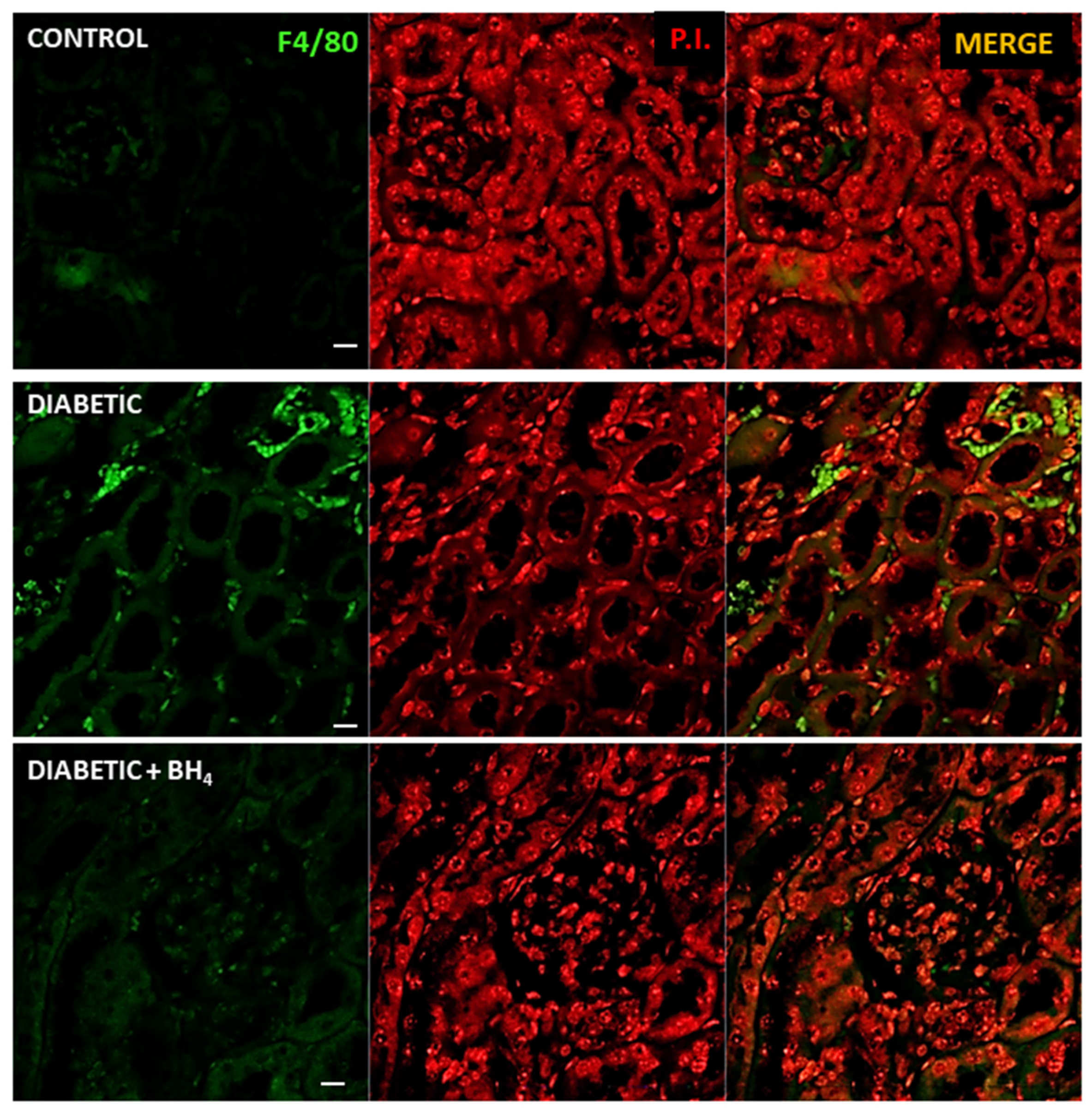
3.2. Apoptosis and Inflammatory Cells
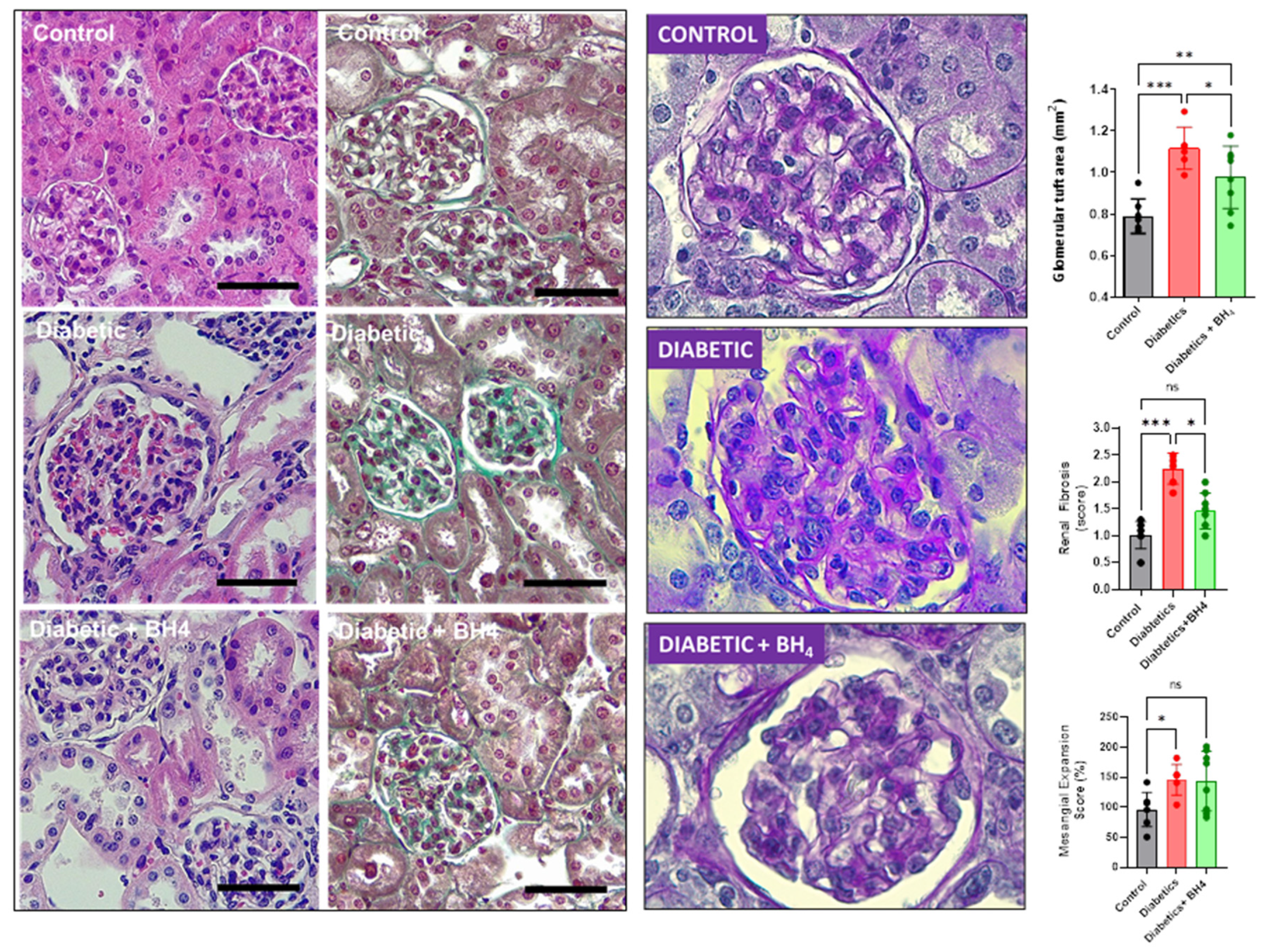
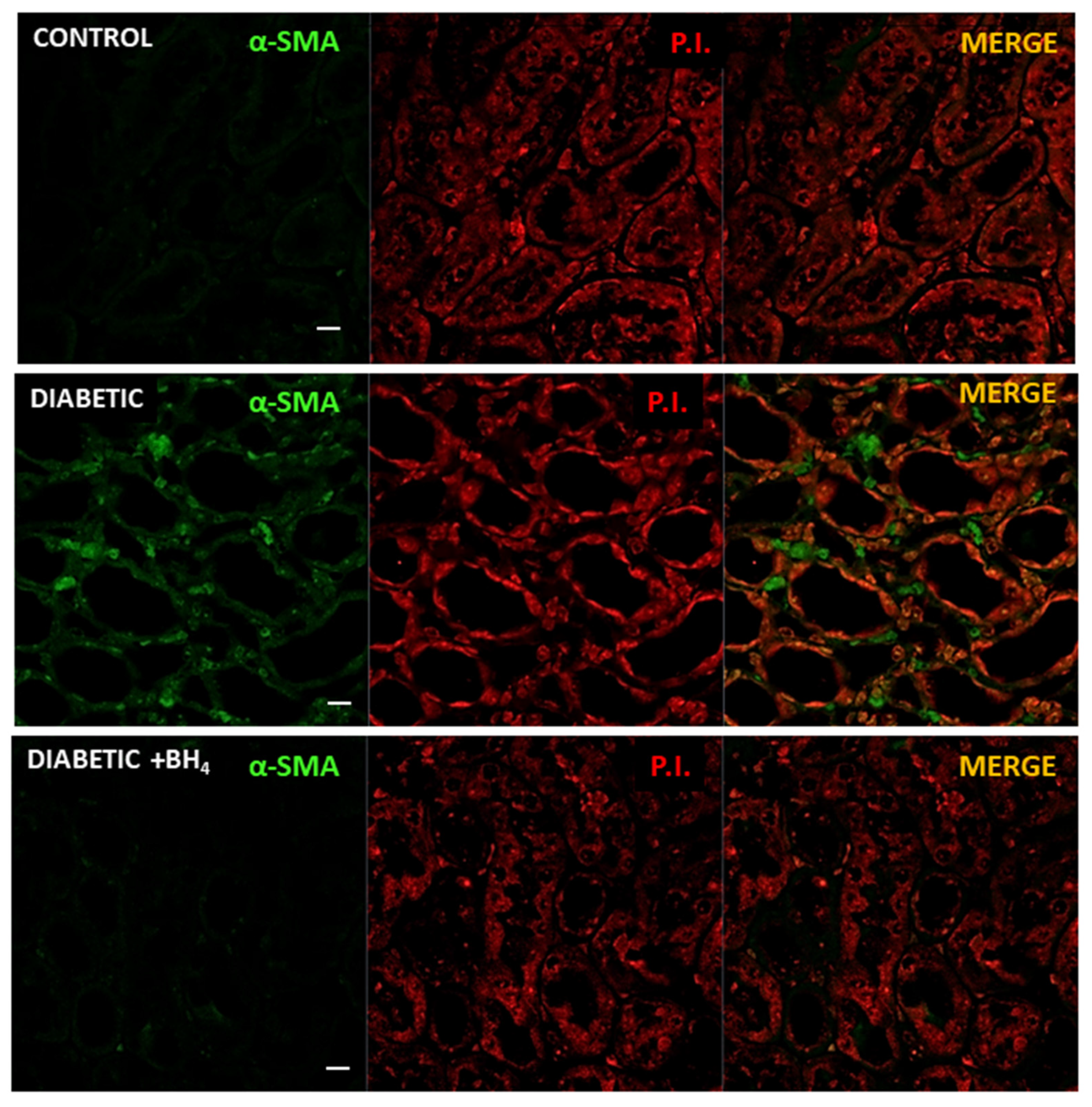
Because there is significant cardiac damage in chronic diabetes, both apoptosis and the presence of inflammatory cells has been described in the diabetic myocardium. Apoptosis was evaluated as the presence of TUNEL-positive cells in cardiac sections (Figure 4A). Diabetes induced an increase in the percentage of TUNEL+ cardiomyocytes compared with normoglycemic hearts, but this increase was not modified in the hearts from sapropterin-treated mice. We also evaluated the presence of infiltrative inflammatory cells by immunofluorescence of F4/80, a cell surface marker that is present in macrophages (Figure 4B). Diabetic hearts showed an increase in the number of macrophages present in the myocardium compared with normoglycemic controls. This number was significantly decreased in the hearts of sapropterin-treated mice.

3.3. Renal Changes

Next, we evaluated the impact of sapropterin treatment on diabetic nephropathy, evaluating glomerular morphology (Figure 5).
Diabetes induced glomerular hypertrophy compared to normoglycemic mice and was prevented by BH4 treatment (0.79 ± 0.08 mm2 in control, 1.12 ± 0.1 in diabetic and 0.98 ± 0.15 mm2 glomerular tuft size in diabetics + BH4, p = 0.0004).
In addition, diabetic mice presented glomerular fibrosis, evaluated by Masson’s trichrome staining, which was prevented in mice that received BH4: 1.01 ± 0.25 in control, 2.25 ± 0.29 in diabetics and 1.46 ± 0.33 score units in diabetics + BH4 (p < 0.0001). Next, we evaluated mesangial expansion, which was increased in diabetic mice compared to controls, but was not reduced by sapropterin treatment (96.1 ± 10.7% control, 145.7 ± 10.4% diabetics, 143.5 ± 17.6% diabetics treated with sapropterin, p < 0.05 diabetics vs. control).
Next, we evaluated the degree of macrophage infiltration and the expression of α-smooth muscle actin. Macrophage infiltration was evaluated by immunofluorescence staining of the cell surface marker F4/80 (Figure 6). Renal sections of control normoglycemic mice did not show the presence of infiltrating inflammatory cells, neither in the glomeruli nor in the peritubular interstitium. On the contrary, F4/80-positive cells were extensively found in the peritubular space of diabetic mice. This infiltration was dramatically reduced in the diabetic kidneys from mice treated with sapropterin.
In addition, we evaluated the presence of myofibroblasts as a marker of initial fibrosis (Figure 7). For this, renal sections were analyzed for α-smooth muscle actin (α-SMA). As expected, in the kidneys from normoglycemic control mice, there was no evidence of the presence of myofibroblasts in the peritubular space. On the contrary, the renal sections of diabetic mice showed the presence of these cells in the peritubular space mainly. Kidneys from sapropterin-treated mice showed almost no signal for α-SMA.
These results suggested that sapropterin treatment reduced macrophage infiltration and peritubular fibrosis in the diabetic kidneys. Overall, these results suggested that chronic treatment with BH4 in mice ameliorates the cardiorenal effects of diabetes, probably by restoring the nitric oxide production. This offers a possible new alternative to explore a BH4-based treatment for the organ damage of diabetes.

4. Discussion

Our present results showed that oral administration of sapropterin (BH4) for one month to diabetic mice was able to ameliorate some pathological changes in both the heart and kidneys, two of the main organ targets of diabetes. Notably, BH4 administration reduced cardiac hypertrophy and fibrosis, while preventing glomerular hypertrophy in the kidney. These effects were associated with the reduction in oxidative stress, but apparently independent of NOS1 recoupling. Previous reports have shown that eNOS uncoupling is an important source of ROS in the diabetic kidney [30,31,32,33,34].
Reduced levels of tetrahydrobiopterin were attributed to the reduced expression of guanosine5′-triphosphate cyclohydrolase I (GTPCH), a rate-limiting enzyme in the synthesis of BH4 [35]. Experiments where GTPCH was overexpressed then reverted the phenotypes associated with diabetic nephropathy [32] and cardiomyopathy [36,37].
In the diabetic heart, NOS uncoupling has emerged as an important source of ROS [36,37,38,39], in a way that appears as an important therapeutic target to prevent the development of diabetic cardiomyopathy. Our results agree with those obtained by recent studies regarding the role of NOS1 uncoupling in the heart, which indicate that BH4 supplementation or genetic modifications that lead to the increased intracellular production of BH4 has a beneficial impact on the left ventricular function of diabetic mice. This effect is achieved by improving intracellular calcium handling, hence left ventricular systolic and diastolic mechanics. Interestingly, some of the positive effects of BH4 supplementation in the diabetic heart appear to be independent of NOS recoupling [39]. Our results are consistent with those findings because we did not observe significant recoupling of NOS1 in the diabetic heart after BH4 treatment, although we observed reduction in cardiac oxidative stress. It was suggested that this protective effect may be exerted by a metabolic action of NOS1, increasing the expression of insulin-independent glucose transporters (GLUT-1), which improved myocardial energetics [39]. In addition, it was also shown that BH4 exerts its beneficial effects in diabetic cardiomyopathy by activating peroxisome proliferator-activated receptor-γ coactivator 1-α (PGC-1α) signaling by interacting with calcium/calmodulin-dependent protein kinase kinase 2 (CaMKK2). These effects are also independent of NOS1 activity [40].
It was described in a model of cardiac hypertrophy by transverse aortic constriction that BH4 supplementation inhibits macrophage infiltration in the myocardium, probably by reducing the inflammatory signaling [41]. Interestingly, this protection is also independent of NOS uncoupling, which is consistent with a role of BH4 in macrophages biology. The exact mechanism through which BH4 mediates these anti-inflammatory effects remains to be determined, but it has been reported that BH4 is important for the macrophage functions, dependent and independent of iNOS [42,43].
We also observed a reduction in the number of macrophages present in the diabetic myocardium after treatment with BH4. Nevertheless, we did not evaluate the origin of these macrophages. The recent literature indicates the presence of at least four types of macrophages in the heart, with one being the infiltrating-monocytes-derived macrophages [44,45,46]. Particularly, a study using streptozotocin-induced diabetes in mice showed that cardiac macrophages producing interleukin 1β play an important role in the generation of arrhythmias in the diabetic heart [47]. A recent study documented that BH4 deficiency in macrophages increased the production of interleukin 1β and the inflammatory profile of these cells [48].
In the context of the diabetic patient, it is relevant to consider both the development of cardiomyopathy and nephropathy. Here, we showed that BH4 treatment was able to prevent the cardiac damage associated with the initial stages of diabetic cardiomyopathy, reducing macrophage infiltration, fibrosis, and hypertrophy, and similar effects were observed in the kidney at the glomerular and tubular levels. Interestingly, even though we observed a general beneficial effect of BH4 supplementation on renal morphology in diabetic mice, BH4 mesangial expansion was not modified. This is consistent with a previous report that suggested that BH4 may induce mesangial proliferation [49]. Plasma BH4 concentration was postulated as a predictor of renal function in diabetic patients [50].
Fibrosis has been identified as a common factor in cardiorenal syndrome [51,52], which is a reciprocal interaction between cardiac and renal dysfunction in several pathological states, including diabetes [53,54]. Here we verified that fibrosis affected both organs and was prevented by BH4 administration.
Importantly, sapropterin, a form of BH4, is available and approved for use in humans in phenylketonuric patients [55]. This opens the possibility that this drug may be considered for use in diabetes clinical trials.

5. Conclusions

The results of the present study indicated that chronic oral administration of sapropterin (BH4) in mice ameliorates the morphological changes produced by diabetes in the heart and the kidney, probably by reducing oxidative stress, reducing inflammation, and the fibrotic processes that occur in the myocardium and at the glomerular and peritubular space in the kidney. This offers a possible new alternative to explore a BH4-based treatment for the organ damage caused by diabetes.

Author Contributions

Conceptualization, D.R.G. and K.S.; methodology, K.S.; validation, D.R.G., J.V. and U.N.; formal analysis, D.R.G.; investigation, C.V.; data curation, D.R.G.; writing—original draft preparation, D.R.G.; writing—review and editing, D.R.G.; visualization, A.V.T.X; supervision, J.V.; project administration, D.R.G.; funding acquisition, D.R.G. All authors have read and agreed to the published version of the manuscript.

Funding

This work was supported by FONDECYT grant 1150662.

Institutional Review Board Statement

The protocol involving animal experimentation was approved by the Animal Care Committee of Universidad de Talca (# 2015-087-DG).

Informed Consent Statement

Not applicable.

Data Availability Statement

Data is available upon request.

Conflicts of Interest

The authors declare no conflict of interest.

References

  1. Laditka, S.B.; Laditka, J.N. Active life expectancy of Americans with diabetes: Risks of heart disease, obesity, and inactivity. Diabetes Res. Clin. Pract. 2015, 107, 37–45. [Google Scholar] [CrossRef] [PubMed]
  2. Miki, T.; Yuda, S.; Kouzu, H.; Miura, T. Diabetic cardiomyopathy: Pathophysiology and clinical features. Heart Fail. Rev. 2013, 18, 149–166. [Google Scholar] [CrossRef] [Green Version]
  3. Tan, Y.; Zhang, Z.; Zheng, C.; Wintergerst, K.A.; Keller, B.B.; Cai, L. Mechanisms of diabetic cardiomyopathy and potential therapeutic strategies: Preclinical and clinical evidence. Nat. Rev. Cardiol. 2020, 17, 585–607. [Google Scholar] [CrossRef] [PubMed]
  4. Jia, G.; Hill, M.A.; Sowers, J.R. Diabetic Cardiomyopathy: An Update of Mechanisms Contributing to This Clinical Entity. Circ. Res. 2018, 122, 624–638. [Google Scholar] [CrossRef] [PubMed]
  5. Salvatore, T.; Pafundi, P.C.; Galiero, R.; Albanese, G.; Di Martino, A.; Caturano, A.; Vetrano, E.; Rinaldi, L.; Sasso, F.C. The Diabetic Cardiomyopathy: The Contributing Pathophysiological Mechanisms. Front. Med. (Lausanne) 2021, 8, 695792. [Google Scholar] [CrossRef] [PubMed]
  6. Kaur, N.; Guan, Y.; Raja, R.; Ruiz-Velasco, A.; Liu, W. Mechanisms and Therapeutic Prospects of Diabetic Cardiomyopathy Through the Inflammatory Response. Front. Physiol. 2021, 12, 694864. [Google Scholar] [CrossRef]
  7. Shah, M.S.; Brownlee, M. Molecular and Cellular Mechanisms of Cardiovascular Disorders in Diabetes. Circ. Res. 2016, 118, 1808–1829. [Google Scholar] [CrossRef] [Green Version]
  8. Fu, J.; Lee, K.; Chuang, P.Y.; Liu, Z.; He, J.C. Glomerular endothelial cell injury and cross talk in diabetic kidney disease. Am. J. Physiol. Renal. Physiol. 2015, 308, F287–F297. [Google Scholar] [CrossRef] [Green Version]
  9. Hung, P.H.; Hsu, Y.C.; Chen, T.H.; Lin, C.L. Recent Advances in Diabetic Kidney Diseases: From Kidney Injury to Kidney Fibrosis. Int. J. Mol. Sci. 2021, 22, 11857. [Google Scholar] [CrossRef]
  10. Fakhruddin, S.; Alanazi, W.; Jackson, K.E. Diabetes-Induced Reactive Oxygen Species: Mechanism of Their Generation and Role in Renal Injury. J. Diabetes Res. 2017, 2017, 8379327. [Google Scholar] [CrossRef]
  11. Sifuentes-Franco, S.; Padilla-Tejeda, D.E.; Carrillo-Ibarra, S.; Miranda-Diaz, A.G. Oxidative Stress, Apoptosis, and Mitochondrial Function in Diabetic Nephropathy. Int. J. Endocrinol. 2018, 2018, 1875870. [Google Scholar] [CrossRef] [PubMed]
  12. Chen, C.; Yuan, S.; Zhao, X.; Qiao, M.; Li, S.; He, N.; Huang, L.; Lyu, J. Metformin Protects Cardiovascular Health in People with Diabetes. Front. Cardiovasc. Med. 2022, 9, 949113. [Google Scholar] [CrossRef]
  13. Wu, L.; Gunton, J.E. The Changing Landscape of Pharmacotherapy for Diabetes Mellitus: A Review of Cardiovascular Outcomes. Int. J. Mol. Sci. 2019, 20, 5853. [Google Scholar] [CrossRef] [PubMed] [Green Version]
  14. Van Ruiten, C.C.; Hesp, A.C.; van Raalte, D.H. Sodium glucose cotransporter-2 inhibitors protect the cardiorenal axis: Update on recent mechanistic insights related to kidney physiology. Eur. J. Intern. Med. 2022, 100, 13–20. [Google Scholar] [CrossRef]
  15. Wiviott, S.D.; Raz, I.; Bonaca, M.P.; Mosenzon, O.; Kato, E.T.; Cahn, A.; Silverman, M.G.; Zelniker, T.A.; Kuder, J.F.; Murphy, S.A.; et al. Dapagliflozin and Cardiovascular Outcomes in Type 2 Diabetes. N. Engl. J. Med. 2019, 380, 347–357. [Google Scholar] [CrossRef] [PubMed]
  16. Kluger, A.Y.; Tecson, K.M.; Lee, A.Y.; Lerma, E.V.; Rangaswami, J.; Lepor, N.E.; Cobble, M.E.; McCullough, P.A. Class effects of SGLT2 inhibitors on cardiorenal outcomes. Cardiovasc. Diabetol. 2019, 18, 99. [Google Scholar] [CrossRef] [PubMed] [Green Version]
  17. Faria, A.; Persaud, S.J. Cardiac oxidative stress in diabetes: Mechanisms and therapeutic potential. Pharmacol. Ther. 2017, 172, 50–62. [Google Scholar] [CrossRef] [Green Version]
  18. Schmidt, T.S.; Alp, N.J. Mechanisms for the role of tetrahydrobiopterin in endothelial function and vascular disease. Clin. Sci. 2007, 113, 47–63. [Google Scholar] [CrossRef] [Green Version]
  19. Kim, H.L.; Park, Y.S. Maintenance of cellular tetrahydrobiopterin homeostasis. BMB Rep. 2010, 43, 584–592. [Google Scholar] [CrossRef]
  20. Okumura, M.; Masada, M.; Yoshida, Y.; Shintaku, H.; Hosoi, M.; Okada, N.; Konishi, Y.; Morikawa, T.; Miura, K.; Imanishi, M. Decrease in tetrahydrobiopterin as a possible cause of nephropathy in type II diabetic rats. Kidney Int. 2006, 70, 471–476. [Google Scholar] [CrossRef]
  21. Raij, L.; Azar, S.; Keane, W. Mesangial immune injury, hypertension, and progressive glomerular damage in Dahl rats. Kidney Int. 1984, 26, 137–143. [Google Scholar] [CrossRef] [PubMed] [Green Version]
  22. Xu, S.; Jiang, B.; Maitland, K.A.; Bayat, H.; Gu, J.; Nadler, J.L.; Corda, S.; Lavielle, G.; Verbeuren, T.J.; Zuccollo, A.; et al. The thromboxane receptor antagonist S18886 attenuates renal oxidant stress and proteinuria in diabetic apolipoprotein E-deficient mice. Diabetes 2006, 55, 110–119. [Google Scholar] [CrossRef] [PubMed]
  23. Guimaraes-Souza, N.K.; Yamaleyeva, L.M.; Lu, B.; Ramos, A.C.; Bishop, C.E.; Andersson, K.E. Superoxide overproduction and kidney fibrosis: A new animal model. Einstein, 2015; 13, 79–88. [Google Scholar]
  24. Zhao, X.F.; Liu, Y.H.; Han, Z.M.; Xu, Y.U. Effect of erythropoietin on the expression of dynamin-related protein-1 in rat renal interstitial fibrosis. Exp. Ther. Med. 2015, 9, 2065–2071. [Google Scholar] [CrossRef] [Green Version]
  25. Novoa, U.; Arauna, D.; Moran, M.; Nunez, M.; Zagmutt, S.; Saldivia, S.; Valdes, C.; Villasenor, J.; Zambrano, C.G.; Gonzalez, D.R. High-Intensity Exercise Reduces Cardiac Fibrosis and Hypertrophy but Does Not Restore the Nitroso-Redox Imbalance in Diabetic Cardiomyopathy. Oxid. Med. Cell Longev. 2017, 2017, 7921363. [Google Scholar] [CrossRef] [PubMed] [Green Version]
  26. Vielma, A.Z.; Leon, L.; Fernandez, I.C.; Gonzalez, D.R.; Boric, M.P. Nitric Oxide Synthase 1 Modulates Basal and beta-Adrenergic-Stimulated Contractility by Rapid and Reversible Redox-Dependent S-Nitrosylation of the Heart. PLoS One 2016, 11, e0160813. [Google Scholar] [CrossRef] [Green Version]
  27. Soto, G.; Rodriguez, M.J.; Fuentealba, R.; Treuer, A.V.; Castillo, I.; Gonzalez, D.R.; Zuniga-Hernandez, J. Maresin 1, a Proresolving Lipid Mediator, Ameliorates Liver Ischemia-Reperfusion Injury and Stimulates Hepatocyte Proliferation in Sprague-Dawley Rats. Int. J. Mol. Sci. 2020, 22, 540. [Google Scholar] [CrossRef] [Green Version]
  28. Valdes, C.; Arauna, D.; Gonzalez, D.; Villasenor, J. Simplified HPLC methodology for quantifying biological pterins by selective oxidation. J. Chromatogr. B Analyt. Technol. Biomed. Life Sci. 2017, 1055–1056, 113–118. [Google Scholar] [CrossRef]
  29. Vielma, A.Z.; Boric, M.P.; Gonzalez, D.R. Apocynin Treatment Prevents Cardiac Connexin 43 Hemichannels Hyperactivity by Reducing Nitroso-Redox Stress in Mdx Mice. Int. J. Mol. Sci. 2020, 21, 5415. [Google Scholar] [CrossRef]
  30. Satoh, M.; Fujimoto, S.; Arakawa, S.; Yada, T.; Namikoshi, T.; Haruna, Y.; Horike, H.; Sasaki, T.; Kashihara, N. Angiotensin II type 1 receptor blocker ameliorates uncoupled endothelial nitric oxide synthase in rats with experimental diabetic nephropathy. Nephrol. Dial. Transplant 2008, 23, 3806–3813. [Google Scholar] [CrossRef] [Green Version]
  31. Cheng, H.; Wang, H.; Fan, X.; Paueksakon, P.; Harris, R.C. Improvement of endothelial nitric oxide synthase activity retards the progression of diabetic nephropathy in db/db mice. Kidney Int. 2012, 82, 1176–1183. [Google Scholar] [CrossRef]
  32. Kidokoro, K.; Satoh, M.; Channon, K.M.; Yada, T.; Sasaki, T.; Kashihara, N. Maintenance of endothelial guanosine triphosphate cyclohydrolase I ameliorates diabetic nephropathy. J. Am. Soc. Nephrol. 2013, 24, 1139–1150. [Google Scholar] [CrossRef] [PubMed] [Green Version]
  33. Satoh, M.; Fujimoto, S.; Haruna, Y.; Arakawa, S.; Horike, H.; Komai, N.; Sasaki, T.; Tsujioka, K.; Makino, H.; Kashihara, N. NAD(P)H oxidase and uncoupled nitric oxide synthase are major sources of glomerular superoxide in rats with experimental diabetic nephropathy. Am. J. Physiol. Renal. Physiol. 2005, 288, F1144–F1152. [Google Scholar] [CrossRef] [PubMed]
  34. Faria, A.M.; Papadimitriou, A.; Silva, K.C.; Lopes de Faria, J.M.; Lopes de Faria, J.B. Uncoupling endothelial nitric oxide synthase is ameliorated by green tea in experimental diabetes by re-establishing tetrahydrobiopterin levels. Diabetes 2012, 61, 1838–1847. [Google Scholar] [CrossRef] [PubMed] [Green Version]
  35. Xu, J.; Wu, Y.; Song, P.; Zhang, M.; Wang, S.; Zou, M.H. Proteasome-dependent degradation of guanosine 5′-triphosphate cyclohydrolase I causes tetrahydrobiopterin deficiency in diabetes mellitus. Circulation 2007, 116, 944–953. [Google Scholar] [CrossRef] [PubMed] [Green Version]
  36. Wu, H.E.; Baumgardt, S.L.; Fang, J.; Paterson, M.; Liu, Y.; Du, J.; Shi, Y.; Qiao, S.; Bosnjak, Z.J.; Warltier, D.C.; et al. Cardiomyocyte GTP Cyclohydrolase 1 Protects the Heart Against Diabetic Cardiomyopathy. Sci. Rep. 2016, 6, 27925. [Google Scholar] [CrossRef] [Green Version]
  37. Carnicer, R.; Hale, A.B.; Suffredini, S.; Liu, X.; Reilly, S.; Zhang, M.H.; Surdo, N.C.; Bendall, J.K.; Crabtree, M.J.; Lim, G.B.; et al. Cardiomyocyte GTP cyclohydrolase 1 and tetrahydrobiopterin increase NOS1 activity and accelerate myocardial relaxation. Circ. Res. 2012, 111, 718–727. [Google Scholar] [CrossRef] [Green Version]
  38. Jo, H.; Otani, H.; Jo, F.; Shimazu, T.; Okazaki, T.; Yoshioka, K.; Fujita, M.; Kosaki, A.; Iwasaka, T. Inhibition of nitric oxide synthase uncoupling by sepiapterin improves left ventricular function in streptozotocin-induced diabetic mice. Clin. Exp. Pharmacol. Physiol. 2011, 38, 485–493. [Google Scholar] [CrossRef]
  39. Carnicer, R.; Duglan, D.; Ziberna, K.; Recalde, A.; Reilly, S.; Simon, J.N.; Mafrici, S.; Arya, R.; Rosello-Lleti, E.; Chuaiphichai, S.; et al. BH4 Increases nNOS Activity and Preserves Left Ventricular Function in Diabetes. Circ. Res. 2021, 128, 585–601. [Google Scholar] [CrossRef]
  40. Kim, H.K.; Ko, T.H.; Song, I.S.; Jeong, Y.J.; Heo, H.J.; Jeong, S.H.; Kim, M.; Park, N.M.; Seo, D.Y.; Kha, P.T.; et al. BH4 activates CaMKK2 and rescues the cardiomyopathic phenotype in rodent models of diabetes. Life Sci. Alliance 2020, 3. [Google Scholar] [CrossRef]
  41. Hashimoto, T.; Sivakumaran, V.; Carnicer, R.; Zhu, G.; Hahn, V.S.; Bedja, D.; Recalde, A.; Duglan, D.; Channon, K.M.; Casadei, B.; et al. Tetrahydrobiopterin Protects Against Hypertrophic Heart Disease Independent of Myocardial Nitric Oxide Synthase Coupling. J. Am. Heart Assoc. 2016, 5, e003208. [Google Scholar] [CrossRef]
  42. McNeill, E.; Crabtree, M.J.; Sahgal, N.; Patel, J.; Chuaiphichai, S.; Iqbal, A.J.; Hale, A.B.; Greaves, D.R.; Channon, K.M. Regulation of iNOS function and cellular redox state by macrophage Gch1 reveals specific requirements for tetrahydrobiopterin in NRF2 activation. Free Radic. Biol. Med. 2015, 79, 206–216. [Google Scholar] [CrossRef] [Green Version]
  43. McNeill, E.; Stylianou, E.; Crabtree, M.J.; Harrington-Kandt, R.; Kolb, A.L.; Diotallevi, M.; Hale, A.B.; Bettencourt, P.; Tanner, R.; O’Shea, M.K.; et al. Regulation of mycobacterial infection by macrophage Gch1 and tetrahydrobiopterin. Nat. Commun. 2018, 9, 5409. [Google Scholar] [CrossRef] [PubMed] [Green Version]
  44. Lavine, K.J.; Epelman, S.; Uchida, K.; Weber, K.J.; Nichols, C.G.; Schilling, J.D.; Ornitz, D.M.; Randolph, G.J.; Mann, D.L. Distinct macrophage lineages contribute to disparate patterns of cardiac recovery and remodeling in the neonatal and adult heart. Proc. Natl. Acad. Sci. USA 2014, 111, 16029–16034. [Google Scholar] [CrossRef] [PubMed] [Green Version]
  45. Heidt, T.; Courties, G.; Dutta, P.; Sager, H.B.; Sebas, M.; Iwamoto, Y.; Sun, Y.; Da Silva, N.; Panizzi, P.; van der Laan, A.M.; et al. Differential contribution of monocytes to heart macrophages in steady-state and after myocardial infarction. Circ. Res. 2014, 115, 284–295. [Google Scholar] [CrossRef] [PubMed] [Green Version]
  46. Bajpai, G.; Bredemeyer, A.; Li, W.; Zaitsev, K.; Koenig, A.L.; Lokshina, I.; Mohan, J.; Ivey, B.; Hsiao, H.M.; Weinheimer, C.; et al. Tissue Resident CCR2- and CCR2+ Cardiac Macrophages Differentially Orchestrate Monocyte Recruitment and Fate Specification Following Myocardial Injury. Circ. Res. 2019, 124, 263–278. [Google Scholar] [CrossRef]
  47. Monnerat, G.; Alarcon, M.L.; Vasconcellos, L.R.; Hochman-Mendez, C.; Brasil, G.; Bassani, R.A.; Casis, O.; Malan, D.; Travassos, L.H.; Sepulveda, M.; et al. Macrophage-dependent IL-1beta production induces cardiac arrhythmias in diabetic mice. Nat. Commun. 2016, 7, 13344. [Google Scholar] [CrossRef] [Green Version]
  48. Bailey, J.D.; Diotallevi, M.; Nicol, T.; McNeill, E.; Shaw, A.; Chuaiphichai, S.; Hale, A.; Starr, A.; Nandi, M.; Stylianou, E.; et al. Nitric Oxide Modulates Metabolic Remodeling in Inflammatory Macrophages through TCA Cycle Regulation and Itaconate Accumulation. Cell Rep. 2019, 28, 218–230.e7. [Google Scholar] [CrossRef] [Green Version]
  49. Wang, J.; Yang, Q.; Nie, Y.; Guo, H.; Zhang, F.; Zhou, X.; Yin, X. Tetrahydrobiopterin contributes to the proliferation of mesangial cells and accumulation of extracellular matrix in early-stage diabetic nephropathy. J. Pharm. Pharmacol. 2017, 69, 182–190. [Google Scholar] [CrossRef]
  50. Deng, C.; Wang, S.; Niu, Z.; Ye, Y.; Gao, L. Newly established LC-MS/MS method for measurement of plasma BH4 as a predictive biomarker for kidney injury in diabetes. Free Radic. Biol. Med. 2022, 178, 1–6. [Google Scholar] [CrossRef]
  51. Zannad, F.; Rossignol, P. Cardiorenal Syndrome Revisited. Circulation 2018, 138, 929–944. [Google Scholar] [CrossRef]
  52. Delgado-Valero, B.; Cachofeiro, V.; Martinez-Martinez, E. Fibrosis, the Bad Actor in Cardiorenal Syndromes: Mechanisms Involved. Cells 2021, 10, 1824. [Google Scholar] [CrossRef] [PubMed]
  53. Zhang, J.; Bottiglieri, T.; McCullough, P.A. The Central Role of Endothelial Dysfunction in Cardiorenal Syndrome. Cardiorenal. Med. 2017, 7, 104–117. [Google Scholar] [CrossRef] [PubMed] [Green Version]
  54. Boudoulas, K.D.; Triposkiadis, F.; Parissis, J.; Butler, J.; Boudoulas, H. The Cardio-Renal Interrelationship. Prog. Cardiovasc. Dis. 2017, 59, 636–648. [Google Scholar] [CrossRef] [PubMed]
  55. Sanford, M.; Keating, G.M. Sapropterin: A review of its use in the treatment of primary hyperphenylalaninaemia. Drugs 2009, 69, 461–476. [Google Scholar] [CrossRef] [PubMed]
Figure 1. Oral administration of sapropterin restores levels of tetrahydrobiopterin (BH4). Left, plasma levels of BH4 in control, diabetics, and diabetic mice that received sapropterin (BH4) in drinking water. Right, intracardiac BH4 levels in control (black), diabetic mice (red), and diabetic mice that received BH4 (green). n = 6 in each group. * p < 0.05 vs. control and diabetics.
Biomedicines 10 02479 g001
Figure 2. Oral administration of sapropterin reduces the intracardiac levels of oxidative stress. (A) Western blot analysis of nitrotyrosine in cardiac protein extracts from control, diabetic mice, and diabetic mice that received sapropterin (BH4) in drinking water. (B) Western blot for the levels of NOS1 and NOS3 in homogenates from control, diabetics and diabetics mice that received sapropterin (BH4) in drinking water. * p < 0.05, ***, p < 0.001. ns = not significant.
Biomedicines 10 02479 g002
Figure 3. Cardiac remodeling in diabetes is ameliorated by oral administration of BH4. Left panel, representative hematoxylin and eosin stained cross-sections of hearts from control, diabetic, and diabetic mice that received BH4. Middle panel, representative Masson´s trichrome staining for collagen. Right panel, bar graphs depicting cardiac hypertrophy and fibrosis in control (black), diabetic mice (red), and diabetic mice treated with BH4 (green). * p < 0.05; ** p < 0.005 vs. the other groups. ns = not significant.
Biomedicines 10 02479 g003
Figure 4. Impact of sapropterin in macrophage infiltration and cardiac apoptosis in diabetic mice. (A), representative images of TUNEL positive cells in cardiac sections obtained from control, diabetic and diabetic mice that received BH4. Arrows indicate TUNEL positive nuclei. (B), representative confocal microscopy immunofluorescence images of F4/80 (green) in cardiac sections from control, diabetic and diabetic mice treated with sapropterin (BH4). Asterisk * p < 0.05, *** p < 0.001 compared to the group indicated by brackets. ANOVA followed by Tukey post hoc test. Scale bar indicates 30 µm. ns = not significant.
Biomedicines 10 02479 g004
Figure 5. Renal morphological changes of diabetes are ameliorated by oral administration of BH4. Left panel, representative images of hematoxylin–eosin staining in renal sections from kidneys of control, diabetic, and diabetic mice that received BH4. Middle panel, representative Masson’s trichrome staining for collagen IV. Right panel, periodic acid-Schiff (PAS)-stained renal sections from kidneys of control, diabetic, and diabetic mice that received BH4. * p < 0.05; ** p < 0.01; *** p < 0.001 vs. the groups indicated by brackets. Bar indicates 50 µm. ns = not significant.
Biomedicines 10 02479 g005
Figure 6. Renal tubular macrophage infiltration is prevented by oral administration of BH4 to diabetic mice. Representative confocal images of renal sections probed for F4/80 (green), a marker of macrophages, in normoglycemic control, diabetic, and diabetic mice treated with sapropterin (BH4). Middle panel, corresponding section counterstained with propidium iodide (P.I., red) for nuclei. Right panel, merge of both F4/80 and P.I: signals. Images were obtained at 40× magnification. Scale bar indicates 10 µm.
Biomedicines 10 02479 g006
Figure 7. Renal tubular fibrosis was prevented by oral administration of BH4 to diabetic mice. Representative confocal images of renal sections (medulla) enriched in tubules probed for alpha smooth muscle actin (α-SMA, green), a marker of myofibroblast, in normoglycemic control, diabetic, and diabetic mice treated with sapropterin (BH4). Middle panel, corresponding section counterstained with propidium iodide (P.I., red) for nuclei. Right panel, merge of both α-SMA and P.I: signals. Images were obtained at 40× magnification. Scale bar indicates 10 µm.
Biomedicines 10 02479 g007
Table 1. Morphometric and plasmatic parameters of control, diabetic and diabetic mice treated with tetrahydrobiopterin (BH4). BNP; brain natriuretic peptide. ANOVA followed by Tukey as post-hoc test. * p < 0.05 vs. control, ** p < 0.005 vs. control.
ControlDiabeticsDiabetics + BH4p Value
n9910
Body weight (g)40.7 ± 1.336.8 ± 1.034.0 ± 1.3 *0.0026
Heart weight (g)0.157 ± 0.0100.161 ± 0.0070.139 ± 0.0030.0616
Heart weight/tibia length (g/mm)7.57 ± 0.398.59 ± 0.34 *7.39 ± 0.160.0495
Insulin (ng/mL)1.09 ± 0.30 *0.29 ± 0.120.21 ± 0.080.0037
BNP (pg/mL)227.9 ± 25.3 **96.1 ±13.566.6 ± 12.60.0495
Glucose (mg/dL)137 ± 7.7 *316 ± 69.7247.3 ± 29.70.0132
Publisher’s Note: MDPI stays neutral with regard to jurisdictional claims in published maps and institutional affiliations.

