Abstract
In situ administration of vaginal probiotics has been proposed as an effective prevention strategy against gynecological diseases caused by microecological disorders. In this study, a thermosensitive in situ gel formulation was prepared for intravaginal delivery of Lactobacillus gasseri (L. gasseri). The optimized formulation was characterized for the rheological properties, in vitro release properties, and microencapsulation efficiency. The mixtures of poloxamer 407 (26.0% w/w) and 188 (9.0% w/w) produced an increase in gelation extent at 37 °C after dilution in simulated vaginal fluid (SVF). The presence of a low concentration of hyaluronic acid (HA, 0.3% w/w) improved the mucoadhesive properties and the capability to gel at 37 °C. Additionally, the viability of L. gasseri encapsulated with alginate or via co-extrusion technique with fructooligosaccharide (FOS, 0.5% w/w) was maintained at 11 log CFU/mL for eight weeks at 4 °C. In conclusion, the evaluation of the in situ thermosensitive gel formulation was shown to be efficacious for intravaginal delivery of L. gasseri with suitable textural and rheological properties.
1. Introduction
Within the vagina, the intense interaction between a variety of microorganisms and hosts provides the first line of defense against the migration of opportunistic pathogens. Under healthy conditions, the microbiome maintains a stable balance known as eubiosis. When the microecological balance is disrupted, known as dysbiosis, the pathogenic microorganisms continue to multiply and cause vaginal-related diseases [1]. Women with abnormal proliferation of anaerobic bacteria are at an increased risk of sexually transmitted infections, such as human immunodeficiency virus (HIV), Mycoplasma genitalium, human papillomavirus (HPV), and herpes simplex virus (HSV), etc. [2,3,4,5,6]. Although antibiotics have been proven to be an effective treatment strategy in eliminating bacterial pathogens in the short term, relapse of infection, further dysbiosis, and side effects remain serious problems [7].
In situ administration of lactobacillus has a great potential to restore vaginal health, as the vaginal microflora of healthy women is typically dominated by one of the four Lactobacilli species: Lactobacillus gasseri (L. gasseri), Lactobacillus crispatus (L. crispatus), Lactobacillus iners (L. iners), and Lactobacillus jensenii (L. jensenii) [8]. L. gasseri has been approved by European Food Safety Authority (EFSA) and listed as a microorganism suitable for use in food and feed production [9]. As a member of the Gram-positive lactobacillus genus, L. gasseri protects the vaginal epithelial barrier from pathogen colonization and invasion by producing antimicrobial peptides, lactic acid, and hydrogen peroxide [10] and adhering to vaginal epithelial cells (VECs) to form a protective film to competitively inhibit pathogen adhesion [11]. L. gasseri E has been demonstrated to effectively prevent vaginal bacterial infections through direct inhibition of pathogen colonization in the vaginal lumen [12]. Moreover, pretreatment of L. gasseri JM1 attenuated the lipopolysaccharide-induced inflammatory response as evidenced by the downregulation of genes encoding proinflammatory cytokines and the upregulation of genes encoding anti-inflammatory cytokines [13]. The clinical isolation of L. gasseri LG1 was identified to have the potential to inhibit vulvovaginal candidiasis (VVC) by anti-adhesion and anti-biofilm formation [14]. L. gasseri HMV18 from a healthy vagina could inhibit the growth of food-borne pathogens such as Escherichia coli (E. coli), Salmonella enterica (S. aureus), and Klebsiella oxytoca (K. oxytoca) [15]. Moreover, the vaginal administration of L. gasseri LN40 resulted in vaginal colonization, resulting in lower recurrences and less malodorous discharge [16]. In general, the use of L. gasseri for the vaginal administration could be a practical strategy for vaginal disease prevention. Therefore, L. gasseri was selected in the present study as the therapeutically effective probiotic for topical replenishment of vaginal microflora.
Local and direct vaginal administration has been investigated as a more effective route of drug delivery due to the accessible surface area of the vaginal mucosa with rich blood supply, avoidance of gastrointestinal tract disturbance, and hepatic first-pass metabolism [17]. However, effective vaginal administration is challenged by the high variability in anatomy, physiology, and microbiology in addition to the presence of secretions and vaginal fluids that reduce the residence time of the formulation [18]. In this regard, in situ hydrogels have been proposed as a more convenient topical dosage form that tolerates the changes of the microenvironment and has good mucoadhesion, retention, and flowing properties to spread over the vaginal mucosa [19]. The application of polymers in hydrogel formulations has attracted extensive attention, and among these promising polymers, poloxamer compositions are suitable for vaginal gel formulations own to their thermosensitive self-assembly characteristics, sufficient resistance to dilution in presence of vaginal fluids, low biotoxicity, low stimulation for organisms, and good biocompatibility [20]. The low viscosity of the formulation consisting of poloxamers is beneficial for the drug delivery to the vaginal cavity and spreading on the mucosa at room temperature of 25 °C. An increase of the temperature from 25 °C to 37 °C promotes the in situ gelation of the administered formulation, thereby resisting the removal process of the vaginal environment, which in turn ensures long-term retention of the loaded therapeutic agent at the infection site. Poloxamers have been approved by U.S. Food and Drug Administration (FDA) and listed as pharmaceutical excipients in the United States and European Pharmacopoeias [21]. Poloxamer-based thermosensitive vaginal gel has been used in clinics as the delivery agent of L. gasseri (ATCC 33323) [22] and L. crispatus (ATCC 33197) [20]. Vaginal gel formulation with poloxamer 407 (P407), poloxamer 188 (P188), and polycarbophil-based delivery of clotrimazole were found to significantly prolong the antifungal activity of clotrimazole in vivo [23].
Additionally, the lack of adhesion is the main drawback of poloxamer, which can be improved by other adhesive additives [24]. Hyaluronic acid (HA) is a glycosaminoglycan that can be used as a humectant and adhesive to improve the physicochemical properties of the gel by incorporation into poloxamer gels [25]. The addition of HA to the poloxamer temperature-sensitive gel is able to slow down gel erosion, alter the microstructure of the gel, and make the gel more compact with smaller pore sizes, thereby improving the performance of the gel [25].
Given the aforementioned premises, this study aims to develop an in situ thermosensitive gel formulation for intravaginal delivery of L. gasseri. Firstly, the mixtures of P407 and P188 were assessed as the basic gel matrix for L. gasseri in order to achieve the suitable thermosensitive property. In addition, the gel formulation was optimized, and the release of the formulation in vitro was simulated using rheology and viscoelasticity at 25 °C before administration and at 37 °C with the dilution in simulated vaginal fluid (SVF) to mimic the in vivo administration. In the next step, the viability and stability of probiotic lactobacillus was improved by the adjunction of prebiotics and encapsulation. Moreover, the long-term stability during storage was further evaluated.
2. Materials and Methods
2.1. Chemicals
P407 and P188 were purchased from Kolliphor, BASF SE, Ludwigshafen, Germany. HA, lactose, glucomannan, lactulose alginate, and soluble starch were obtained from Shanghai Maclean Biochemical Technology Co., Ltd., Shanghai, China. Lactic acid, urea, acetic acid, and glucose were acquired from Sinopharm Chemical Reagent Co., Ltd., Shanghai, China. Bovine serum albumin was purchased from Shanghai Huixing Biochemical Reagent Co., Ltd., Shanghai, China. Fructooligosaccharide (FOS) was provided by Shanghai Yuanye Bio-Technology Co., Ltd., Shanghai, China. Syto9 and propidium iodide (PI) were offered by KeyGEN Bio TECH, Nanjing, China.
2.2. Microorganism and Cultivation
Lactobacillus gasseri M01 GDMCC60781 (L. gasseri M01) was a laboratory-conserved strain that was deposited in GDMCC (Guangdong Microbial Culture Collection Center, Guangzhou, China). L. rhamnosus ATCC7469, L. delbrueckii ATCC11842, L. reuteri ATCC23272, and L. jannaschii ATCC25258 were purchased from American Type Culture Collection (ATCC, Rockville, MD, USA). Lactobacillus plantarum (L. plantarum) NRRL B-14768 was acquired from ARS Culture Collection (NRRL, Peoria, IL, USA). Lactobacillus strains were grown in Man Rogosa Sharpe (MRS) broth at 37 °C.
2.3. Freeze-Drying Procedure
The overnight cultures of lactobacillus were centrifuged (5000 rpm, 10 min, 4 °C) and washed twice with sterile water. The lactobacillus was resuspended in 5% (w/v) skimmed milk. The emulsion with lactobacillus was frozen overnight in at −80 °C freezer and then freeze-dried using freeze drier (SCIENTZ-10ND, Xinzhi Freeze Drying Equipment Co., Ningbo, China) for 48 h (h). The freeze-dried lactobacillus powders were stored in a sealed container and immediately analyzed for the post-processing viability, and then, the storage trials commenced.
2.4. Preparation of the Thermosensitive Gel Formulations Containing L. gasseri
The vaginal thermosensitive gel formulations of L. gasseri with poloxamers were prepared through cold method. Lactic acid/sodium lactate buffer (0.01 M, pH 3.2) was cooled to 4 °C. Poloxamers, HA, prebiotics, and freeze-dried bacterial powder (prepared in Section 2.3) were then slowly added to the lactic acid/sodium lactate buffer with continuous agitation. L. gasseri was added at the concentration of 11 log CFU/mL. The gels were left at 4 °C until a clear solution was obtained. A final pH of 3.8 was determined. Blank gels were prepared according to the above process without adding L. gasseri.
2.5. Preparation of Hydrogels Diluted by Simulated Vaginal Fluid
SVF was prepared according to the reported with 3.5 g/L NaCl, 1.4 g/L KOH, 0.2 g/L Ca(OH)2, 0.02 g/L bovine serum albumin, 2.0 g/L lactic acid, 1.0 g/L acetic acid, 0.2 g/L glycerol, 0.4 g/L urea, and 5.0 g/L glucose [26]. This mixture was adjusted to a pH of 4.2 using HCl and NaOH. Then, 9 mL of each of the abovementioned formulations were diluted with 1 mL of SVF.
2.6. Characterization of Gels
An aliquot (1 mL) of each gel was put into a glass tube. The tube was kept in cold water and then gradually heated to 37 °C at a speed of 0.5 °C/min. The flowability of the gels in the tube was observed with frequent flip-flopping. The gelation temperature was defined as the temperature at which the state of complete gelation was achieved. The final gel was placed in a water bath at 37 °C, and the gelation time was defined as the time at which the state of complete gelation was reached.
2.7. Response Surface Methodology (RSM)
RSM model was applied to simultaneously optimize the levels of variables to attain the best system performance to make statistical predictions. Design Expert 8.0.6 software (Stat-Ease, Inc., Minneapolis, MN, USA).was applied to formulate a suitable poloxamer-based hydrogel. To optimize the preparation variables with the gelation temperature of resulting hydrogel, the central composite design (CCD) was applied for systemic study of joint influence of the effect of independent variables. During the preparation process, P407, P188, HA, and pH were chosen as four variables. Their levels and ranges were listed in Table 1.

Table 1.
The actual value of design variables.
To optimize the best condition to release the L. gasseri from the poloxamer-based hydrogel, Design Expert 8.0.6 software was applied. The CCD consists of 2 independent variables with 5 replications and center points, yielding 13 experiments in total (Table 2). The ranges of 24% to 30% P407 and 6% to 11% P188 were used according to the four-factor experiment. Formulations with suitable gelation temperatures were obtained from the above optimization. The volume of lactic acid/sodium lactate buffer (0.01 M, pH 3.2) of 10 mL was kept constant during the tests. Design Expert 8.0.6 software showed the optimum ratio of the mixture required to obtain the optimum gelation temperature of the hydrogels.
Table 2.
Complete design layout corresponding runs.
2.8. Rheological Characterization of the Hydrogels
The rheological properties of the gel were tested by the Anton-Paar’ s Modular Compact Rheometer 302 (MCR) rotational rheometer equipped with a cone-plate geometry. The samples were heated at a rate of 1 °C every 60 s (s) and the temperature changed from 25 °C to 45 °C during the procedure with angular frequency omega of 100 rad/s. The complex viscosity, the shear storage/elastic modulus (G’), and the shear loss/viscous modulus (G’’) were recorded as a function of the temperature. The gelation temperature was determined as the sample exhibited a switch from G” > G’ to G’ > G” [27].
2.9. Assessment of the Distribution of L. gasseri within the Gel
The distribution and viability of L. gasseri in the gel were assessed by using fluorescent stains. The fluorescent dye Syto9 stains both living and dead bacteria green, whereas PI stains dead bacteria red only [28]. Shortly, working solutions were prepared by adding 20 µL of Syto9 stock solution (1 mM) and 30 µL of PI (20 mM) to 1 mL of sterile-filtered water. The working solutions were added to the gel at a ratio of 1/1000 (v/w) and mixed thoroughly by pipetting up and down. The mixed samples were incubated in the dark at 37 °C for 30 min. Fluorescence was microphotographed under a 485 nm wavelength excitation using a fluorescence microscope (Nikon ECLIPSE Ti2E, Tokyo, Japan).
2.10. In Vitro Release of L. gasseri
A non-film dissolution method was used to evaluate L. gasseri release from gels [29]. Gel (5 mL) was added into the SVFs (10 mL) in a 25 mL glass tube and oscillated at 50 rpm and 37 °C in the dark. At predetermined time points, an aliquot (3 mL) of supernatants was withdrawn, and preheated fresh SVFs were supplemented. The gels were maintained at the bottom of the tube. The number of bacteria in the supernatants was recorded.
The mechanism of L. gasseri release was investigated using log-transformation and least squares regression analysis, and the data generated from the study were fitted to a power-law equation (known as the Korsmeyer–Peppas equation):
Mt/M∞ is the fraction of drug released at the time t, k is the constant related to the structural and geometrical features of the delivery system, and n is the release exponent.
2.11. Evaluation of Different Prebiotics Added into the Gel
MRS broth was reconstituted to 0.5% prebiotic-supplemented dextrose-free media. Then, 1 mL of an overnight culture of each LAB strain was inoculated in 100 mL of the above-mentioned media and cultured at 37 °C. OD600 and pH were measured to indicate whether prebiotics were fermented and if this process could restore acidity.
2.12. Microencapsulation of L. gasseri
Calcium alginate beads of L. gasseri were prepared by emulsification/external gelation technique. The probiotic was prepared for the microencapsulation, and the propagated culture was harvested after around 12 h by centrifugation process (10,000 rpm, 5 min), washed twice, and resuspended in sterile physiological saline until the encapsulation step. The applied initial cell counts were between 9 and 11 log CFU/mL. The alginate (2%, 3%, 4%, 5% (w/v)) and alginate (2% (w/v)) + soluble starch (2% (w/v)) were chosen as the encapsulation material. All solutions used for entrapment were sterilized using an autoclave (at 121 °C for 15 min). Probiotic bacteria in 1 mL physiological saline were added to the encapsulation liquid (9 mL) and dispersed evenly by gentle agitation. Afterwards, 10 mL alginate–cell mixture was subsequently emulsified in 50 mL liquid paraffin containing 0.5% (v/v) Span 85 under magnetic agitation at 200 rpm for 30 min. The gelation process was started by adding 10 mL of 0.05 M calcium chloride (CaCl2) solution slowly and evenly along the wall of the cup into the emulsion. During one hour of continuous stirring, alginate microcapsules were gradually formed. The oil phase and the water phase were separated using a separatory funnel. The target beads were located in the water phase. The beads were collected after gentle centrifugation at 1000 rpm for 5 min.
2.13. Morphology and Particle Size Measurements
Particle size was measured in de-ionized water. Immediately after microencapsulation, the morphology of the probiotic-filled calcium alginate microbeads was examined with a light microscope ensuring uniform microbead shape and size distribution.
2.14. Stability Studies of the Gel
The gels with the addition of prebiotics or treated with alginate (3%, 4%, 5% (w/v))-encapsulated bacteria were stored over eight weeks at 4 °C and 25 °C, respectively, in order to assess their stability. L. gasseri distribution and viability were evaluated using fluorescent microscope. As mentioned before, the bacterial activity was illustrated by staining with Syto9 and PI.
2.15. Statistical Analysis
Statistical analysis was made with GraphPad Prism Version 8.0.1 (GraphPad Software, San Diego, CA, USA). The difference of the two groups was verified by Student’s t-test, and the difference of multiple groups was defined through one-way analysis of variance (ANOVA) followed by Tukey’s multiple comparison test. All data were presented as mean values ± standard deviation (SD). Differences were considered statistically significant at p < 0.05.
3. Results
3.1. Effect of Hydrogels Composition on Gelation Temperature
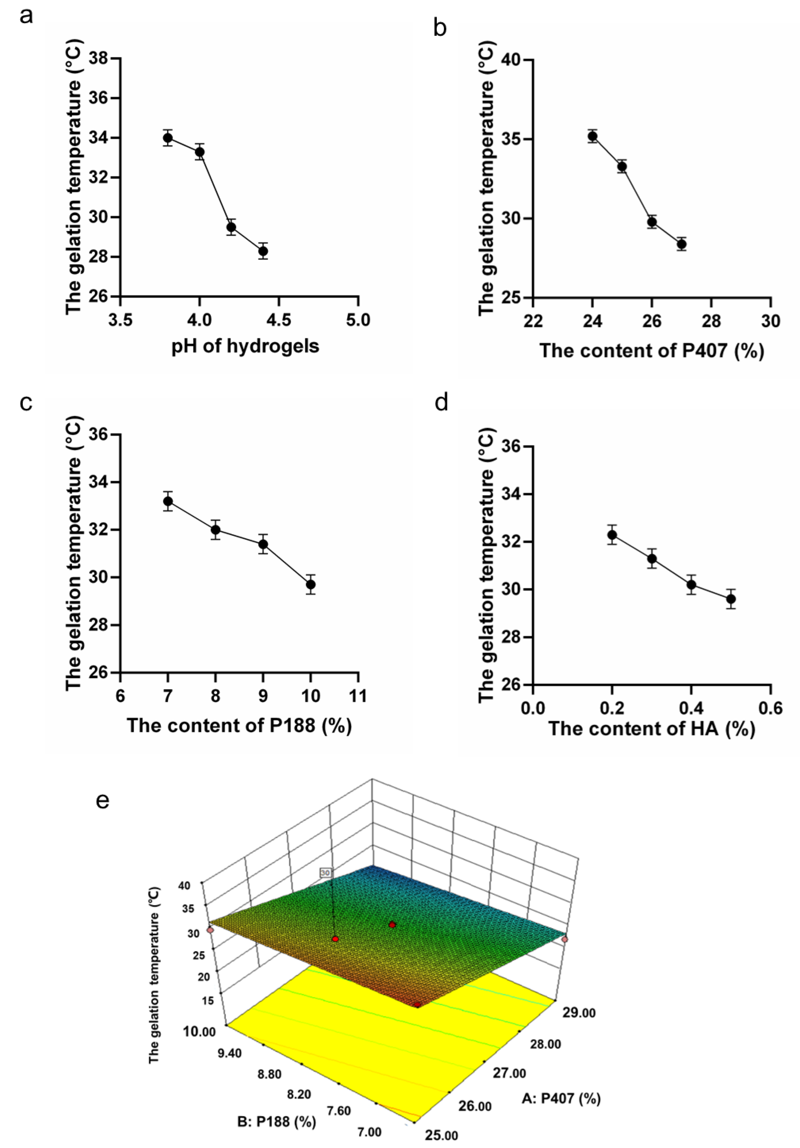
According to the preliminary experiments on polymer formulation, the range of poloxamer mixing with P407 was stated as 24% to 30%. The percentage of P188 ranging from 6% to 11% was proved to form the hydrogel sufficiently. Considering the maintenance of suitable adhesion and moisturizing effect of the gel, 0.3% HA was also chosen [27,30,31] (Mayol et al. 2008; Ur-Rehman et al. 2011; Nappi et al. 2021). In the bacterial infection sites, there was a growth in vaginal pH (>4.5) [32]. Keeping gel formulations at a stable pH of 3.8 was beneficial for maintaining of normal vaginal pH value. In this study, the gelation temperature of L. gasseri hydrogels showed to decrease with the increasing of pH value (Figure 1a). Moreover, with the increase of P407, P188, and HA, the gelation temperature also decreased, respectively (Figure 1b–d).

Figure 1.
Formulation optimization by RSM test. (a). The lactic acid/sodium lactate buffer’s (0.01 M) pH effect on the gelation temperature (Mean ± SD, n = 3). (b). The effect of P407 on the gelation temperature. (c). The effect of P188 on the gelation temperature. (d). The effect of HA on the gelation temperature. (e). The 3D image of the response surface optimization model.
3.2. RSM Model and Target Formulation
RSM model was applied to make statistical predictions through fitting with experimental data by polynomial equation. According to the Design Expert software, an optimization choice could be applied to solve problems of optimal formulations to obtain the desired hydrogel gelation temperature of 30 °C. The experimental design and statistical analysis are shown in Table 2. The mathematical model is shown in Equation (2) (below) and Figure 1e.
The Equation (2) and Figure 1e suggested by the software indicated the change of the gelation temperature of the L. gasseri hydrogel in relation to the content of poloxamers. In Table 3 and Figure 1e, the regression analysis with an R2 = 0.9405 indicated a satisfactory correlation between the independent variables and the response. An ANOVA test was applied to evaluate the adequacy of the empirical second-order polynomial model (Table 3); the F-value of 79.10 (p < 0.0001) implied that the model was significant. In the present study, P407 and P188 were identified as significant model terms. Three-dimensional surfaces and contours (Figure 1e), as the graphical representations of the regression equation, showed the considerable influences of P407 and P188 on the gelation temperature. The gelation temperature decreased smoothly with the increase of P188 from 6.0% to 11.0% and the increase of P407 from 24.0% to 30.0%.
Table 3.
The model parameters.
The optimal formula was suggested by the software according to the optimal gelation temperature (30 °C) (Table 4). The experiment was carried out to validate the gelation temperature of the formulation. The formula with the composition of P407 (26%) and P188 (8.86%) as well as the tested gelation temperature are shown in Table 4. The difference between the gelation temperature obtained in the solvent and the predicted gelation temperature was less than 15%, indicating that the optimization process of the computational design was feasible. After the diluted in SVF, the gelation temperature of the formulation was lower than 37℃, implying that the formulation could be used for further analysis.
Table 4.
The predicted and actual value of optimized desired formulation.
3.3. Rheological Characterization
Further investigation of the rheological properties of the gel revealed the changes in the complex viscosity, storage, and modulus of loss of the gel with the alteration of temperatures, as shown in Figure 2a and b. With the increase of temperature, the viscosity of the gel gradually improved (Figure 2a), and the difference between the storage modulus and the loss modulus constantly increased (Figure 2b), indicating that the gel had a high sensitivity to the change of temperature. At 37 °C, the complex viscosity reached a relatively high level (Figure 2a), which was beneficial because the gel could be retained at the site of administration for a longer period to prolong the residence time of L. gasseri in the vaginal cavity. The storage modulus was significantly higher than the loss modulus of the gel (Figure 2b), suggesting that the gel exhibited elastic behavior predominantly.
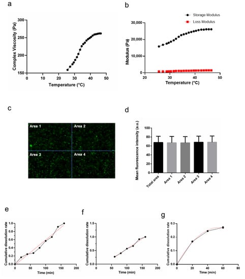
Figure 2.
Physical–chemical properties of the gels. (a). The complex viscosity of the gels. (b). Flow curves at elevated temperatures from 25 °C and 46 °C. (c). The representative image of the distribution of L. gasseri within the gel and a schematic representation of the zoning. (d). The mean fluorescence intensity analysis of the regions in Figure 2C (Mean ± SD, n = 3). (e). The release curve and the simulated fitted line with a nonlinear fit to the entire curve. (f). The plot of nonlinear fit to the points of the release curve after 60 min and the fitted line. (g). The plot of a nonlinear fit to the points of the release curve before 60 min and the fitted line.
3.4. Distribution of L. gasseri within the Gel
The Syto9 and PI stains were considered to be the effective indicators in the evaluation of cell metabolic activity and membrane integrity [33]. The distribution of L. gasseri was essentially the same along the x and y axes as observed in the gel (Figure 2c). The images were divided into four parts, and the mean fluorescence intensity of each part was counted separately (Figure 2c). ANOVA test confirmed the results and showed that there was no significant difference between the four parts (p > 0.05, Figure 2d). The results confirmed that the distribution of L. gasseri in the gel is uniform, which might be beneficial for the uniform distribution of L. gasseri in vaginal administration.
3.5. In Vitro Release of L. gasseri
The release curve of L. gasseri in Figure 2e shows that L. gasseri cells of the gel released continuously for 160 min until the gel was completely dissolved. The obtained data were fitted to the equations described in Section 2.10, which simulated the presumed release process and principles. The overall curve was analyzed using a nonlinear fit method with an R-squared of 0.9798 (Figure 2e). Separate nonlinear fitting of the curves would obtain a better fit. Particularly, it was showed that the linear fitting after 60 min had a better curve fit with an overall R-squared of 0.9862 (Figure 2f). The fitting formula was as follows:
Mt/M∞ is the fraction of drug released at time t; k is a constant, related to the structural and geometrical features of the delivery system; n is the release exponent.
The nonlinear fitting before 60 min had a better curve fitting with an overall R-squared of 0.9944 (Figure 2g). The fitting formula was as follows:
The release process could be divided into two stages with 60 min as the node. The slope of the curve gradually decreased in the first 60 min, indicating a sudden release phenomenon, and the subsequent release rate slowed down; 60 min later, the slope of the curve was more stable, and the release rate was more constant. The release curve indicated that the sustained-release properties of the gel were remarkable, which facilitated the colonization of L. gasseri in vivo.
3.6. Fermentation Profiles of Prebiotics by Vaginal Microbes
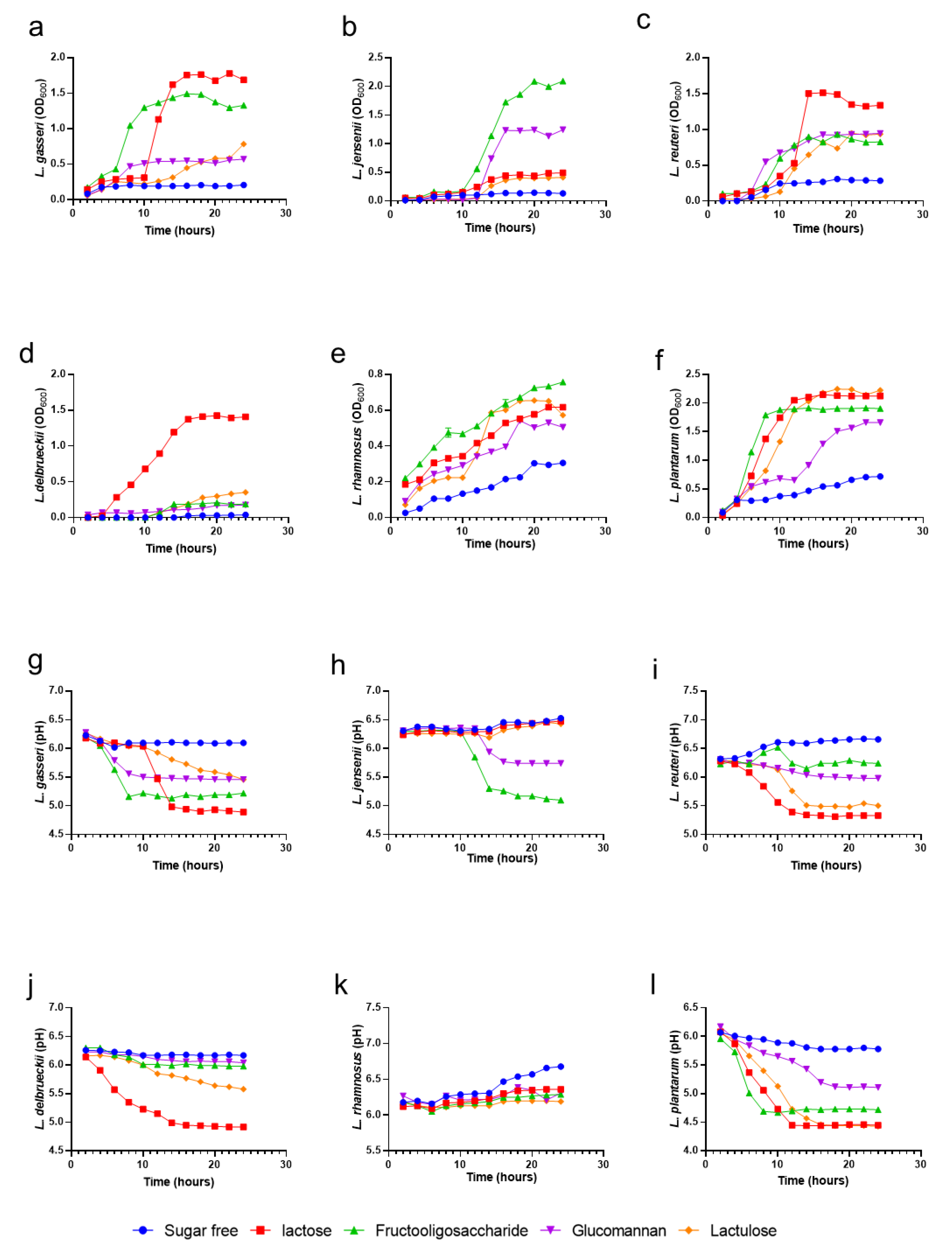
Prebiotics were selected for their beneficial effects on L. gasseri activity in the hydrogel. Here, we compared the effects of different prebiotics on various vaginal beneficial microbes and on the maintenance of a proper pH. In the growth curve experiments with different prebiotics in Figure 3a–f, it is shown that lactose increased the maximal growth of L. gasseri (p < 0.0001), L. jannaschii (p < 0.0001), L. reuteri (p < 0.0001), L. delbrueckii (p < 0.0001), L. rhamnosus (p < 0.0001), and L. plantarum (p < 0.0001) compared to the media without prebiotic. FOS, glucomannan, and lactulose also increased the maximal growth of all the experimental strains (p < 0.0001). For pH maintenance tests (Figure 3g–l), lactose and galactose made the pH-lowering effect of L. jannaschii less significant, with p-values of 0.0017 and 0.0001. During the fermentation process, except for L. rhamnosus, the augmented acidity of the medium was directly related to the increment in the growth rate of the lactobacilli tested, which can effectively maintain normal acidic vaginal pH. Considering the effects of lactose intolerance [34] as well as the effect of lowering pH and the combination of multiple probiotic growth conditions, the use of FOS as the prebiotics was a practical solution.
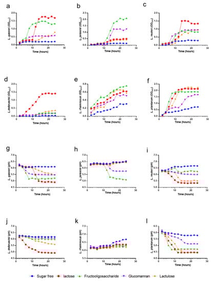
Figure 3.
Growth curves and pH changes of vaginal lactobacilli cultured with different prebiotics. (a–f). Growth curves of various probiotics in medium with different prebiotics. (g–l). The pH changes of various probiotics in medium with different prebiotics.
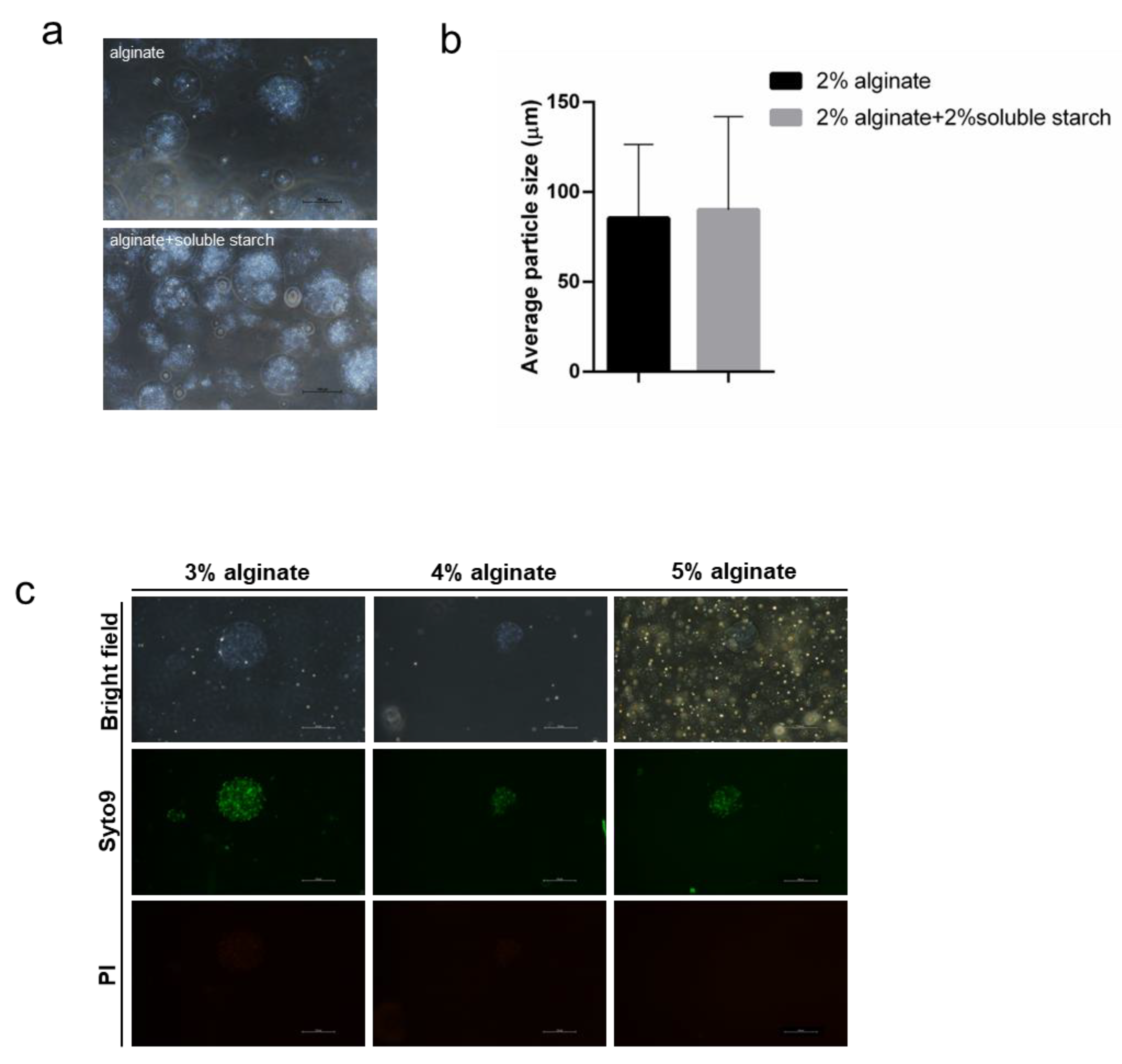
3.7. Morphology and Particle Size of Microspheres
Microencapsulation has been shown to be an effective method to enhance bacterial activity. Microcapsules are generally spherical under the microscope (Figure 4a). The size of the distributed microbeads had a wide range, between approximately 20–220 µm. The addition of soluble starch had no significant effect on the morphological particle size of the L. gasseri-embedded spheres (p > 0.5, Figure 4b). The beads’ size enlarged as the concentration of sodium alginate in 0.05 M calcium chloride solution increased. The augmented alginate concentration led to higher viscosity, resulting in larger microspheres and greater entrapment of bacteria, thus causing a high degree of cross-linking [35]. Moreover, L. gasseri showed high activity at the experimental sodium alginate concentrations of 3%, 4%, and 5% (Figure 4c).
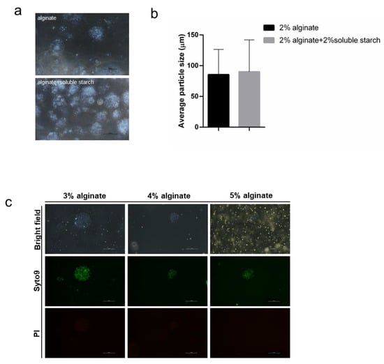
Figure 4.
Morphology and mean particle size of the microspheres. (a). Morphology of sodium alginate microspheres embedded with L. gasseri. (b). Morphology of sodium-alginate-soluble starch composite microspheres encapsulated with L. gasseri. The scale bar corresponds to 100 µm. (c). Effect of encapsulant on the size of droplet with L. gasseri. The scale bar corresponds to 100 µm.
3.8. Influence of the Storage Conditions
To investigate the storage conditions, the gels with FOS were stored at 4 °C and 25 °C, respectively, for eight weeks. There was no significant decrease in bacterial activity, while extremely few bacterial mortalities occurred when the gels were stored at 25 °C (Figure 5). The results suggested that the addition of probiotics could provide protection of activity similar to encapsulation. However, for the gels with alginate-encapsulated L. gasseri, 2% sodium alginate fragmented after eight weeks, while other concentrations (3% and 4%) of encapsulated spheres were likely to precipitate after a long-term storage. Therefore, it was preferable to add probiotics for strain protection.
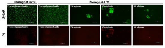
Figure 5.
Viability of L. gasseri contained in the gel. The concentration of the encapsulant sodium alginate was 3%, 4%, and 5%. Living and dead bacteria were stained with Syto9 (green), and dead bacteria were stained with PI (red). The scale bar corresponds to 100 µm.
4. Discussion
In recent years, researchers have realized that vagina is an underutilized route for drug administration. The local administration of vaginal probiotics, especially lactobacilli, are proposed as an effective prevention strategy against vaginal infectious diseases. N’Guessan Gnaman et al. developed a thermo-sensitive hydrogel by evaluation of the rheological properties and stability, which contains 21.5% of poloxamer 407, 1% of sodium alginate, and 9 log CFU of L. crispatus per gel sample (5 g) [20]. Vigani et al. developed a mucoadhesive in situ gelling formulation for the vaginal administration of L. gasseri with the main mixture of two thermosensitive polymers, i.e., P407 and methylcellulose (MC), containing 6 log CFU/mL of L. gasseri. The rheological properties and the cytocompatibility towards the HeLa cell line were assessed in their work [22]. In Oerlemans’ study, L. plantarum WCFS1, L. pentosus KCA 1, and L. rhamnosus GG were formulated in an innovative silicone gel at a dose of 9–10 log CFU/g. The lactobacilli remained viable over multiple months of storage at 5 °C and 25 °C. Moreover, the formulation was proven to be effective on VVC in clinic [36]. The thermosensitive gel formulation was evidenced to enhance the viability of epithelial cells without affecting the morphology of the vaginal mucosa [23]. In this study, a thermosensitive in situ gel formulation was developed for intravaginal delivery of L. gasseri. The final gel consisted of P407 at 26%, P188 at 8.86%, HA at 0.3%, FOS at 0.5%, and L. gasseri at 11 log CFU/mL. L. gasseri was proven to stay highly active for at least eight weeks in the formulation at 4 °C. This vaginal gel has potential as an adjuvant medication or daily prevention for vaginal infection by maintaining and restoring vaginal microecology.
Optimizing the pharmaceutical characterization of vaginal gels is essential in optimizing safety, efficacy, and acceptability. In the current work, the model for formulation optimization was established to obtain the most optimal composition and proportions; then, the optimized formulation was characterized for the rheological properties and in vitro release properties. Overall release behavior is important for a therapeutic formulation, which can be predict by mathematical models fitted by measuring some physical parameters and experimental release data. However, there is still a lack of study about the in vitro release of bacteria in the in situ gel solutions. In this research, the release was shifted from diffusion as the main driving force to swelling and relaxation of the polymer-blend matrix. The Korsmeyer–Peppas equation was applied in the piecewise fitting of release curves, and the “n” value in Equation (1) predicts the mechanism of drug release. Likewise, 0.45 ≤ n corresponds to the Fickian diffusion mechanism, 0.45 < n < 0.89 corresponds to non-Fickian transport, n = 0.89 corresponds to Case II transport, and n > 0.89 corresponds to super case II transport. In the first half of the fitting, “n” value was 0.4259 (<0.45), indicating that the release of bacteria in the gel acted as the Fickian transport, and the high initial osmolarity difference led to a high release rate of L. gasseri, which was slowly reduced due to the decrease in osmolarity. This was consistent with the vancomycin-release mechanism [37]; the fitting “n” value of the second half was 1.332 (>0.89), indicating that the release of bacteria in the gel was attributed to the super case II transport. This was consistent with the drug release trend of metformin HCl from seed mucilage (FSM)-alginate mucoadhesive beads [38]. In all, the sustained-release properties of the gel indicated by the release curve facilitated the delivery of L. gasseri into vaginal cavity.
To further improve the viability of the lactobacillus, the addition of prebiotics and microencapsulation were applied. It was shown that the viabilities of L. gasseri encapsulated with alginate and via co-extrusion technique with FOS were stably maintained for eight weeks at 4 °C, which suggested that the addition of probiotics could provide protection of activity similar to encapsulation. However, in the 2% alginate-encapsulated gel, sodium alginate fragmented after eight weeks. In 3% and 4% alginate-encapsulated gels, as the particle size increased, the microspheres precipitated after standing and did not form a good suspension in the gel. The addition of prebiotics to the formulation was significantly better than encapsulation in the improvement of the stability of the gel. Therefore, the addition of prebiotics was preferred for protection of the viability and stability of lactobacillus. In addition, the gel prepared with FOS exhibited improved storage stability compared to that reported [20]. Nevertheless, the homogeneity of the gel, the appropriate embedding method to improve the storage stability, and the stability of the bacteria in the gel still need to be further explored.
FOS is one of the most widely studied and reported prebiotics. FOS had become a probiotic functional food supplement with GRAS (Generally Recognized as Safe) status. FOS fermentation produces short-chain fatty acids and other organic acids that lower the pH of the lumen and maintain a healthy vaginal environment [39]. FOS addition can increase the viscosity and viscoelasticity of the gel [40]. In addition, FOS is able to improve the activity of lactobacillus during storage [40]. Therefore, in this study, L. gasseri was encapsulated by coextrusion technique with FOS (0.5% w/w) to enhance the viability.
In this study, there was a certain difference between the gelation temperature detected by the test-tube-inversion method and the rheometer test. The rheometer test results showed that the gelation temperature of the formulation was less than 25 °C, while the test-tube-inversion result was above 30 °C. This apparent discrepancy may be due to the fact that the inversion method was based on the observation of the flow state of the gel, while the rheological method determined the sol-gel transition temperature by calculation. The end points of the two methods for determination of gel state were inconsistent. Even so, it was reported that the gelation temperature obtained by the tube inversion method was positively correlated with the rheometer temperature [41]. Figure 2b showed that the storage modulus at 25° was significantly higher than the loss modulus, indicating that there was a great tendency to gelation at 25 °C after long-term storge. In the formula design, the gelation was expected to occur after administration. Therefore, based on the rheological results, 4 °C was chosen as the preferred storage temperature.
The influence of HA on the gelation properties of poloxamers blends has been well-studied. HA addition affected the self-assembly process of the poloxamer and was able to significantly enhance the rheological properties of poloxamers, thus suggesting that HA might interact with the micelles through secondary bonds (e.g., hydrogen bonds) to enhance the gel structure [27]. In addition, HA and sodium lactate buffers have a significant effect on the gelation temperature by modulating the hydrogen bonding between the polymers. It was reported that the addition of HA delayed the gelation temperature by a few degrees Celsius [27]. The same trend was also confirmed in the gel of this study. The mucoadhesion experiments demonstrated that the rheological synergy between poloxamer/HA gels lead to the changes in flow behavior from a rather Newtonian solution alone to a pseudoplastic mixture. Moreover, the strong interaction between HA-P407 and mucin enhanced the adhesion of the tissue [30]. Furthermore, HA also provided moisturizing effects [31], which was beneficial for vaginal administration.
In conclusion, the evaluation of in situ thermosensitive gel formulations was shown to be efficacious for intravaginal delivery of L. gasseri, with suitable textural and rheological properties. Further mucoadhesion experiments and pharmacological experiments are needed to verify the safety and effectiveness of the gel.
Author Contributions
Y.Z. (Ye Zhao) and C.T. conceived and designed the research; Y.Z. (Ye Zhao), T.W., Z.C. and S.L. conducted the experiments; Y.Z. (Ye Zhao), T.W., H.R., P.S. and Y.Z. (Yingying Zhu) analyzed the data; Y.Z. (Ye Zhao) and T.W. wrote the manuscript. All authors have read and agreed to the published version of the manuscript.
Funding
This work was supported by grants from the National Natural Science Foundation of China (No. 31701279) and Xiamen Municipal Science and Technology Bureau-Xiamen City Division of Funding Management Project (No. 3502Z20203016).
Institutional Review Board Statement
Not applicable.
Informed Consent Statement
This study was approved by the ethics committee of Nanjing Tech University with the number of SCXK(Su) 2017-0001.
Data Availability Statement
All data generated or analyzed during this study are included in this published article.
Conflicts of Interest
The authors declare no competing interests.
References
- Kalia, N.; Singh, J.; Kaur, M. Microbiota in vaginal health and pathogenesis of recurrent vulvovaginal infections: A critical review. Ann. Clin. Microbiol. Antimicrob. 2020, 19, 5. [Google Scholar] [CrossRef] [PubMed]
- Bagnall, P.; Rizzolo, D. Bacterial vaginosis: A practical review. JAAPA 2017, 30, 15–21. [Google Scholar] [CrossRef] [PubMed]
- Brotman, R.M.; Klebanoff, M.A.; Nansel, T.R.; Yu, K.F.; Andrews, W.W.; Zhang, J.; Schwebke, J.R. Bacterial vaginosis assessed by gram stain and diminished colonization resistance to incident gonococcal, chlamydial, and trichomonal genital infection. J. Infect. Dis. 2010, 202, 1907–1915. [Google Scholar] [CrossRef] [PubMed]
- Cohen, C.R.; Lingappa, J.R.; Baeten, J.M.; Ngayo, M.O.; Spiegel, C.A.; Hong, T.; Donnell, D.; Celum, C.; Kapiga, S.; Delany, S.; et al. Bacterial vaginosis associated with increased risk of female-to-male HIV-1 transmission: A prospective cohort analysis among African couples. PLoS Med. 2012, 9, e1001251. [Google Scholar] [CrossRef] [PubMed]
- Sobel, R.; Sobel, J.D. Metronidazole for the treatment of vaginal infections. Expert Opin. Pharmacother. 2015, 16, 1109–1115. [Google Scholar] [CrossRef]
- Taylor, B.D.; Darville, T.; Haggerty, C.L. Does bacterial vaginosis cause pelvic inflammatory disease? Sex Transm Dis. 2013, 40, 117–122. [Google Scholar] [CrossRef]
- Zhang, Q.-Q.; Liu, Z.-H.; Liu, L.-L.; Hu, G.; Lei, G.-L.; Wang, Y.; Cao, Y.; Wu, W.; Zhang, L.; Liao, Q.-P. Prebiotic Maltose Gel Can Promote the Vaginal Microbiota From BV-Related Bacteria Dominant to Lactobacillus in Rhesus Macaque. Front. Microbiol. 2020, 11, 594065. [Google Scholar] [CrossRef]
- Ravel, J.; Gajer, P.; Abdo, Z.; Schneider, G.M.; Koenig, S.S.; McCulle, S.L.; Karlebach, S.; Gorle, R.; Russell, J.; Tacket, C.O.; et al. Vaginal microbiome of reproductive-age women. Proc. Natl. Acad. Sci. USA 2011, 108 (Suppl. 1), 4680–4687. [Google Scholar] [CrossRef]
- Ricci, A.; Allende, A.; Bolton, D.; Chemaly, M.; Davies, R.; Girones, R.; Koutsoumanis, K.; Lindqvist, R.; Nørrung, B.; Robertson, L.; et al. Update of the list of QPS-recommended biological agents intentionally added to food or feed as notified to EFSA 6: Suitability of taxonomic units notified to EFSA until March 2017. Efsa J. 2017, 15, e04884. [Google Scholar] [CrossRef]
- Selle, K.; Klaenhammer, T.R. Genomic and phenotypic evidence for probiotic influences of Lactobacillus gasseri on human health. FEMS Microbiol. Rev. 2013, 37, 915–935. [Google Scholar] [CrossRef] [Green Version]
- Parolin, C.; Marangoni, A.; Laghi, L.; Foschi, C.; Ñahui Palomino, R.A.; Calonghi, N.; Cevenini, R.; Vitali, B. Isolation of Vaginal Lactobacilli and Characterization of Anti-Candida Activity. PLoS ONE 2015, 10, e0131220. [Google Scholar] [CrossRef]
- Maldonado-Barragán, A.; Caballero-Guerrero, B.; Martín, V.; Ruiz-Barba, J.L.; Rodríguez, J.M. Purification and genetic characterization of gassericin E, a novel co-culture inducible bacteriocin from Lactobacillus gasseri EV1461 isolated from the vagina of a healthy woman. BMC Microbiol. 2016, 16, 37. [Google Scholar] [CrossRef] [PubMed]
- Sun, L.; Tian, W.; Guo, X.; Zhang, Y.; Liu, X.; Li, X.; Tian, Y.; Man, C.; Jiang, Y. Lactobacillus gasseri JM1 with potential probiotic characteristics alleviates inflammatory response by activating the PI3K/Akt signaling pathway in vitro. J. Dairy Sci. 2020, 103, 7851–7864. [Google Scholar] [CrossRef] [PubMed]
- Itapary Dos Santos, C.; Ramos França, Y.; Duarte Lima Campos, C.; Quaresma Bomfim, M.R.; Oliveira Melo, B.; Assunção Holanda, R.; Santos, V.L.; Gomes Monteiro, S.; Buozzi Moffa, E.; Souza Monteiro, A.; et al. Antifungal and Antivirulence Activity of Vaginal Lactobacillus Spp. Products against Candida Vaginal Isolates. Pathogens 2019, 8, 150. [Google Scholar] [CrossRef] [PubMed]
- Gao, X.; Wang, Z.; Li, X.; Zhang, X.; Du, S.; Jia, M.; Hu, D.; Jia, X.; Cong, B.; Zhang, Y. A new Lactobacillus gasseri strain HMV18 inhibits the growth of pathogenic bacteria. Food Sci. Hum. Wellness 2022, 11, 247–254. [Google Scholar] [CrossRef]
- Ehrström, S.; Daroczy, K.; Rylander, E.; Samuelsson, C.; Johannesson, U.; Anzén, B.; Påhlson, C. Lactic acid bacteria colonization and clinical outcome after probiotic supplementation in conventionally treated bacterial vaginosis and vulvovaginal candidiasis. Microbes Infect. 2010, 12, 691–699. [Google Scholar] [CrossRef]
- Thapa, R.; Gurung, S.; Parat, M.-O.; Parekh, H.S.; Pandey, P. Application of Sol-Gels for Treatment of Gynaecological Conditions-Physiological Perspectives and Emerging Concepts in Intravaginal Drug Delivery. Gels 2022, 8, 99. [Google Scholar] [CrossRef]
- Dos Santos, A.M.; Carvalho, S.G.; Araujo, V.H.S.; Carvalho, G.C.; Gremião, M.P.D.; Chorilli, M. Recent advances in hydrogels as strategy for drug delivery intended to vaginal infections. Int. J. Pharm. 2020, 590, 119867. [Google Scholar] [CrossRef]
- Karavana, S.Y. A new in-situ gel formulation of itraconazole for vaginal administration. Pharmacol. Pharm. 2012, 3, 417. [Google Scholar] [CrossRef]
- N’Guessan Gnaman, K.C.; Bouttier, S.; Yeo, A.; Aka Any-Grah, A.A.S.; Geiger, S.; Huang, N.; Nicolas, V.; Villebrun, S.; Faye-Kette, H.; Ponchel, G.; et al. Characterization and in vitro evaluation of a vaginal gel containing Lactobacillus crispatus for the prevention of gonorrhea. Int. J. Pharm. 2020, 588, 119733. [Google Scholar] [CrossRef]
- Wang, X.; Liu, G.; Ma, J.; Guo, S.; Gao, L.; Jia, Y.; Li, X.; Zhang, Q. In situ gel-forming system: An attractive alternative for nasal drug delivery. Crit. Rev. Ther. Drug Carrier Syst. 2013, 30, 411–434. [Google Scholar] [CrossRef] [PubMed]
- Vigani, B.; Faccendini, A.; Rossi, S.; Sandri, G.; Bonferoni, M.C.; Grisoli, P.; Ferrari, F. Development of a Mucoadhesive in Situ Gelling Formulation for the Delivery of Lactobacillus gasseri into Vaginal Cavity. Pharmaceutics 2019, 11, 511. [Google Scholar] [CrossRef] [PubMed]
- Chang, J.Y.; Oh, Y.K.; Kong, H.S.; Kim, E.J.; Jang, D.D.; Nam, K.T.; Kim, C.K. Prolonged antifungal effects of clotrimazole-containing mucoadhesive thermosensitive gels on vaginitis. J. Control Release 2002, 82, 39–50. [Google Scholar] [CrossRef]
- Giuliano, E.; Paolino, D.; Fresta, M.; Cosco, D. Drug-Loaded Biocompatible Nanocarriers Embedded in Poloxamer 407 Hydrogels as Therapeutic Formulations. Medicines 2018, 6, 7. [Google Scholar] [CrossRef]
- Chen, Y.; Lee, J.-H.; Meng, M.; Cui, N.; Dai, C.; Jia, Q.; Lee, E.-S.; Jiang, H. An Overview on Thermosensitive Oral Gel Based on Poloxamer 407. Materials 2021, 14, 4522. [Google Scholar] [CrossRef]
- Owen, D.H.; Katz, D.F. A vaginal fluid simulant. Contraception 1999, 59, 91–95. [Google Scholar] [CrossRef]
- Mayol, L.; Quaglia, F.; Borzacchiello, A.; Ambrosio, L.; La Rotonda, M.I. A novel poloxamers/hyaluronic acid in situ forming hydrogel for drug delivery: Rheological, mucoadhesive and in vitro release properties. Eur. J. Pharm. Biopharm. 2008, 70, 199–206. [Google Scholar] [CrossRef]
- Moore, S.; Kailasapathy, K.; Phillips, M.; Jones, M.R. Development of a viability standard curve for microencapsulated probiotic bacteria using confocal microscopy and image analysis software. J. Microbiol. Methods 2015, 114, 16–22. [Google Scholar] [CrossRef]
- Zhang, M.; Zhuang, B.; Du, G.; Han, G.; Jin, Y. Curcumin solid dispersion-loaded in situ hydrogels for local treatment of injured vaginal bacterial infection and improvement of vaginal wound healing. J. Pharm. Pharmacol. 2019, 71, 1044–1054. [Google Scholar] [CrossRef]
- Ur-Rehman, T.; Tavelin, S.; Grobner, G. Chitosan in situ gelation for improved drug loading and retention in poloxamer 407 gels. Int. J. Pharm. 2011, 409, 19–29. [Google Scholar] [CrossRef]
- Nappi, R.E.; Kotek, M.; Brest’ansky, A.; Giordan, N.; Beriotto, I.; Tramentozzi, E. Treatment of vulvo-vaginal atrophy with hyaluronate-based gel: A randomized controlled study. Minerva Obstet. Gynecol. 2021, 11. [Google Scholar] [CrossRef] [PubMed]
- Coudray, M.S.; Madhivanan, P. Bacterial vaginosis-A brief synopsis of the literature. Eur. J. Obstet. Gynecol. Reprod. Biol. 2020, 245, 143–148. [Google Scholar] [CrossRef] [PubMed]
- Zotta, T.; Guidone, A.; Tremonte, P.; Parente, E.; Ricciardi, A. A comparison of fluorescent stains for the assessment of viability and metabolic activity of lactic acid bacteria. World J. Microbiol. Biotechnol. 2012, 28, 919–927. [Google Scholar] [CrossRef] [PubMed]
- Di Costanzo, M.; Berni Canani, R. Lactose Intolerance: Common Misunderstandings. Ann. Nutr. Metab. 2018, 73 (Suppl. 4), 30–37. [Google Scholar] [CrossRef]
- Kaur, N.; Singh, B.; Sharma, S. Hydrogels for potential food application: Effect of sodium alginate and calcium chloride on physical and morphological properties. Pharma Innov. J. 2018, 7, 142–148. [Google Scholar]
- Oerlemans, M.; Bellen, G.; Claes, I.; Henkens, T.; Allonsius, N.; Wittouck, S.; Lebeer, S. Impact of a lactobacilli-containing gel on vulvovaginal candidosis and the vaginal microbiome. Sci. Rep. 2020, 10, 7976. [Google Scholar] [CrossRef]
- Unagolla, M.; Jayasuriya, C. Drug transport mechanisms and in vitro release kinetics of vancomycin encapsulated chitosan-alginate polyelectrolyte microparticles as a controlled drug delivery system. Eur. J. Pharm. Sci. 2018, 114, 199–209. [Google Scholar] [CrossRef]
- Nayak, K.; Pal, D.; Pradhan, J.; Hasnain, S. Fenugreek seed mucilage-alginate mucoadhesive beads of metformin HCl: Design, optimization and evaluation. Int. J. Biol. Macromol. 2013, 4, 144–154. [Google Scholar] [CrossRef]
- Hajar-Azhari, S.; Hafiz Abd Rahim, M.; Razid Sarbini, S.; Muhialdin, B.J.; Olusegun, L.; Saari, N. Enzymatically synthesised fructooligosaccharides from sugarcane syrup modulate the composition and short-chain fatty acid production of the human intestinal microbiota. Food Res. Int. 2021, 149, 110677. [Google Scholar] [CrossRef]
- Silva, K.C.G.; Sato, A.C.K. Biopolymer gels containing fructooligosaccharides. Food Res. Int. 2017, 101, 88–95. [Google Scholar] [CrossRef]
- Shimokawa, K.; Saegusa, K.; Ishii, F. Rheological properties of reversible thermo-setting in situ gelling solutions with the methylcellulose-polyethylene glycol-citric acid ternary system (2): Effects of various water-soluble polymers and salts on the gelling temperature. Colloids Surf. B Biointerfaces 2009, 74, 56–58. [Google Scholar] [CrossRef] [PubMed]


Publisher’s Note: MDPI stays neutral with regard to jurisdictional claims in published maps and institutional affiliations. |
© 2022 by the authors. Licensee MDPI, Basel, Switzerland. This article is an open access article distributed under the terms and conditions of the Creative Commons Attribution (CC BY) license (https://creativecommons.org/licenses/by/4.0/).