Article

Structural, Magnetic, and Magnetothermal Properties of Co100−xNix Nanoparticles for Self-Controlled Hyperthermia

by
Muhammad Hisham Al Nasir
1,2,
Shumaila Siddique
1,
Samson O. Aisida
3,
Yasir Altowairqi
4,
Mohammed M. Fadhali
5,6,
Mohammad Shariq
5,
M. Shakir Khan
7,
Muhammad Azam Qamar
8,*,
Tauseef Shahid
9,
Muhammad Imran Shahzad
2 and
Syed Kashif Ali
10
1
Department of Physics, COMSATS University, Islamabad 45550, Pakistan
2
Department of Applied Physics, Federal Urdu University of Arts, Science and Technology, Islamabad 44000, Pakistan
3
Department of Physics and Astronomy, University of Nigeria, Nsukka 410113, Nigeria
4
Department of Physics, College of Science, Taif University, P.O. Box 11099, Taif 21944, Saudi Arabia
5
Department of Physics, College of Science, Jazan University, Jazan 45142, Saudi Arabia
6
Department of Physics, Faculty of Science, Ibb University, Ibb 70270, Yemen
7
Department of Physics, College of Science, Al-Zulfi, Majmaah University, Al-Majmaah 11952, Saudi Arabia
8
Department of Chemistry, School of Science, University of Management and Technology, Lahore 54770, Pakistan
9
Ningbo Institute of Technology and Engineering, Chinese Academy of Sciences, Ningbo 315201, China
10
Department of Chemistry, College of Science, Jazan University, Jazan 45142, Saudi Arabia
*
Author to whom correspondence should be addressed.
Coatings 2022, 12(9), 1272; https://doi.org/10.3390/coatings12091272
Submission received: 23 June 2022 / Revised: 31 July 2022 / Accepted: 5 August 2022 / Published: 1 September 2022

Abstract

:
In this study, the structural and magnetic properties of a series of functionalized Co100−xNix (x = 20, 30, 40, 50, 60, 80, 85) nanoparticles (NPs) were analyzed with the objective of attaining a high specific absorption rate (SAR). The magnetic nanoparticles (MNPs) obtained by ball milling at 1425 rpm lie in the range of 03–29 nm and are studied as potential candidates for magnetic fluid hyperthermia. Magnetic measurements show that all samples possess soft ferromagnetic properties with the reduction in the Curie temperature (TC) by Ni substitution in Co100−xNix alloys and by ball milling. The specific absorption rate (SAR) and specific loss power (SLP) obtained from magnetothermal measurements for powder and fluid samples lie in the range 4.4–83 W/g and 19–382 W/g, respectively, showing strong dependence on structural and magnetic properties. The SAR/SLP values as a function of the applied field at 425 kHz display a square dependence on the applied magnetic field below 165 Oe, which is expected for single domain ferromagnetic nanoparticles, but deviate from this at higher values of the applied field. We also obtained the effective anisotropy constant K e f f for ball-milled nanoparticles at 1425 rpm for 200, 300, and 500 min. within the framework of linear response theory, showing strong dependence on average crystallite size. Additionally, the toxicity of the prepared nanoparticles in the form of percentage hemolysis was controlled with oleic acid.

1. Introduction

Magnetic nanoparticles (MNPs) and their ferrofluids are advantageous candidates for drug delivery, catalysis, photocatalysis, environmental science, energy conversion, [1,2,3,4,5,6,7] etc. This is because they have a higher surface area to volume ratio than their bulk counterparts. MNPs are extensively employed in biomedical applications such as magnetic particle imaging [8], moderate magnetic fluid hyperthermia [9], and targeted drug delivery systems [10]. Different MNPs have been investigated and tested for biomedical applications that require preclinical physiochemical characterization [11] because of their aggregation. To prevent aggregation, MNPs are often surface-functionalized with materials that are benign, biodegradable, hydrophilic, and biocompatible surfactants [12]. The surface functionalization can prevent the MNPs’ oxidation and reduction [13,14].
Magnetic hyperthermia or thermal therapy is an alternative therapy for cancer with reduced side effects. In thermal therapy, heat is produced by MNPs when they are subjected to external alternating magnetic fields (AMF) at frequencies < 106 Hz to destroy tumor tissues around them by providing relatively precise control of tissue destruction by increasing the local temperature to 315–320 K [15,16,17,18,19,20]. MNPs act as a heating agent and convert the energy absorbed from a time-varying AMF to heat. The ability to achieve maximum power from AMF depends on the different loss mechanisms, which are determined by the structural and magnetic properties of the system [21,22]. The heating ability of the MNPs is often denoted as specific absorption rate (SAR) which is directly related to the power absorbed by a volume of biological tissue, exposed to AMF. The term specific loss power (SLP) is also used interchangeably with SAR, which measures the rate at which energy is dissipated per unit mass of the MNPs for a given frequency [15,22].
SAR / SLP = A   ·   f ρ
where ρ is the density of the sample mass of MNPs, A is the specific losses at the frequency f at which the experiment is conducted [15,23]. Achieving high SAR/SLP values is important to minimize the dosage of the hyperthermia agent and shorten the exposure time [24]. Within the domain validity of the linear response theory (LRT), the area of the hysteresis loop during a complete AC cycle is given by Equation (2)
A = π µ o 2 H m a x 2 M s 2 V   τ N 3 k B T ( 1 + 2 τ N 2 )  
Here, ῳ is the angular frequency of AMF and τ N   is the Néel relaxation time, τ N   = τ o   exp K e f f V k B T   [25].
Among the potential agents of magnetic hyperthermia, various MNPs and their derivatives have reportedly shown large saturation magnetization, high heating efficiency, and biocompatibility [16,26,27,28,29]. Most of them are unsuitable for self-controlled hyperthermia applications because their Curie temperature (TC) lies well above the therapeutic range [30]. For example, Fe3O4 NPs stabilized by polyvinyl alcohol, dextran, hydrogel, and cationic liposomes and maghemite γ-Fe2O3 NPs stabilized by ligand dextran [31,32,33,34,35] have a TC of 738 K [36] and 545 K [37], respectively, are non-toxic, and are extensively used for biomedical applications [38]. Such a significant value of TC makes these MNPs unsuitable for self-controlled hyperthermia applications. For example, if the MNPs’ TC is high and the hyperthermia temperature is not regulated correctly, then the tumor tissues may be overheated and damage the living cells’ culture. Therefore, Curie temperature is much preferred to lie within the therapeutic temperature range by doping and surfactant-assisted ball milling [28,39,40,41,42]. In this context, compounds of bimetallic magnetic nanostructures such as NiCu, FeNi, NiSi, and NiAl [28,43,44,45,46] appear to be extremely attractive owing to their high saturation magnetization and tunable TC. For example, the TC of the Ni1−xCux [28,47] and γ-FeNi [46] NPs were successfully reduced and kept well within the recommended therapeutic range, but this resulted in the reduction in saturation magnetization and lowering of the SAR values [47]. This problem can be handled by using a system that keeps its nanoscale magnetism while maintaining a robust magnetothermal response.
Bulk cobalt and nickel are ferromagnetic with a TC of 1388 K and 631 K, respectively [48]. The TC of bulk Co can be lowered by replacing Co with Ni to bring it within the required range [49]. Similarly, reducing CoNi particle size to the nanoscale leads to the loss of bulk magnetic ordering [49,50]. The CoNi NPs have a high saturation magnetization (Ms) of 106 emu/g with soft magnetic behavior [51], ensuring small dosages of the MNPs and making them cost-effective, and also, they have been applied as a diagnostic tool for targeted cancer therapy in MRI applications [52]. These possibilities have led to more investigations of Co-based NPs for biomedical applications. However, most of these investigations were limited to the magnetic properties of the NPs [51,53,54,55], and little attention was paid to the biomedical uses of these MNPs [52].
In the present work, we synthesized Co100−xNix (x = 20, 30, 40, 50, 60, 80, 85) NPs by the hydrothermal method to evaluate their potency as an agent of self-controlled magnetic hyperthermia with large magnetization. The TC of these particles was successfully reduced by replacing Co with Ni and by surfactant-assisted ball milling. The reduction in TC and particle size was found to be accompanied by a maximum SAR of 83 W/g for Co15Ni85 powder samples and 382 W/g for fluid samples milled for 100 min (@1425 rpm). MNPs those were ball milled for 1000 and 1200 min shows stable suspensions for 03 and 05 min, respectively, after stirring for 5 h. Within the domain validity of linear response theory, we also obtained the effective magnetic anisotropy constant of 5.9 × 10⁴ J/m3 by using experimental SAR values, and finally, hemolysis analysis was carried out on fresh human blood samples to study the toxicity of bare and oleic-acid-coated Co15N85 MNPs as reported in Ref. [56].

2. Experimental Procedure

2.1. Materials

Analytic-grade sodium hydroxide (NaOH), nickel chloride hexahydrate (NiCl2·6H2O), cobalt chloride hexahydrate (CoCl2·6H2O), ethanol, iso-octane and hydrazine hydrate (N2H4·H2O) were procured commercially from within Pakistan and used without further purifications. Deionized water (DIW) was used for all reactions.

2.2. Hydrothermal Method of CoxNi100−x NPs

Co100−xNix (x = 20, 30, 40, 50, 60, 80, 85) NPs were synthesized by using the hydrothermal method [57]. In this procedure, the stoichiometric quantity of both precursors was dissolved in DIW and the estimated amounts of 1 M NaOH and 3 wt.% hydrazine hydrate (H6N2O) were added under constant stirring. A black fluffy powder was observed at the base of the beaker, while the stirring was maintained for the next 30 min. Finally, the slurry solution was placed in the furnace oven at 150 °C for 2 h. The final product of the black fluffy was the CoNi powder. To remove the residual matrix, the powder samples were washed several times with ethanol followed by iso-octane in a centrifuge tube for 5 min. The final product was dried at 40 °C for 4 h following the procedure mentioned in ref. [57].
Powder samples were ball-milled for 100 min (@1425 rpm) in the mass ratio of 5:1 using oleic acid and heptane solvent at 99.9% purity in the presence of an inert atmosphere. All the samples were labeled according to Ni contents and milling time; for example, the BM20-100 and BM40-100 indicate the Co80Ni20 and Co60Ni40 samples milled for 100 min (@1425 rpm). Moreover, a series of powder samples, the composition BM85-100 (Co15Ni85) having MS = 48 emu/g and TC = 653 K, was milled (@1425 rpm) for 200, 300, 500, 1000, and 1200 min to reduce its TC to close to a therapeutic range. The obtained samples after milling were washed with iso-octane followed by drying at 40 °C for 4 h and preserved in an inert-gas container before characterization.

2.3. Hemolytic Activity

To verify the toxicity of the oleic-acid-coated and bare MNPs, a hemolysis assay following the procedure of Nadhman et al. [56] was carried out using fresh human blood washed with phosphate-buffered saline (PBS). For this purpose, 5 mg of ball-milled BM85-200, BM85-300 and BM585-500 (Co15Ni85) NPs were dissolved in 1 mL of chloroform separately. Next, 10 µL chloroform solution was added to 990 µL of washed blood to prepare 1000 µg/mL concentrations. A similar concentration of the dimethyl sulfoxide (DMSO) solution and Triton X-100 (0.5%) was prepared as a negative and positive control, respectively (Table 1). The test tubes were incubated at 37 °C for 5 h. followed by centrifugation at 4000 rpm for 15 min. The optical density (OD)/absorbance of MNPs was calculated at 540 nm using an Elisa plate reader (Elx-800, Bio Tek, Winooski, VT, USA) [58,59]. In all cases, the tolerable limit of hemolysis was 3.2, 3.6, and 4% for oleic-acid-coated samples ball-milled at 200, 300, and 500, respectively, which is less than the tolerated limit of hemolysis for biomedical applications (5%). The hemolytic percentage was calculated as
Hemolysis   % = OD at   540   nm   in   sample   solution   OD at   540   nm   in   PBS OD at   540   nm   in   0.5 %   Triton   X 100   OD at   540   nm   in   PBS × 100

2.4. Characterization of MNPs

The structural analysis of the samples was performed by X-ray diffraction (Philips X’Pert MPD (Panalytical, Malvern Panalytical DV, Almelo, The Netherlands) using Cu-Kα radiation at room temperature in the continuous scanning mode of 2θ = 25°–85°. The analysis of the diffraction pattern was carried out by the High Score Plus software. The morphology and particle-size distribution of the oleic acid-coated NPs were analyzed with the TEM (JEMARM200cF system, JEOL Ltd., Tokyo, Japan) and field-emission SEM (TESCAN MIRA3, Tescan, as., Brno, Czech Republic, JSM-6490A, JEOL Lts., Tokyo, Japan) and EDX attached was used to analyze the elemental composition of the sample. The chemical composition of the prepared samples before surfactant-assisted ball milling was measured using inductively coupled plasma (ICP, Thermo Jarrell Ash RISAP, Franklin, MA, USA). The thermogravimetric analysis (TGA) of as-prepared samples was performed using SDT Q 600, TA instrument (New Castle, DE, USA) in the temperature range of 100–800 °C at the heating rate of 10 °C/min in the presence of an inert atmosphere.

2.5. Magnetic and Magnetothermal Measurements

The magnetization properties of the MNPs were concluded by a Vibrating Sample Magnetometer (VSM, LakeShore, Westerville, OH, USA) Lakeshore 7410 and physical properties measurement system (PPMS Quantum Design, San Diego, CA, USA) at room temperature in the applied field of ±22 kOe, the moderate hyperthermia analysis was conducted using powder and fluid samples with AC magnetic field within the range of 125–180 Oe at a fixed frequency of 425 kHz (COMDEL CLF-5000, USA RF generator, Gloucester, MA, USA).

3. Results and Discussion

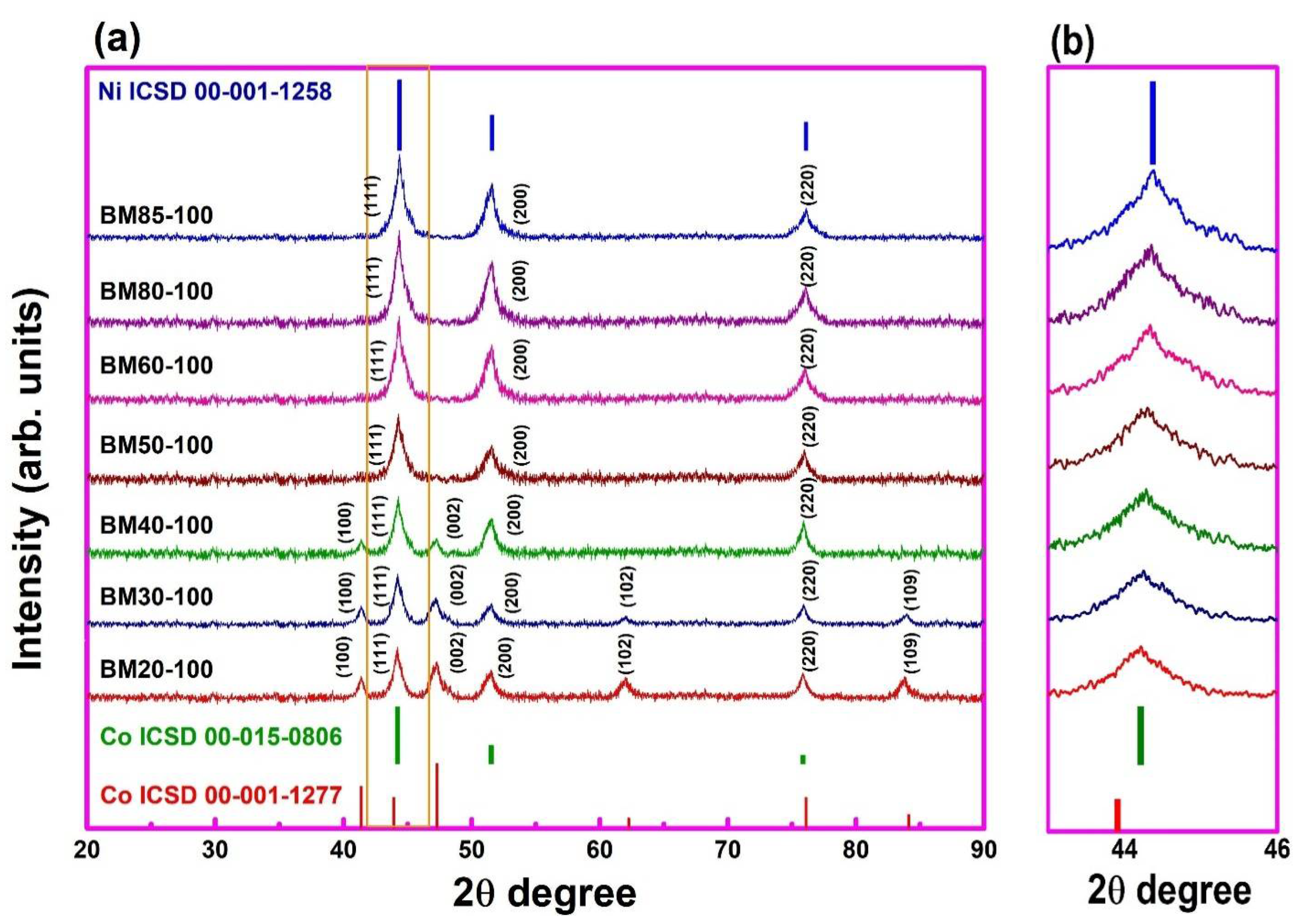
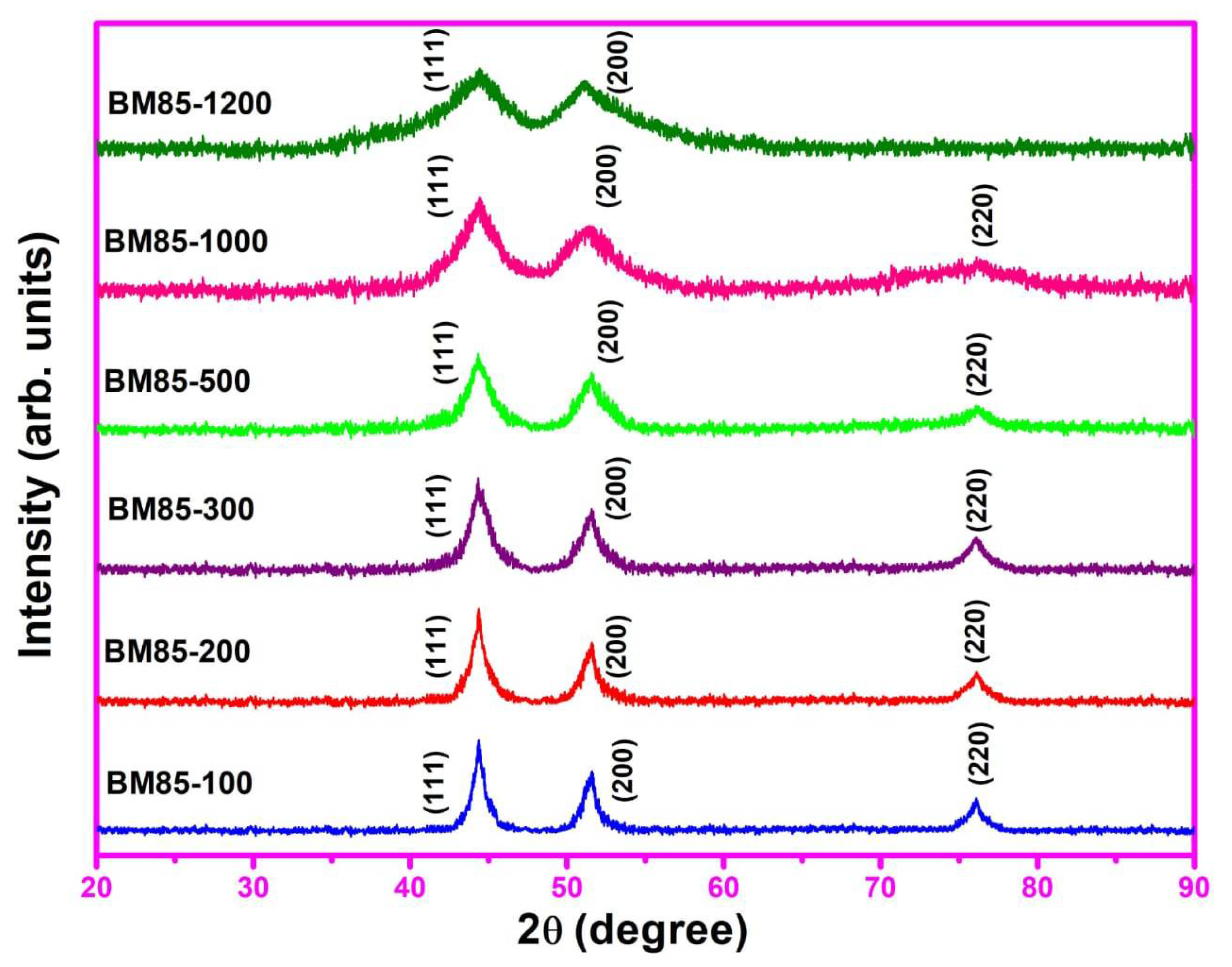
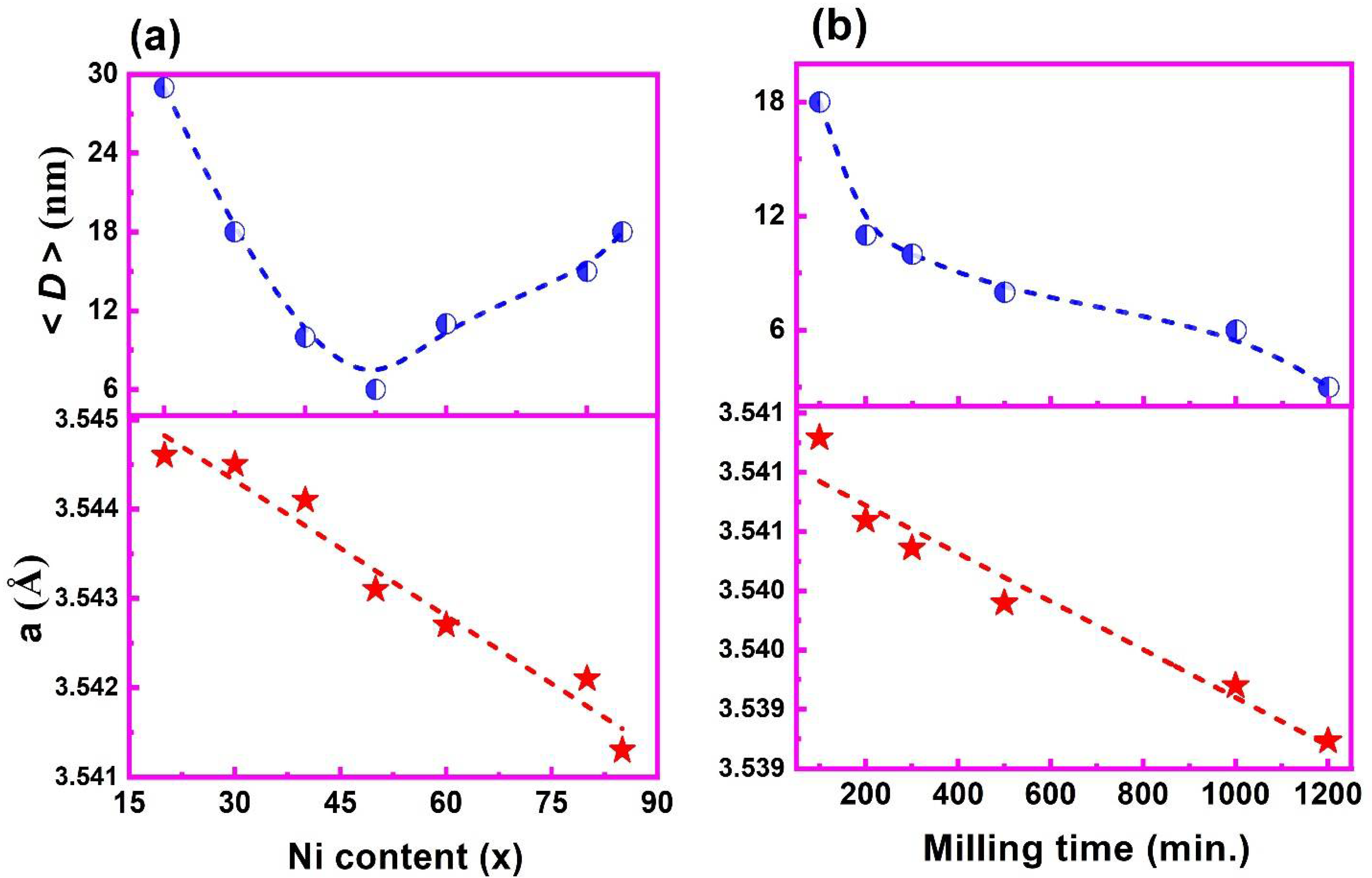
3.1. XRD Spectra Analysis

The XRD spectra of ball-milled Co100−xNix (x = 20, 30, 40, 50, 60, 80, 85) NPs at 1425 rpm for 100 min are shown in Figure 1a. The obtained peaks show the crystallinity of the bimetallic Co100−xNix alloys and no extra peaks of impurities such as Co(OH)2 or Ni(OH)2 are detected. This suggests the completion of the reduction reaction to form CoNi alloy. All the patterns are fitted with the Rietveld method using High Score Plus software. The dominant broad peaks of ferromagnetic Co (ICSD 00-001-1277), (ICSD 00-015-0806), and Ni (ICSD 00-001-1258) show the phase transition from a mixture of face-centered cubic (FCC) and hexagonal closed-packed (HCP) (Co80Ni20, Co70Ni30, Co60Ni40) to FCC (Co50Ni50, Co40Ni60, Co20Ni80, Co15Ni85) [57]. The existence of the HCP phase is because of higher Co content, which is in agreement with the previous reports [49]. The three peaks at 44.37°, 51.59°, and 76.08° corresponding to (111), (200), and (220), respectively, can be easily assigned to FCC CoNi alloy [57]. In Figure 1b, the evolution of the (111), (200), and (220) peaks as a function of composition are shown. When Co is replaced with Ni in Co100−xNix NPs, the peaks change monotonically to large angles, showing a lattice contraction in lattice parameter a. The decrease in the lattice parameter with the increase in Ni contents confirm the CoNi alloy formation [60].
Besides a series of alloy samples, we picked the BM85-100 samples having MS = 48 emu/g and TC = 653 K for ball milling at 1425 rpm for 200, 300, 500, 1000, and 1200 min using the same surfactant. The sample BM85-100 was taken due to its high TC and heating ability for moderate hyperthermia applications. Further ball-milled samples were named as BM85-200, BM85-300, BM85-500, BM85-1000, and BM85-1200 according to their milling times of 200, 300, 500, 1000, and 1200 min, respectively. The XRD spectra of ball-milled NPs are shown in Figure 2. The same phase with the FCC crystal structure similar to the parent compound was observed in spectra. One can also see from the results of the XRD measurements that the intensity of the BM85-100 peaks decreases with the increase in milling time. This was probably due to a reduction in the average crystallite size (as shown in the upper panel of Figure 3b), while there is a significant peak broadening with the increase in full width half maximum peak with increasing milling time from 100 to 1200 min.
The lattice parameter “a” and the average crystallite size < D > of Co100−xNix and ball-milled Co15Ni85 NPs were obtained (using High Score Plus software) from XRD data in Figure 1a and Figure 2, respectively. The results of XRD measurements are presented as a function of Ni content x and milling time as in Figure 3. The lattice parameter determined in this work corresponds to lying between that for Co with 2.544 Å (ICSD 00-015-0806) and Ni with 3.540 Å (ICSD 00-001-1258), respectively. The lattice parameter obtained for milled samples also lies in line with the previous reports on CoNi MNPs [54,57,61].
The lattice parameter for all studied compositions decreases slowly with Ni doping from 3.544 Å in the compound BM40-100 and then it further drops to 3.541 Å in the compound BM85-100, as shown in the lower panel of Figure 3a. This behavior is because of the disappearance of the HCP phase along with the evolution of the FCC phase which leads to a shifting in the peak towards a higher angle, indicating a shrinkage in the lattice parameter a, which is in accordance with Bragg’s law. The increased milling time to 1200 min changed the lattice parameter to 3.539 Å as shown in the lower panel of Figure 3b, whereas the unit cell volume (not shown here) gradually shrank from 44.54 Å3 for BM20-100 to 44.41 Å3 for BM85-100 and then to 44.33 Å3 for BM85-1200, respectively, which is accompanied by the reduction in the lattice parameter.
The average crystallite size < D > obtained for the FCC phase as well as the HCP phase (not shown) shows a decreasing trend with Ni contents as shown in the upper panel of Figure 3a. It can be observed that the crystallite size for the compositions lies in the nanometer regime indicating the nanostructured character of ball-milled samples. However, it is interesting to note that < D > decreases from 29 nm to 6 nm and then increases to 18 nm with the increase in Ni content. Although all Ni-doped samples were prepared as well as milled under identical conditions, the observed variation in the < D > could be due to the different rates of alloy formation for different Ni concentrations. On the other hand, the crystallite size of BM85-100 decreases from 18 to 3 nm for BM85-1200 upon milling as shown in the upper panel of Figure 3b.

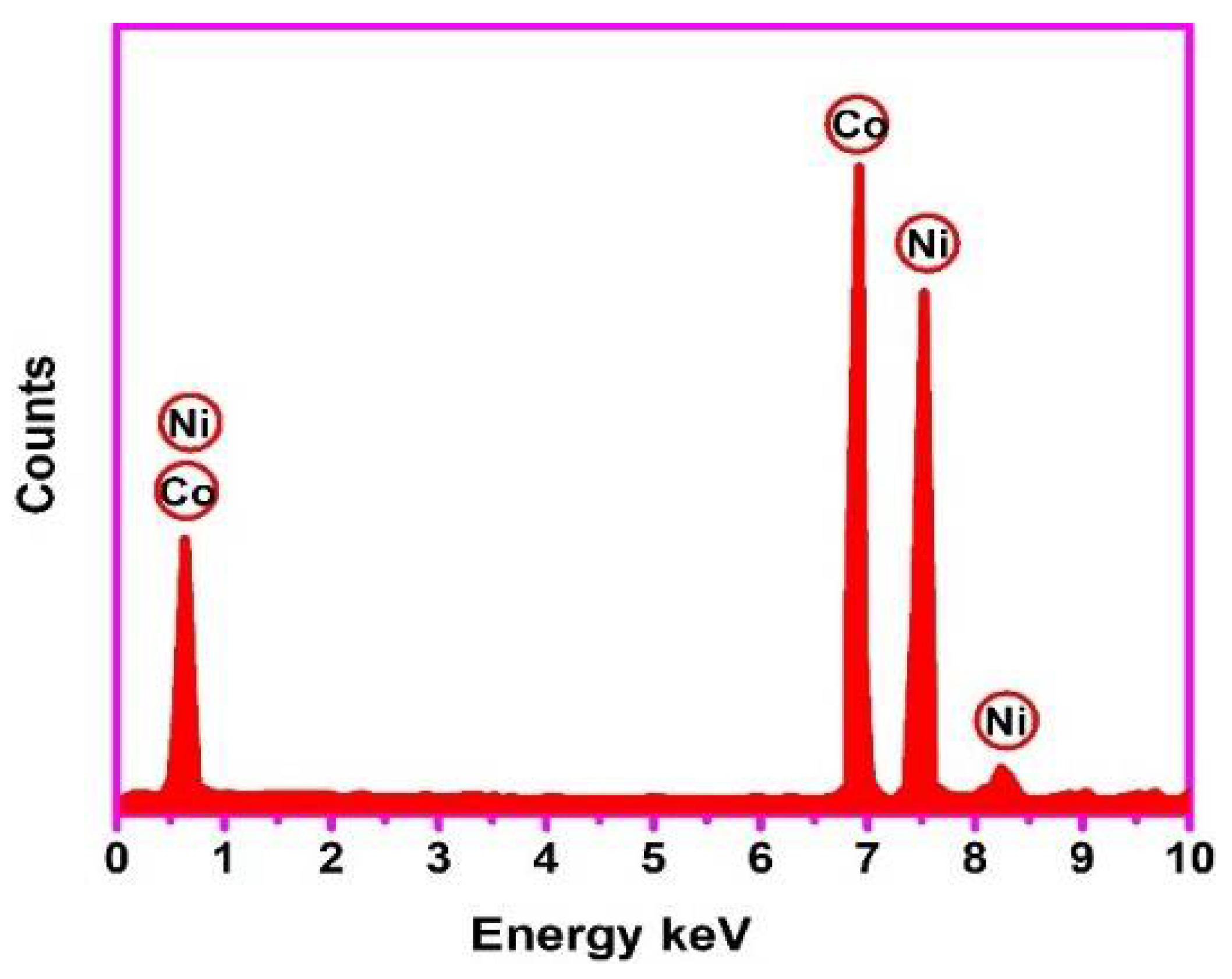
3.2. Elemental Analysis

The EDX spectra analysis gives the elemental compositions of Co100−xNix (x ≥ 50) NPs milled at 1425 rpm for 100 min to confirm the chemical composition of the samples. A representative EDX spectrum for BM50-100 NPs is shown in Figure 4.
The atomic percentages (at.%) of Co and Ni for Co100−xNix NPs and pure Co15Ni85 NPs obtained from EDX and ICP analysis are shown in Table 2. A strong absorption peak at 0.6 keV and 6.9 keV for Co and 0.6 keV, 7.5 keV and 8.2 keV for Ni was observed. For the representative spectrum, the atomic percentage of Co and Ni are 50.26 and 49.74%, respectively, which are close to the initial stoichiometric content of precursors. The result obtained from EDX analysis is in good agreement with the values obtained from ICP.

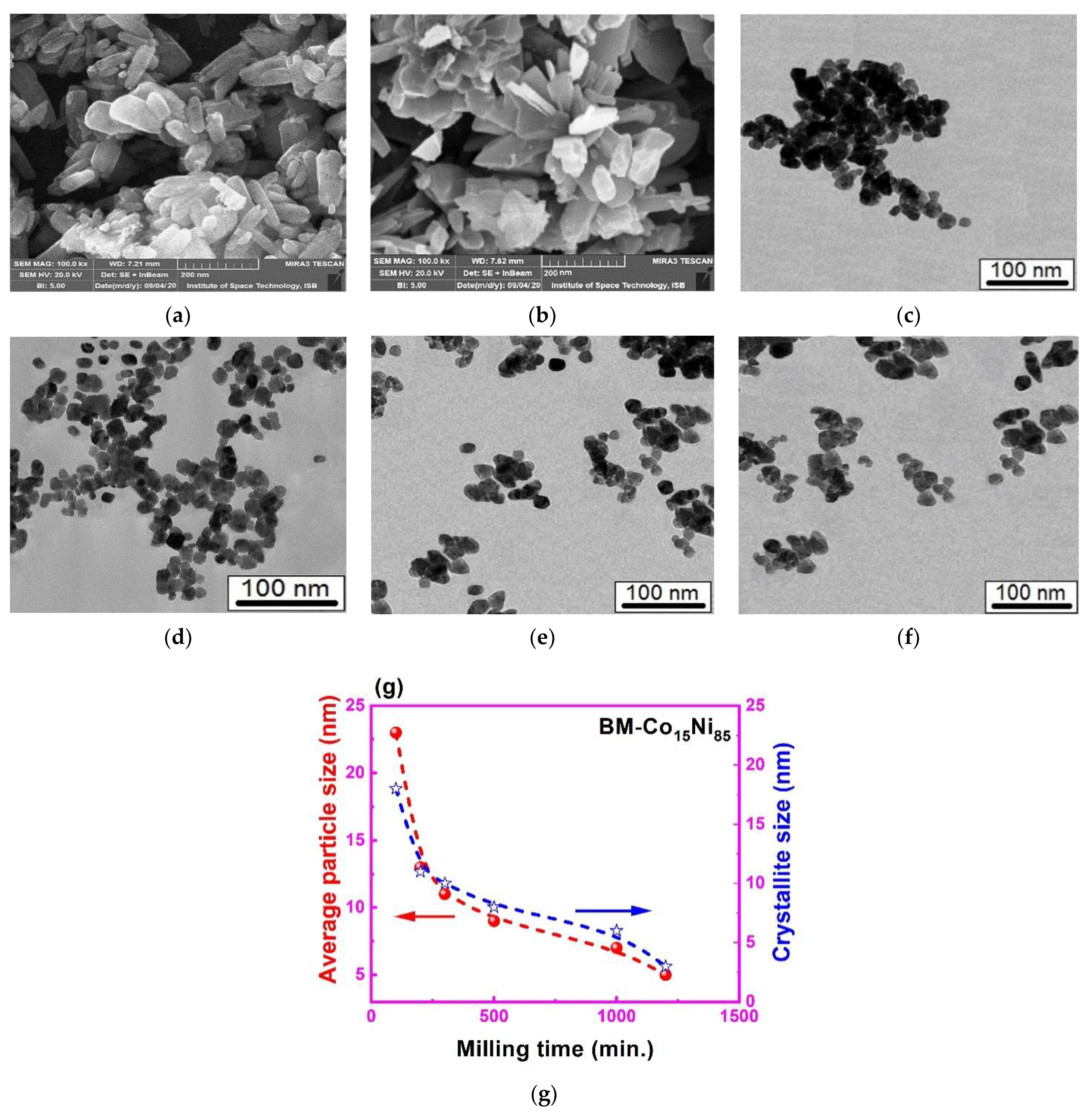
3.3. Surface Morphology

The morphology of Co100−xNix, as well as ball-milled NPs, was investigated with SEM. The SEM micrographs showed different morphologies with the increase in Ni content and similar morphologies for ball-milled samples. To de-agglomerate the MNPs, they were disseminated in n-hexane and sonicated for half an hour. A dried suspension on the glass slide was used for imaging. The SEM micrographs of some representative samples are displayed in Figure 5a,b. It can be seen that the sample morphology changes from rod-like particles for BM20-100 to flower for BM50-100 and then to spherical-like particles for BM85-100 with the increase in Ni contents. These rod-like particles have diameters less than 80 nm and lengths in the range of 50–130 nm (Figure 5a), while flower-like NPs have diameters about 50–150 nm (Figure 5b). The sample with higher Ni content shows agglomeration. This may be due to the ferromagnetic exchange interaction between the Co and Ni NPs [62,63]. The average particle diameters < d > of individual particles were measured from the SEM and TEM data (Figure 5c–f) using image analysis software (Image J). For the Ni-doped samples, < d > falls in the range of 23–120 nm, while for the milled samples, < d > varies between 5 and 23 nm. The average particle size for milled samples (BM85-200 to BM85-500) decreases with the increase in milling time and shows close correspondence with the crystallite sizes obtained from XRD as shown in Figure 5g. We can realize that the crystallite size < D > obtained from XRD for the Ni-doped samples is smaller than those observed from the SEM images. This could be due to the clustered formation of grains, which does not affect diffraction peak formation.

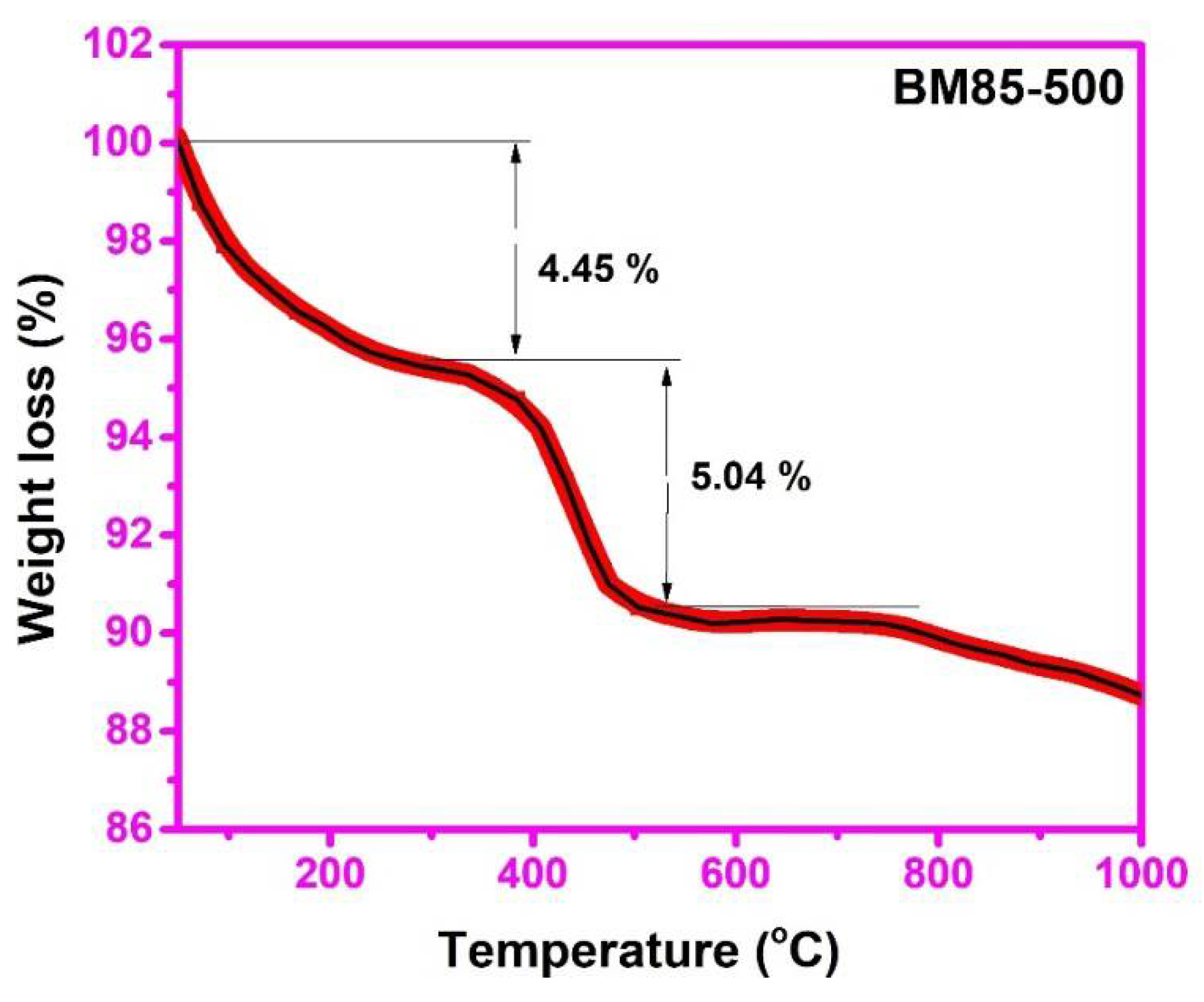
3.4. Thermo-Gravimetric Analysis (TGA)

Thermal analysis was carried out to reveal the presence of oleic acid on ball-milled (BM85-500) NPs. Figure 6 shows the representative TGA analysis of weight loss versus calcination up to 1000 °C (@10 °C/min) in the inert atmosphere. In the TGA curves, there were two phases of decomposition. In the first phase, a 4.45% weight loss was detected in the 50–380 °C range that may be accredited to the vaporization of water molecules from the surface of BM85-500 NPs. The second phase of 5% weight loss in the temperature range of 380–536 °C was ascribed to the decomposition of organic molecules adhered to the surface of the NPs, after which no significant weight loss was seen. Our TGA results are in line with the already reported work [64,65]. Therefore, it could be said that the sample was stable beyond 541 °C.

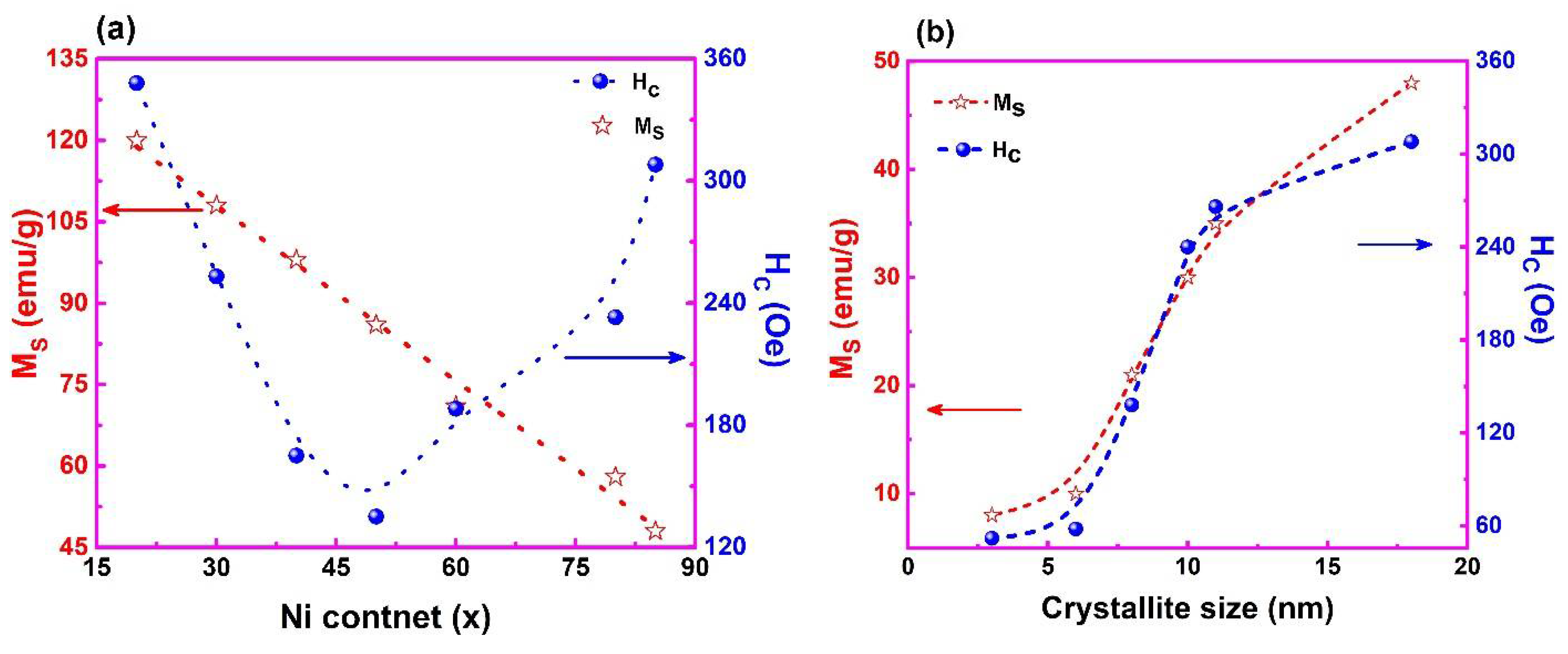
3.5. Field Dependent Magnetization

The results for the magnetic properties of the prepared MNPs are shown in the form of hysteresis loops as shown in Figure 7a,b. These loops were obtained for the samples of Co100−xNix NPs which were milled for 100 min and Co15Ni85 NPs milled for 200, 300, 500, 1000, and 1200 min. All the samples show soft ferromagnetic behavior with coercivities less than 315 Oe. In addition, the magnification of the coercivity is presented in the zoomed inset of Figure 7a. The sample BM80-100 has a saturation magnetization of 120 emu/g which is very near to the published value of 123 emu/g for un-milled samples [57], which indicates that surface degradation for 100 min does not affect the saturation magnetization. However, it is still less than the bulk values of 141 emu/g [57,66]. This reduction in MS can be ascribed to the nanoscale character of particles. The maximum magnetization (Mmax) of BM85-500, BM85-1000, and BM85-1200 does not saturate at ±20 kOe because of a weak paramagnetic component which may be attributed to surface spin disorder during milling for 1200 min [67]. As a result, the magnetization calculated at 20 kOe (M@20 kOe), is the maximum field employed in our tests which can be assumed to be close to MS.
Figure 8 shows the MS and HC values retrieved from the M(H) curves. The MS values decrease from 120 emu/g for BM20-100 to 48 emu/g for BM85-100 with increasing Ni concentration as shown in Figure 8a. The MS values of the CoNi are expected to lie within the bulk MS value of Co and Ni. Thus, the substitution of Ni+2 cations on Co+2 sites would reduce the MS value. Our findings are consistent with those of the prior study [66]. Additionally, the lower values of MS can be attributed to crystal defects, chemical changes on the surface, and magnetic degradation of the surface and the increasing surface area of the NPs [68,69,70]. A significant decrease in MS was observed by milling BM85-100 NPs at 1425 rpm for 200, 300, 500, 1000 and 1200 min, as shown in Figure 8b. Further milling results in surface defects with a decrease in crystallite size <D> as evident from Figure 5g, which decreases the MS value in accordance with ref. [69].
As the Ni content increases, the coercivity decreases from 348 Oe for BM80-100 to 135 Oe for BM50-100 and then increases to a maximum value of 308 Oe for BM85-100, as shown in Figure 8b. The decrease in coercivity with Ni content is accompanied by a decrease in crystallite size. Similarly, the large value of coercivity for higher Co content is related to the large magneto-crystalline anisotropy of the HCP phase. The substitution of Co+2 by Ni+2 reduces the magnetic coupling in the lattice, which may lessen the magneto-crystalline anisotropy with the decrease in the HCP phase and leads to lower coercivity. The enhancement in coercivity with the increase in Ni content can also be caused by the change in shape anisotropy from a rod-like structure to spherical [54,55]. On the other hand, Figure 8b also shows variation in coercivity HC of the ball-milled samples as a function of average crystallite size. The decrease in HC with the decrease in < D > indicates that samples milled at 1425 rpm for 100, 200, 300, 500, 1000 and 1200 min are in the single-domain regime [39,71].

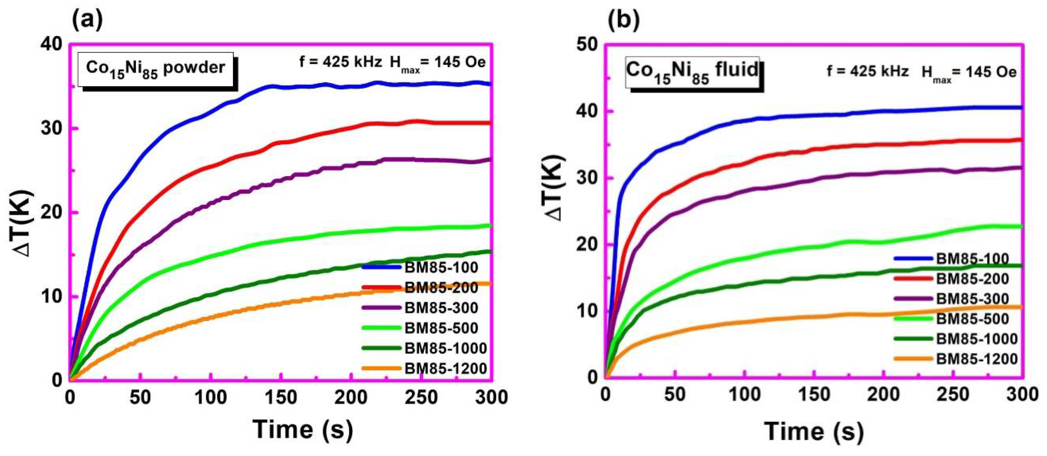
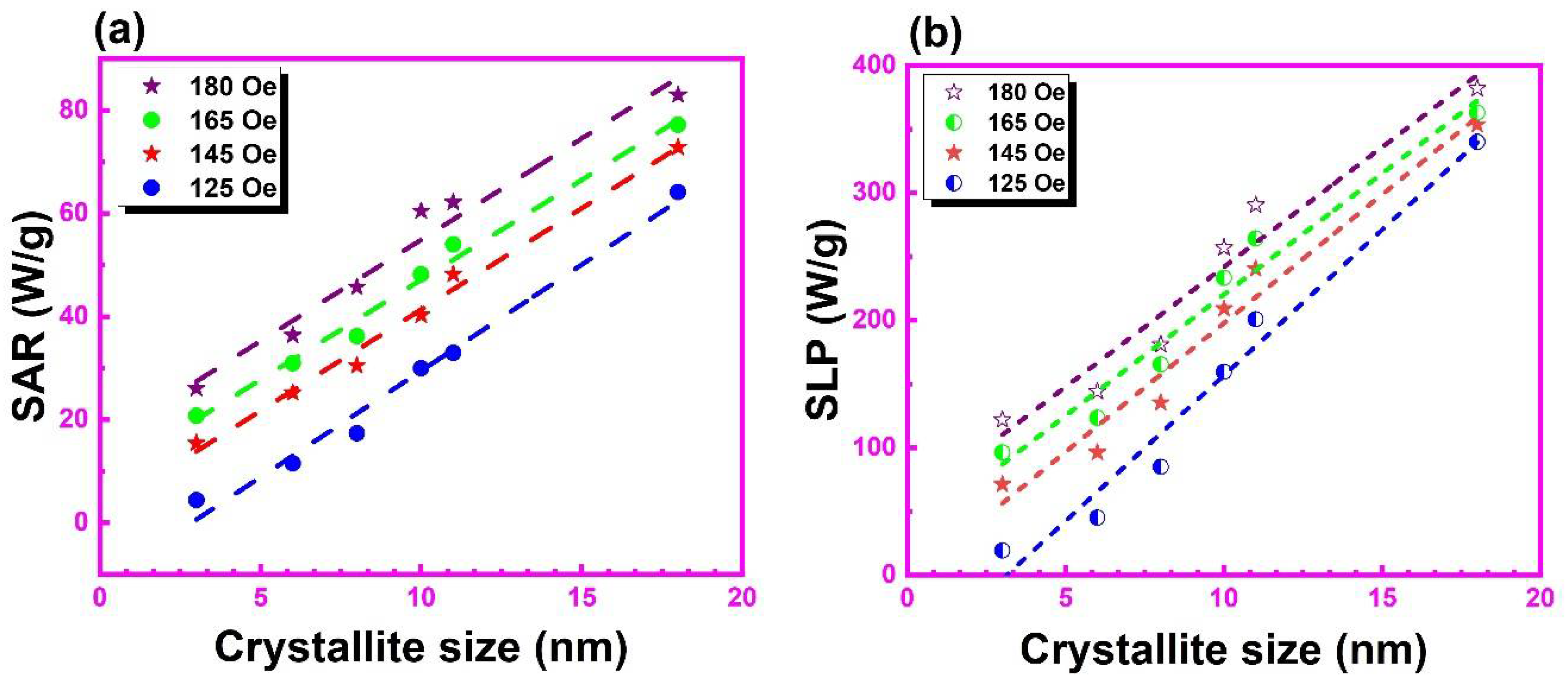
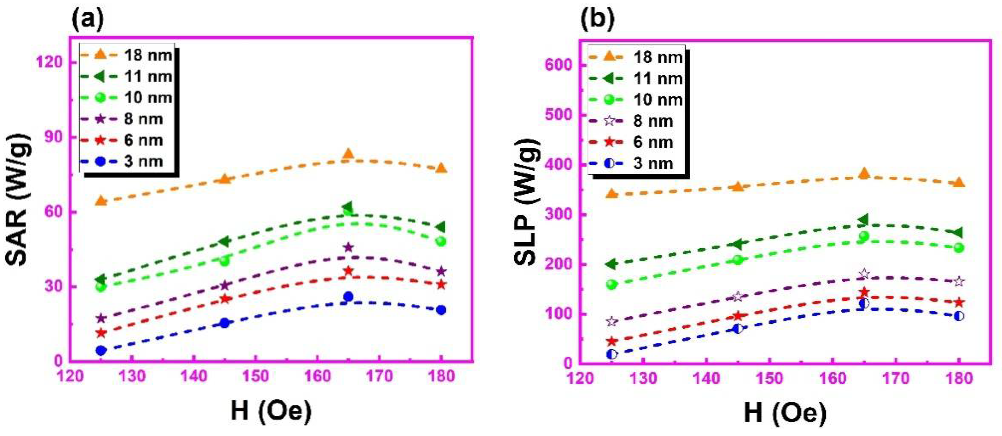
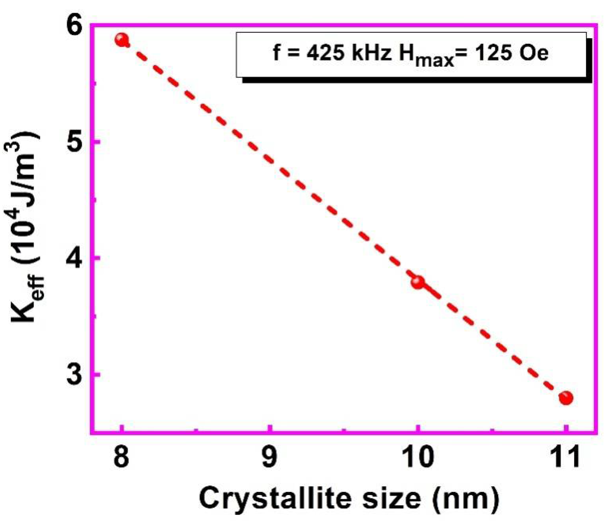
3.6. Temperature-Dependent Magnetization

To investigate the effect of Ni doping as well as milling time on TC, we also measured the temperature dependence of the magnetization M(T) in a static field of 150 Oe. The obtained results for Co100−xNix NPs (x = 20, 30, 40, 50, 60, 80, and 85) and Co15Ni85 NPs milled at a speed of 1425 rpm for 100, 200, 300 and 500 min are shown in Figure 9a,b, respectively. The TC was obtained as the minimum of the (dM/dt) curve [72]. All the samples show a quite sharp ferromagnetic-to-paramagnetic transition at their TC. The TC of the BM20-100 sample is TC~1183 K which is lower than the TC of Co in bulk form (1388 K). This can be reduced by adjusting the Co and Ni ratio. A further decrease in TC was generated by selecting the sample (BM85-100) that had TC~653 K and MS~48 emu/g as shown in Figure 9b. The increase in milling time widens the alteration between ferromagnetic and paramagnetic states while lowering the TC.
The TC values obtained from the above graphs are shown in Figure 10a,b as a function of Ni content and crystallite size, respectively. Ni and Co both have nearly equal atomic masses (58.93 a.u. for Co and 58.69 a.u. for Ni), atomic/ionic radii (1.2 Å for Co and 1.28 Å for Ni) and are ferromagnets; therefore, replacing Co (with higher magnetic moment) with Ni (with lower magnetic moment) reduced the overall moment at the magnetic site. This weakened the ferromagnetic exchange interaction in Co100−xNix alloys and lowered the Curie temperature TC.
Figure 10b shows the variation in TC of ball-milled samples as a function of average crystallite size. The TC decreased as the average crystallite size decreased, probably due to the reduction in dipolar interaction. Increasing milling time extended the transition between ferromagnetic and paramagnetic states while reducing the TC. The lowest TC of 318 K was obtained for BM85-1200 with an average crystallite size of 3 nm. The Curie temperature could, thus, be adjusted by regulating the dopant composition as well as the size of the MNPs and could be brought in the range (315–320 K) required for self-controlled magnetic hyperthermia. This could also affect the other magnetic as well as magnetothermal properties. However, it is interesting to note that the value of magnetization obtained for these NPs was relatively higher than other bimetallic ferromagnetic alloys which may provide large heating [45,73].

3.7. Temperature-Dependent Magnetization

To clarify the efficacy of the heat generated by the selected composition Co15Ni85 due to high Ms and moderate TC value, magnetothermal analysis was conducted on 20 mg powder and fluid samples in an AC magnetic field of amplitude 125, 145, 165 and 180 kOe using a fixed frequency of 425 kHz. The characteristic heating measurements of ball-milled Co15Ni85 powder and its suspension in an AC magnetic field of amplitude Hmax = 145 Oe and frequency f = 425 kHz are shown in Figure 11a,b, respectively. The specific absorption rate (SAR), also known as specific loss power (SLP), represents the magnetothermal behavior of NPs and is evaluated by calculating initial heating rates d T d t t = 0 and applying the equation: SAR = C s a m p l e d T d t t = 0 m s a m p l e m m a g n . For powder sample [74] and SLP = C w a t e r d T d t t = 0 V s a m p l e m m a g n . For fluid sample [75], where Csample and Cwater are the specific heat capacity of the sample for the powder and fluid sample (assumed to be equal to that of pure water 4185 J/LK), respectively; msample is the mass of the sample; Vsample is the sample volume; and mmag. is the fraction of magnetically active elements. These graphs illustrate a significant and consistent rise in temperature for 20 mg powder and fluid sample because it implies the smaller concentration of magnetic NPs [24].
Figure 12a,b show the SAR and SLP values obtained from all heating measurements in an AC magnetic field with a field amplitude of 125, 145, 165, and 180 Oe at a fixed frequency of 425 kHz as a function of average crystallite size. Of all the fields investigated, the highest SAR and SLP values were obtained at 165 Oe. The highest SAR and SLP values obtained for BM85-100 powder and fluid samples are 83 W/g and 382 W/g, respectively. The SAR/SLP values decrease with the decrease in average crystallite sizes, as has been reported for particle sizes lying in the single-domain regime [76]. Such a decreasing trend in SAR/SLP also resembles the behavior of saturation magnetization and coercivity of this sample as shown in Figure 8b. The SAR/SLP values are much higher than those of Fe3O4 [14,77], CoFe3O4 [23], and MFe3O4 (M = Ni, Cu, Zn) [16], which were measured at comparable or greater values of AC field amplitude and frequency, indicating the potential of Co15Ni85 NPs over other potential candidates for magnetically induced hyperthermia. The value of the SAR/SLP for a given average crystallite size also depends upon the AC field amplitude [15].
To investigate the impact of varying the amplitude of the AC field on single-domain NPs with an average crystallite size of 18 nm, 11 nm, 10 nm, 8 nm, 6 nm and 3 nm, the SAR and SLP values are plotted as a function of AC field amplitudes at a fixed frequency of 425 kHz, as shown in Figure 13a,b, respectively. The SAR/SLP values increase linearly with increasing strength of the applied magnetic field Hmax and deviate from linear behavior at the large field for all samples following the linear response theory (LRT) [15,22].
In the following section, we show how the magnetothermal measurements presented for powder samples can be used to obtain the effective anisotropy parameter K e f f for single-domain NPs within the framework of the linear response theory (LRT) [15,22]. According to Carrey et al., the specific area of the hysteresis loop in a complete AC cycle can be expressed as A = π µ o 2 H m a x 2 M s 2 V   τ N 3 k B T ( 1 + 2 τ N 2 ) [15] within the linear response regime, which is valid when the parameter ξ = μ o M S V H m a x k B T   1 . This condition is satisfied for BM85-200 (13 nm), BM85-300 (11 nm) and BM85-500 (9 nm) milled at 1425 rpm when their magnetothermal response is measured at suitable values of Hmax = 125 Oe and f = 425 Hz. Under these conditions, the parameter ξ = 0.05 0.61 validating the use of the LRT, therefore, lets one calculate the effective anisotropy constants K e f f by using Equation (1). The obtained values of K e f f for BM85-200, BM85-300, and BM85-500 are shown in Figure 14 as a function of the average crystallite size.
Figure 14 shows that the effective anisotropy constant K e f f was observed to increase as the crystallite size decreases. There are several factors that influence the effective anisotropy constant. For example, the reduction in coordination of atoms located at the surface of MNPs which affects the spin-polarized electronic density of states and may also change the spin–orbit coupling energy, leading to an increase in magnetic anisotropy [78,79,80,81]. Additionally, deviation from the spherical shape and breaking of the surface crystallographic symmetry [82], as well as lattice structure expansion and contraction [83], influences K e f f . However, it is not an easy task to separate the parameters that control the K e f f . In addition, the highest value of the effective anisotropy constant is 5.9 × 10⁴ J/m3 for BM85-500 MNPs of size 9 nm, which is higher than the previous report by Rafique et al. [54], and showed magneto crystalline anisotropy of 4.46 × 10⁴ J/m3 for nanorods of CoNi, comparable to 3.8 × 10⁴ J/m3 for BM85-300 MNPs of size 11 nm. The enhancement in effective anisotropy constant in spherical-shape MNPs is due to the large surface spin effects, shape, and stress anisotropy constant [54].
Finally, the possible cytotoxicity of Co15Ni85 NPs ball-milled for 100, 200, 300 and 500 min at 1425 rpm was estimated by preparing 100 µg/mL of MNPs in chloroform. The degree to which the MNPs cause damage to red blood cells (RBCs) was studied in terms of hemolysis [84]. MNPs interacting with RBCs can damage the cell membrane and interact with the hemoglobin, causing its structural and conformational changes [85]. The higher percentage of hemolysis indicates higher cytotoxicity of the MNPs [86]. The results from the cytotoxicity assay in the form of percentage of hemolysis for oleic-acid-coated and uncoated Co15Ni85 MNPs are shown in Table 1. It has been shown by Purohit et al. [87] that coating gold NPs with bovine serum albumin reduces the hemolysis effect. Additionally, oleic acid is a well-known biocompatible material [88,89]. In our case, oleic-acid-coated MNPs show a significant reduction in the percentage of hemolysis by reducing the direct contact of MNPs with the surface of the RBCs. Therefore, oleic-acid-coated Co15Ni85 MNPs showed significantly lower hemolysis at similar concentrations, suggesting that these MNPs could be of great importance in biomedical applications such as self-controlled magnetic hyperthermia.

4. Conclusions

The properties of oleic-acid-coated Co100−xNix NPs, synthesized using the hydrothermal method and subsequent ball milling, were measured. A reduction in the particle size by ball milling was observed which led to successfully reducing the TC. All the ball-milled NPs exhibited a linear dependence of the SAR and SLP on average crystallite size. The largest SAR and SLP obtained were 83 W/g and 382 W/g for powder and fluid samples, respectively. Moreover, these SAR/SLP values were much larger than those of NPs of maghemite, magnetite, and various mixed ferrites, despite their lower Curie temperatures. We also used magnetothermal measurements using the linear response theory to obtain the K e f f for ball-milled Co15Ni85 NPs with respect to the average crystallite size. Our work indicates that magnetothermal measurements can be used as a quick and easier alternative to conventional magnetometry techniques to determine effective anisotropy and is in line with the linear response theory. From the point of view of biomedical applications, coating these MNPs with oleic acid significantly reduces their cytotoxicity, making them appropriate contenders for self-controlled magnetic hyperthermia, MRI contrast agents, drug delivery, magnetic bio detection, magnetic self-healing composites, nanoparticle-based biosensors and other biomedical applications.

Author Contributions

Conceptualization, M.H.A.N. and S.O.A.; methodology, S.S.; software, M.H.A.N.; validation, M.I.S. and M.A.Q.; formal analysis S.K.A.; investigation, M.H.A.N.; resources, M.I.S.; data curation, M.S.K.; writing—original draft preparation, M.H.A.N.; writing—review and editing, M.A.Q. and M.I.S.; visualization, M.M.F.; supervision, M.S.; project administration, T.S.; funding acquisition, Y.A. All authors have read and agreed to the published version of the manuscript.

Funding

This work is funded by Taif University Researchers Supporting Project number (TURSP-2020/249), Taif University, Taif, Saudi Arabia.

Institutional Review Board Statement

Not applicable.

Informed Consent Statement

Not applicable.

Data Availability Statement

Not applicable.

Acknowledgments

The authors thank Taif University Researchers Supporting Project number (TURSP-2020/249), Taif University, Taif, Saudi Arabia.

Conflicts of Interest

The authors declare no conflict of interest.

References

  1. Zeng, N.; He, L.; Jiang, L.; Shan, S.; Su, H. Synthesis of magnetic/pH dual responsive dextran hydrogels as stimuli-sensitive drug carriers. Carbohydr. Res. 2022, 520, 108632. [Google Scholar] [CrossRef] [PubMed]
  2. Taheri-Ledari, R.; Maleki, A. Magnetic nanocatalysts utilized in the synthesis of aromatic pharmaceutical ingredients. New J. Chem. 2021, 45, 4135–4146. [Google Scholar] [CrossRef]
  3. Kheiri, K.; Sohrabi, N.; Mohammadi, R.; Amini-Fazl, M.S. Preparation and characterization of magnetic nanohydrogel based on chitosan for 5-fluorouracil drug delivery and kinetic study. Int. J. Biol. Macromol. 2022, 202, 191–198. [Google Scholar] [CrossRef]
  4. Tabasi, H.; Mosavian, M.T.H.; Darroudi, M.; Khazaei, M.; Hashemzadeh, A.; Sabouri, Z. Synthesis and characterization of amine-functionalized Fe3O4/Mesoporous Silica Nanoparticles (MSNs) as potential nanocarriers in drug delivery systems. J. Porous Mater. 2022, 1–12. [Google Scholar] [CrossRef]
  5. Taheri-Ledari, R.; Zhang, W.; Radmanesh, M.; Mirmohammadi, S.S.; Maleki, A.; Cathcart, N.; Kitaev, V. Multi-stimuli nanocomposite therapeutic: Docetaxel targeted delivery and synergies in treatment of human breast cancer tumor. Small 2020, 16, 2002733. [Google Scholar] [CrossRef] [PubMed]
  6. Dik, G.; Ulu, A.; Ates, B. Medicinal and Biological Application of Magnetic Alloy Nanoparticles and Their Polymer Nanocomposites. In Handbook of Magnetic Hybrid Nanoalloys and their Nanocomposites; Springer: Berlin/Heidelberg, Germany, 2022; pp. 1–27. [Google Scholar]
  7. Taheri-Ledari, R.; Maleki, A. Antimicrobial therapeutic enhancement of levofloxacin via conjugation to a cell-penetrating peptide: An efficient sonochemical catalytic process. J. Pept. Sci. 2020, 26, e3277. [Google Scholar] [CrossRef]
  8. Gleich, B.; Weizenecker, J. Tomographic imaging using the nonlinear response of magnetic particles. Nature 2005, 435, 1214. [Google Scholar] [CrossRef]
  9. Jordan, A. Hyperthermia classic commentary: Inductive heating of ferrimagnetic particles and magnetic fluids: Physical evaluation of their potential for hyperthermia by Andreas Jordan et al., International Journal of Hyperthermia, 1993;9:51–68. Int. J. Hyperth. 2009, 25, 512–516. [Google Scholar] [CrossRef]
  10. Arruebo, M.; Fernández-Pacheco, R.; Ibarra, M.R.; Santamaría, J. Magnetic nanoparticles for drug delivery. Nano Today 2007, 2, 22–32. [Google Scholar] [CrossRef]
  11. Grossman, J.H.; McNeil, S.E. Nanotechnology in cancer medicine. Phys. Today 2012, 65, 38. [Google Scholar] [CrossRef] [Green Version]
  12. Čampelj, S.; Makovec, D.; Drofenik, M. Functionalization of magnetic nanoparticles with 3-aminopropyl silane. J. Magn. Magn. Mater. 2009, 321, 1346–1350. [Google Scholar] [CrossRef]
  13. Kami, D.; Takeda, S.; Itakura, Y.; Gojo, S.; Watanabe, M.; Toyoda, M. Application of magnetic nanoparticles to gene delivery. Int. J. Mol. Sci 2011, 12, 3705–3722. [Google Scholar] [CrossRef]
  14. Larumbe, S.; Gomez-Polo, C.; Pérez-Landazábal, J.; Pastor, J. Effect of a SiO2 coating on the magnetic properties of Fe3O4 nanoparticles. J. Phys. Condens. Matter. 2012, 24, 266007. [Google Scholar] [CrossRef] [PubMed]
  15. Carrey, J.; Mehdaoui, B.; Respaud, M. Simple models for dynamic hysteresis loop calculations of magnetic single-domain nanoparticles: Application to magnetic hyperthermia optimization. J. Appl. Phys. 2011, 109, 083921. [Google Scholar] [CrossRef]
  16. Verde, E.; Landi, G.T.; Carrião, M.; Drummond, A.L.; Gomes, J.; Vieira, E.; Sousa, M.; Bakuzis, A.F. Field dependent transition to the non-linear regime in magnetic hyperthermia experiments: Comparison between maghemite, copper, zinc, nickel and cobalt ferrite nanoparticles of similar sizes. AIP Adv. 2012, 2, 032120. [Google Scholar] [CrossRef]
  17. Kallumadil, M.; Tada, M.; Nakagawa, T.; Abe, M.; Southern, P.; Pankhurst, Q.A. Corrigendum to “Suitability of commercial colloids for magnetic hyperthermia”. J. Magn. Magn. Mater. 2009, 321, 3650–3651. [Google Scholar] [CrossRef]
  18. Baker, I.; Zeng, Q.; Li, W.; Sullivan, C.R. Heat deposition in iron oxide and iron nanoparticles for localized hyperthermia. J. Appl. Phys. 2006, 99, 08H106. [Google Scholar] [CrossRef]
  19. Hergt, R.; Dutz, S.; Müller, R.; Zeisberger, M. Magnetic particle hyperthermia: Nanoparticle magnetism and materials development for cancer therapy. J. Phys. Condens. Matter 2006, 18, S2919. [Google Scholar] [CrossRef]
  20. Xia, Q.-S.; Liu, X.; Xu, B.; Zhao, T.-D.; Li, H.-Y.; Chen, Z.-H.; Xiang, Q.; Geng, C.-Y.; Pan, L.; Hu, R.-L. Feasibility study of high-temperature thermoseed inductive hyperthermia in melanoma treatment. Oncol. Rep. 2011, 25, 953–962. [Google Scholar]
  21. Jordan, A.; Scholz, R.; Wust, P.; Fähling, H.; Felix, R. Magnetic fluid hyperthermia (MFH): Cancer treatment with AC magnetic field induced excitation of biocompatible superparamagnetic nanoparticles. J. Magn. Magn. Mater. 1999, 201, 413–419. [Google Scholar] [CrossRef]
  22. Rosensweig, R.E. Heating magnetic fluid with alternating magnetic field. J. Magn. Magn. Mater. 2002, 252, 370–374. [Google Scholar] [CrossRef]
  23. Verde, E.L.; Landi, G.T.; Gomes, J.A.; Sousa, M.H.; Bakuzis, A.F. Magnetic hyperthermia investigation of cobalt ferrite nanoparticles: Comparison between experiment, linear response theory, and dynamic hysteresis simulations. J. Appl. Phys. 2012, 111, 123902. [Google Scholar] [CrossRef]
  24. Dutz, S.; Hergt, R. Magnetic nanoparticle heating and heat transfer on a microscale: Basic principles, realities and physical limitations of hyperthermia for tumour therapy. Int. J. Hyperth. 2013, 29, 790–800. [Google Scholar] [CrossRef] [PubMed]
  25. Vázquez, M. Handbook of magnetism and advanced magnetic materials. Nov. Mater. 2007, 4, 1–29. [Google Scholar]
  26. Nguyen, H.D.; Nguyen, T.D.; Nguyen, D.H.; Nguyen, P.T. Magnetic properties of Cr doped Fe3O4 porous nanoparticles prepared through a co-precipitation method using surfactant. Adv. Nat. Sci. Nanosci. Nanotechnol. 2014, 5, 035017. [Google Scholar] [CrossRef]
  27. Zhao, X.; Wang, W.; Zhang, Y.; Wu, S.; Li, F.; Liu, J.P. Synthesis and characterization of gadolinium doped cobalt ferrite nanoparticles with enhanced adsorption capability for Congo Red. Chem. Eng. J. 2014, 250, 164–174. [Google Scholar] [CrossRef]
  28. Alnasir, M.H.; Khan, M.Y.; Ali, W.B.; Ansar, M.; Manzoor, S. Magnetic and hyperthermia properties of Ni1− xCux nanoparticles coated with oleic acid and silica prepared via sol–gel method. Appl. Phys. A 2019, 125, 1–13. [Google Scholar] [CrossRef]
  29. Alnasir, M.H.; Awan, M.; Manzoor, S.; Materials, M. Magnetic and magnetothermal studies of pure and doped gadolinium silicide nanoparticles for self-controlled hyperthermia applications. J. Magn. Magn. Mater. 2018, 449, 137–144. [Google Scholar] [CrossRef]
  30. Idrissi, L.; Tahiri, N.; El Bounagui, O.; Ez-Zahraouy, H. Magnetic properties of NiFe2O4 compound: Ab Initio calculation and Monte Carlo simulation. J. Supercond. Nov. Magn. 2020, 33, 1369–1375. [Google Scholar] [CrossRef]
  31. Dennis, C.; Jackson, A.; Borchers, J.; Ivkov, R.; Foreman, A.; Hoopes, P.; Strawbridge, R.; Pierce, Z.; Goerntiz, E.; Lau, J. The influence of magnetic and physiological behaviour on the effectiveness of iron oxide nanoparticles for hyperthermia. J. Phys. D Appl. Phys. 2008, 41, 134020. [Google Scholar] [CrossRef]
  32. Kawai, N.; Futakuchi, M.; Yoshida, T.; Ito, A.; Sato, S.; Naiki, T.; Honda, H.; Shirai, T.; Kohri, K. Effect of heat therapy using magnetic nanoparticles conjugated with cationic liposomes on prostate tumor in bone. Prostate 2008, 68, 784–792. [Google Scholar] [CrossRef] [PubMed]
  33. Suto, M.; Hirota, Y.; Mamiya, H.; Fujita, A.; Kasuya, R.; Tohji, K.; Jeyadevan, B. Heat dissipation mechanism of magnetite nanoparticles in magnetic fluid hyperthermia. J. Magn. Magn. Mater. 2009, 321, 1493–1496. [Google Scholar] [CrossRef]
  34. Lévy, M.; Wilhelm, C.; Siaugue, J.-M.; Horner, O.; Bacri, J.-C.; Gazeau, F. Magnetically induced hyperthermia: Size-dependent heating power of γ-Fe2O3 nanoparticles. J. Phys. Condens. Matter 2008, 20, 204133. [Google Scholar] [CrossRef] [PubMed]
  35. Blonska-Tabero, A.; Bosacka, M.; Filipek, E.; Piz, M.; Kochmanski, P. High-temperature synthesis and unknown properties of M3Cr4 (PO4)6, where M=Zn or Mg and a new solid solution Zn1.5Mg1.5Cr4 (PO4)6. J. Therm. Anal. Calorim. 2020, 140, 2625–2631. [Google Scholar] [CrossRef]
  36. Thapa, D.; Palkar, V.; Kurup, M.; Malik, S. Properties of magnetite nanoparticles synthesized through a novel chemical route. Mater. Lett. 2004, 58, 2692–2694. [Google Scholar] [CrossRef]
  37. Nikiforov, V.; Koksharov, Y.A.; Polyakov, S.; Malakho, A.; Volkov, A.; Moskvina, M.; Khomutov, G.; Irkhin, V.Y. Magnetism and Verwey transition in magnetite nanoparticles in thin polymer film. J. Alloys Compd. 2013, 569, 58–61. [Google Scholar] [CrossRef]
  38. Hergt, R.; Andra, W.; d′Ambly, C.G.; Hilger, I.; Kaiser, W.A.; Richter, U.; Schmidt, H.-G. Physical limits of hyperthermia using magnetite fine particles. IEEE Trans. Magn. 1998, 34, 3745–3754. [Google Scholar] [CrossRef]
  39. Nauman, M.; Alnasir, M.H.; Hamayun, M.A.; Wang, Y.; Shatruk, M.; Manzoor, S. Size-dependent magnetic and magnetothermal properties of gadolinium silicide nanoparticles. RSC Adv. 2020, 10, 28383–28389. [Google Scholar] [CrossRef]
  40. Ennassiri, N.; Tahiri, N.; El Bounagui, O.; Ez-Zahraouy, H.; Benyoussef, A. Structural, electronic, magnetic, and magnetocaloric properties in metallic antiperovskite compound Mn3GaC. Mater. Res. Bull. 2018, 98, 335–339. [Google Scholar] [CrossRef]
  41. Ennassiri, N.; Tahiri, N.; El Bounagui, O.; Ez-Zahraouy, H.; Benyoussef, A. Magnetic, magnetocaloric and transport properties in AlCMn3 antiperovskite compound. J. Alloys Compd. 2018, 741, 1196–1202. [Google Scholar] [CrossRef]
  42. Bazine, W.; Tahiri, N.; El Bounagui, O.; Ez-Zahraouy, H.; Benyoussef, A. Structural, electronic, magnetic, and magnetocaloric properties in intermetallic compound TbCu2Si2. J. Magn. Magn. Mater. 2019, 481, 72–76. [Google Scholar] [CrossRef]
  43. Cui, B.; Marinescu, M.; Liu, J. High magnetization Fe-Co and Fe-Ni submicron and nanosize particles by thermal decomposition and hydrogen reduction. J. Appl. Phys. 2014, 115, 17A315. [Google Scholar] [CrossRef]
  44. Yi, Z.; Chahine, R.; Bose, T.K. Magnetic entropy change in the Ge-rich alloys Gd-Si-Ge. IEEE Trans. Magn. 2003, 39, 3358–3360. [Google Scholar] [CrossRef]
  45. Pandey, S.; Quetz, A.; Aryal, A.; Dubenko, I.; Mazumdar, D.; Stadler, S.; Ali, N. Magnetic, structural and magnetocaloric properties of Ni-Si and Ni-Al thermoseeds for self-controlled hyperthermia. Int. J. Hyperth. 2017, 33, 779–784. [Google Scholar] [CrossRef]
  46. Ucar, H.; Ipus, J.J.; Laughlin, D.; McHenry, M. Tuning the Curie temperature in γ-FeNi nanoparticles for magnetocaloric applications by controlling the oxidation kinetics. J. Appl. Phys. 2013, 113, 17A918. [Google Scholar] [CrossRef]
  47. Ferk, G.; Stergar, J.; Makovec, D.; Hamler, A.; Jagličić, Z.; Drofenik, M.; Ban, I. Synthesis and characterization of Ni–Cu alloy nanoparticles with a tunable Curie temperature. J. Alloys Compd. 2015, 648, 53–58. [Google Scholar] [CrossRef]
  48. Mohn, P.; Wohlfarth, E. The Curie temperature of the ferromagnetic transition metals and their compounds. J. Phys. F Met. Phys. 1987, 17, 2421. [Google Scholar] [CrossRef]
  49. Nishizawa, T.; Ishida, K. The Co−Ni (Cobalt-Nickel) system. Bull. Alloy. Phase Diagr. 1983, 4, 390–395. [Google Scholar] [CrossRef]
  50. Respaud, M.; Broto, J.; Rakoto, H.; Fert, A.; Thomas, L.; Barbara, B.; Verelst, M.; Snoeck, E.; Lecante, P.; Mosset, A.J.P.R.B. Surface effects on the magnetic properties of ultrafine cobalt particles. Phys. Rev. B 1998, 57, 2925. [Google Scholar] [CrossRef]
  51. Zhang, Y.; Or, S.-W.; Zhang, Z. Synthesis, structure and magnetic properties of CoNi submicrospherical chains. Adv. Mater. Phys. Chem. 2011, 1, 7. [Google Scholar] [CrossRef]
  52. Neamtu, J.; Verga, N. Magnetic nanoparticles for magneto-resonance imaging and targeted drug delivery. Dig. J. Nanomater. Biostruct. 2011, 6, 969–978. [Google Scholar]
  53. Serizawa, K.; Kawai, T.; Ohtake, M.; Futamoto, M.; Kirino, F.; Inaba, N. Magnetostriction behaviors of Ni100-xFex and Ni100-yCoy (001) single-crystal films with fcc structure under rotating magnetic fields. IEEE Trans. Magn. 2017, 53, 1–4. [Google Scholar] [CrossRef]
  54. Rafique, M.Y.; Pan, L.; Khan, W.S.; Iqbal, M.Z.; Qiu, H.; Farooq, M.H.; Ellahi, M.; Guo, Z.J.C. Controlled synthesis, phase formation, growth mechanism, and magnetic properties of 3-D CoNi alloy microstructures composed of nanorods. CrystEngComm 2013, 15, 5314–5325. [Google Scholar] [CrossRef]
  55. Nie, D.; Xu, C.; Chen, H.; Wang, Y.; Li, J.; Liu, Y.J.M.L. Chain-like CoNi alloy microstructures fabricated by a PVP-assisted solvothermal process. Mater. Lett. 2014, 131, 306–309. [Google Scholar] [CrossRef]
  56. Nadhman, A.; Nazir, S.; Khan, M.I.; Ayub, A.; Muhammad, B.; Khan, M.; Shams, D.F.; Yasinzai, M. Visible-light-responsive ZnCuO nanoparticles: Benign photodynamic killers of infectious protozoans. Int. J. Nanomed. 2015, 10, 6891. [Google Scholar]
  57. Bensouilah, A.; Guittoum, A.; Hemmous, M.; Bouremana, A.; Rahal, B.; Yavru, C.; Öksüzoglu, R.; Kechouane, M.; Magnetism, N. Structure, Microstructure and Magnetic Properties of CoxNi100−x Powders Synthesized by Hydrothermal Method. J. Supercond. Nov. Magn. 2017, 30, 2219–2225. [Google Scholar] [CrossRef]
  58. Gondwal, M.; Joshi nee Pant, G. Synthesis and catalytic and biological activities of silver and copper nanoparticles using Cassia occidentalis. Int. J. Biomater. 2018, 2018, 6735426. [Google Scholar] [CrossRef]
  59. Kapusetti, G.; Misra, N.; Singh, V.; Kushwaha, R.; Maiti, P. Bone cement/layered double hydroxide nanocomposites as potential biomaterials for joint implant. J. Biomed. Mater. Res. Part A 2012, 100, 3363–3373. [Google Scholar] [CrossRef]
  60. Mattei, G.; de Julián Fernández, C.; Mazzoldi, P.; Sada, C.; De, G.; Battaglin, G.; Sangregorio, C.; Gatteschi, D. Synthesis, Structure, and Magnetic Properties of Co, Ni, and Co−Ni Alloy Nanocluster-Doped SiO2 Films by Sol-Gel Processing. Chem. Mater. 2002, 14, 3440–3447. [Google Scholar] [CrossRef]
  61. Hu, M.J.; Lin, B.; Yu, S.H.J.N.r. Magnetic field-induced solvothermal synthesis of one-dimensional assemblies of Ni-Co alloy microstructures. Nano Res. 2008, 1, 303–313. [Google Scholar] [CrossRef]
  62. Jayakumar, O.; Salunke, H.; Tyagi, A.J. Synthesis and characterization of stoichiometric NiCo nano particles dispersible in both aqueous and non aqueous media. Solid State Commun. 2009, 149, 1769–1771. [Google Scholar] [CrossRef]
  63. Li, H.; Jin, Z.; Song, H.; Liao, S. Synthesis of Co submicrospheres self-assembled by Co nanosheets via a complexant-assisted hydrothermal approach. J. Magn. Magn. Mater. 2010, 322, 30–35. [Google Scholar] [CrossRef]
  64. Willis, A.L.; Turro, N.J.; O′Brien, S. Spectroscopic characterization of the surface of iron oxide nanocrystals. Chem. Mater. 2005, 17, 5970–5975. [Google Scholar] [CrossRef]
  65. Roca, A.; Morales, M.; O’Grady, K.; Serna, C.J.N. Structural and magnetic properties of uniform magnetite nanoparticles prepared by high temperature decomposition of organic precursors. Nanotechnology 2006, 17, 2783. [Google Scholar] [CrossRef]
  66. Toneguzzo, P.; Viau, G.; Acher, O.; Guillet, F.; Bruneton, E.; Fievet-Vincent, F.; Fievet, F. CoNi and FeCoNi fine particles prepared by the polyol process: Physico-chemical characterization and dynamic magnetic properties. J. Mater. Sci. 2000, 35, 3767–3784. [Google Scholar] [CrossRef]
  67. Iqbal, Y.; Bae, H.; Rhee, I.; Hong, S. Nanotechnology. Intensive analysis of core–shell silica-coated iron-oxide nanoparticles for magnetic hyperthermia. J. Nanosci. Nanotechnol. 2016, 16, 11862–11867. [Google Scholar] [CrossRef]
  68. Kodama, R.H.; Berkowitz, A.E.; McNiff, E., Jr.; Foner, S. Surface spin disorder in NiFe2O4 nanoparticles. Phys. Rev. Lett. 1996, 77, 394. [Google Scholar] [CrossRef]
  69. Zheng, M.; Wu, X.; Zou, B.; Wang, Y.J. Magnetic properties of nanosized MnFe2O4 particles. J. Magn. Magn. Mater. 1998, 183, 152–156. [Google Scholar] [CrossRef]
  70. George, M.; John, A.M.; Nair, S.S.; Joy, P.; Anantharaman, M.R.; Materials, M. Finite size effects on the structural and magnetic properties of sol–gel synthesized NiFe2O4 powders. J. Magn. Magn. Mater. 2006, 302, 190–195. [Google Scholar] [CrossRef]
  71. O′handley, R.C. Modern Magnetic Materials: Principles and Applications; Wiley: New York, NY, USA, 2000. [Google Scholar]
  72. Rostamnejadi, A.; Salamati, H.; Kameli, P.; Ahmadvand, H. Superparamagnetic behavior of La0.67Sr0.33MnO3 nanoparticles prepared via sol–gel method. J. Magn. Magn. Mater. 2009, 321, 3126–3131. [Google Scholar] [CrossRef]
  73. Mornet, S.; Vasseur, S.; Grasset, F.; Duguet, E. Magnetic nanoparticle design for medical diagnosis and therapy. J. Mater. Chem. 2004, 14, 2161–2175. [Google Scholar] [CrossRef]
  74. Hamayun, M.A.; Abramchuk, M.; Alnasir, H.; Khan, M.; Pak, C.; Lenhert, S.; Ghazanfari, L.; Shatruk, M.; Manzoor, S.; Materials, M. Magnetic and magnetothermal studies of iron boride (FeB) nanoparticles. J. Magn. Magn. Mater. 2018, 451, 407–413. [Google Scholar] [CrossRef]
  75. Hanini, A.; Lartigue, L.; Gavard, J.; Kacem, K.; Wilhelm, C.; Gazeau, F.; Chau, F.; Ammar, S.J.J.o.M.; Materials, M. Zinc substituted ferrite nanoparticles with Zn0.9Fe2.1O4 formula used as heating agents for in vitro hyperthermia assay on glioma cells. J. Magn. Magn. Mater. 2016, 416, 315–320. [Google Scholar] [CrossRef]
  76. Vasseur, S.; Duguet, E.; Portier, J.; Goglio, G.; Mornet, S.; Hadová, E.; Knížek, K.; Maryško, M.; Veverka, P.; Pollert, E. Lanthanum manganese perovskite nanoparticles as possible in vivo mediators for magnetic hyperthermia. J. Magn. Magn. Mater. 2006, 302, 315–320. [Google Scholar] [CrossRef]
  77. Wuang, S.C.; Neoh, K.G.; Kang, E.-T.; Pack, D.W.; Leckband, D.E. Synthesis and functionalization of polypyrrole-Fe3O4 nanoparticles for applications in biomedicine. J. Mater. Chem. 2007, 17, 3354–3362. [Google Scholar] [CrossRef]
  78. Bruno, P. Tight-binding approach to the orbital magnetic moment and magnetocrystalline anisotropy of transition-metal monolayers. Phys. Rev. B 1989, 39, 865. [Google Scholar] [CrossRef] [PubMed]
  79. Ballou, R.; Deportes, J.; Gorges, B.; Lemaire, R.; Ousset, J. Anomalous thermal variation of the bulk anisotropy in GdCo5. J. Magn. Magn. Mater. 1986, 54, 465–466. [Google Scholar] [CrossRef]
  80. Drzazga, Z.; Bia, K.; Borgie, W. Effective magnetic anisotropy constant for B-substituted RCo5 (R= Y, Gd) compounds. J. Magn. Magn. Mater. 1991, 101, 399–400. [Google Scholar] [CrossRef]
  81. Szade, J. Magnetism of (Gd1-xYx) 2In. J. Magn. Magn. Mater. 1997, 170, 228–234. [Google Scholar] [CrossRef]
  82. Obaidat, I.; Mohite, V.; Issa, B.; Tit, N.; Haik, Y. Predicting a major role of surface spins in the magnetic properties of ferrite nanoparticles. Cryst. Res. Technol. J. Exp. Ind. Crystallogr. 2009, 44, 489–494. [Google Scholar] [CrossRef]
  83. Dorfbauer, F.; Evans, R.; Kirschner, M.; Chubykalo-Fesenko, O.; Chantrell, R.; Schrefl, T. Effects of surface anisotropy on the energy barrier in cobalt–silver core–shell nanoparticles. J. Magn. Magn. Mater. 2007, 316, e791–e794. [Google Scholar] [CrossRef]
  84. Barshtein, G.; Arbell, D.; Yedgar, S. Hemolytic effect of polymeric nanoparticles: Role of albumin. IEEE Trans. NanoBiosci. 2011, 10, 259–261. [Google Scholar] [CrossRef] [PubMed]
  85. Groopman, J.E.; Itri, L.M. Chemotherapy-induced anemia in adults: Incidence and treatment. J. Natl. Cancer Inst. 1999, 91, 1616–1634. [Google Scholar] [CrossRef] [PubMed]
  86. Mocan, T. Hemolysis as expression of nanoparticles-induced cytotoxicity in red blood cells. BMBN 2013, 1, 7–12. [Google Scholar]
  87. Purohit, R.; Vallabani, N.S.; Shukla, R.K.; Kumar, A.; Singh, S. Effect of gold nanoparticle size and surface coating on human red blood cells. Bioinspired Biomim. Nanobiomater. 2016, 5, 121–131. [Google Scholar] [CrossRef]
  88. Kalhapure, R.S.; Akamanchi, K.G. A novel biocompatible bicephalous dianionic surfactant from oleic acid for solid lipid nanoparticles. Colloids Surf. B Biointerfaces 2013, 105, 215–222. [Google Scholar] [CrossRef]
  89. Zhang, J.; Chen, X.G.; Huang, L.; Han, J.T.; Zhang, X.F. Self-assembled polymeric nanoparticles based on oleic acid-grafted chitosan oligosaccharide: Biocompatibility, protein adsorption and cellular uptake. J. Mater. Sci. Mater. Med. 2012, 23, 1775–1783. [Google Scholar] [CrossRef]
Figure 1. (a) XRD spectra of ball-milled Co100−xNix (x = 20, 30, 40, 50, 60, 80, 85) NPs at 1425 rpm for 100 min (b) The evolution of the (111) reflection in more detail. The base panels show the reference spectra for Co using the reference data (ICSD 00-001-1277) and (ICSD 00-015-0806). The uppermost panels show the reference spectra for Ni (ICSD 00-001-1258).
Coatings 12 01272 g001
Figure 2. XRD patterns of Co15Ni85 NPs milled at 1425 rpm for 100, 200, 300, 500, 1000 and 1200 min.
Coatings 12 01272 g002
Figure 3. (a) Average crystallite size and (b) the lattice constant as a function of (a) Ni content and (b) milling time for FCC Co100−xNix NPs.
Coatings 12 01272 g003
Figure 4. EDX spectra analysis of BM50-100 sample.
Coatings 12 01272 g004
Figure 5. Field-emission scanning micrograph (FE-SEM) of (a) BM20-100, (b) BM50-100, transmission electron microscope (TEM) images of (c) BM80-300, (d) BM85-500, (e) BM85-1000, (f) BM85-1200 NPs and (g) The average particle size of milled NPs against the milling time.
Coatings 12 01272 g005
Figure 6. Thermo-gravimetric analysis of oleic acid-coated BM85-500 NPs.
Coatings 12 01272 g006
Figure 7. Room temperature hysteresis loops of (a) Co100−xNix NPs milled for 100 min and (b) Co15Ni85 NPs milled for 100, 200, 300, 500, 1000, 1200 min. The inset shows the zoomed-in pictures of low-applied-magnetic-field region.
Coatings 12 01272 g007
Figure 8. Saturation magnetization MS (measured at 20 kOe) and coercivity obtained from hysteresis loops of (a) Co100−xNix NPs milled for 100 min and (b) Co15Ni85 NPs milled for 200, 300, 500, 1000, 1200 min as a function of Ni content and average crystallite size, respectively.
Coatings 12 01272 g008
Figure 9. Zero-field-cooled thermal-demagnetization measurements of (a) Co100−xNix NPs milled for 100 min and (b) Co15Ni85 NPs milled for 200, 300, 500, 1000, and 1200 min in a measuring field of 150 Oe.
Coatings 12 01272 g009
Figure 10. Curie temperature against Ni content and crystallite size for (a) Co100−xNix NPs as a function of Ni content and for (b) Co15Ni85 NPs milled at a speed of 1425 rpm for 100, 200, 300, 500, 1000 and 1200 min.
Coatings 12 01272 g010
Figure 11. Heating curves of representative Co15Ni85 NPs milled at 1425 rpm for 100, 200, 300, 500, 1000 and 1200 min. (a) 20 mg of powder and (b) 1000 µL of the fluid sample measured in an AC magnetic field of amplitude Hmax = 145 Oe and frequency f = 425 kHz.
Coatings 12 01272 g011
Figure 12. (a) SAR and (b) SLP of Co15Ni85 NPs milled at 1425 rpm for 100, 200, 300, 500, 1000 and 1200 min as a function of average crystallite size. The straight lines guide the eye.
Coatings 12 01272 g012
Figure 13. (a) SAR and (b) SLP of Co15Ni85 NPs milled at 1425 rpm for 100, 200, 300, 500, 1000, and 1200 min as a function of AC field amplitude at a fixed frequency of 425 kHz.
Coatings 12 01272 g013
Figure 14. Dependence of effective anisotropy constant on average crystallite size of Co15Ni85 NPs milled at 1425 rpm for 200, 300, and 500 min.
Coatings 12 01272 g014
Table 1. Hemolysis activities of bare (as prepared) and oleic-acid-coated Co15Ni85 NPs milled for 100, 200, 300, and 500 min at 1425 rpm.
Oleic-Acid-Coated Co15Ni85 MNPsOptical DensityHemolysis (%)
BM-2000.06524
BM-3000.06814.4
BM-5000.07244.9
As prepared (uncoated)0.245328
Positive control0.7805100
Negative control0.03560
Table 2. Atomic percentages (at.%) of Co and Ni for Co100−xNix (x ≥ 50) NPs.
SampleEDXICP
Co (at.%)Ni (at.%)Co (at.%)Ni (at.%)
BM50-10050.2649.7449.8650.14
BM60-10041.5958.4140.4959.51
BM80-10021.5878.4220.6879.32
BM85-100
Co15Ni85
16.5783.4315.77
15.12
84.23
84.97
Publisher’s Note: MDPI stays neutral with regard to jurisdictional claims in published maps and institutional affiliations.

