
Heart Failure and Pancreas Exocrine Insufficiency: Pathophysiological Mechanisms and Clinical Point of View

 , , , and
Abstract
1. Introduction
2. Pancreatic Circulation and Hemodynamics
3. Pancreatic Structure and Function/Dysfunction
Pancreatic Exocrine Insufficiency
- Injury to pancreatic exocrine parenchyma, resulting in reduced synthetic capacity (e.g., [recurrent] pancreatitis, cystic fibrosis, ageing)
- Reduced stimulation of pancreatic enzyme production (e.g., celiac disease, autonomic dysfunction)
- Pancreatic duct obstruction (e.g., malignancy)
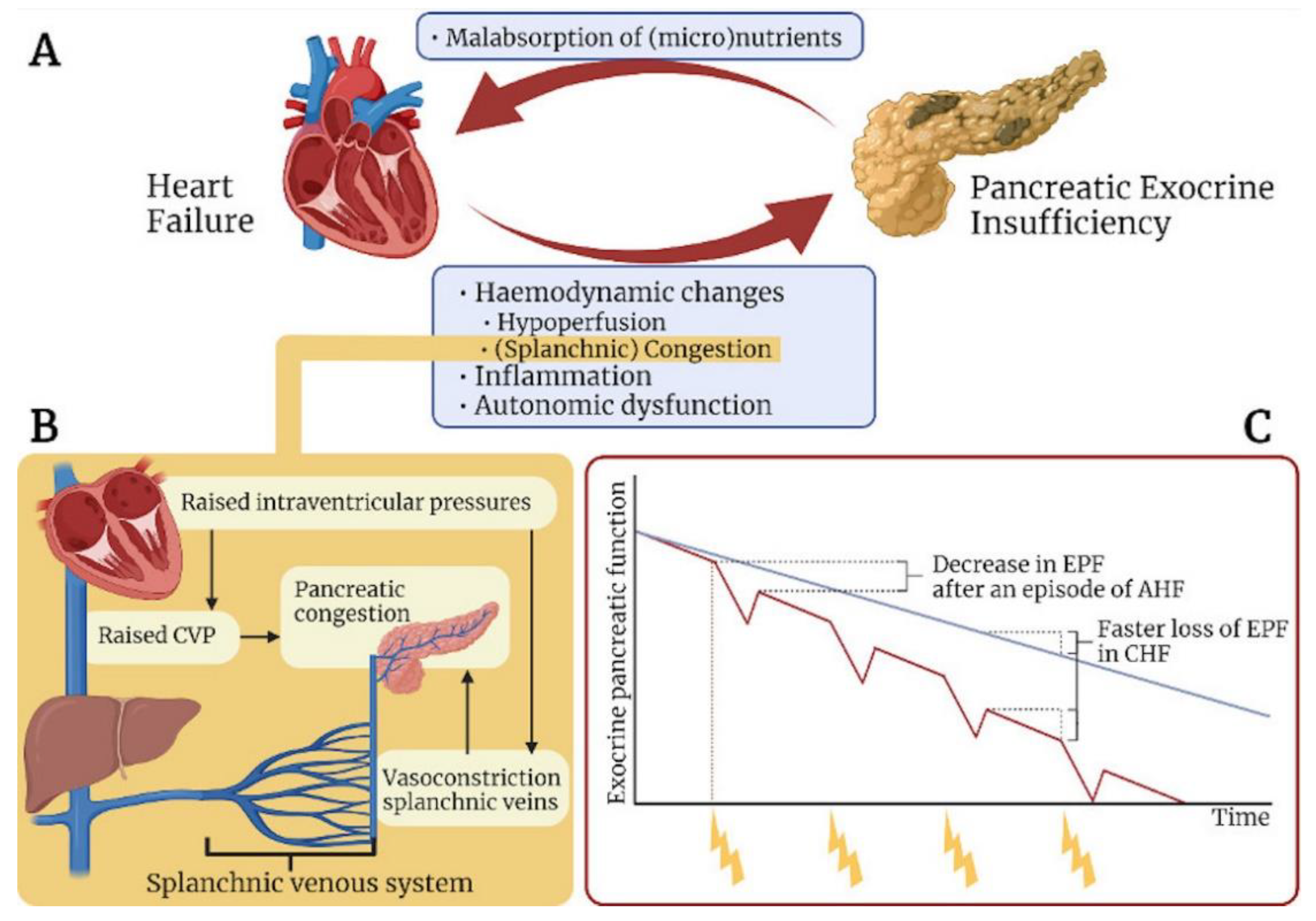
4. Studies on Pancreatic Injury in Heart Failure
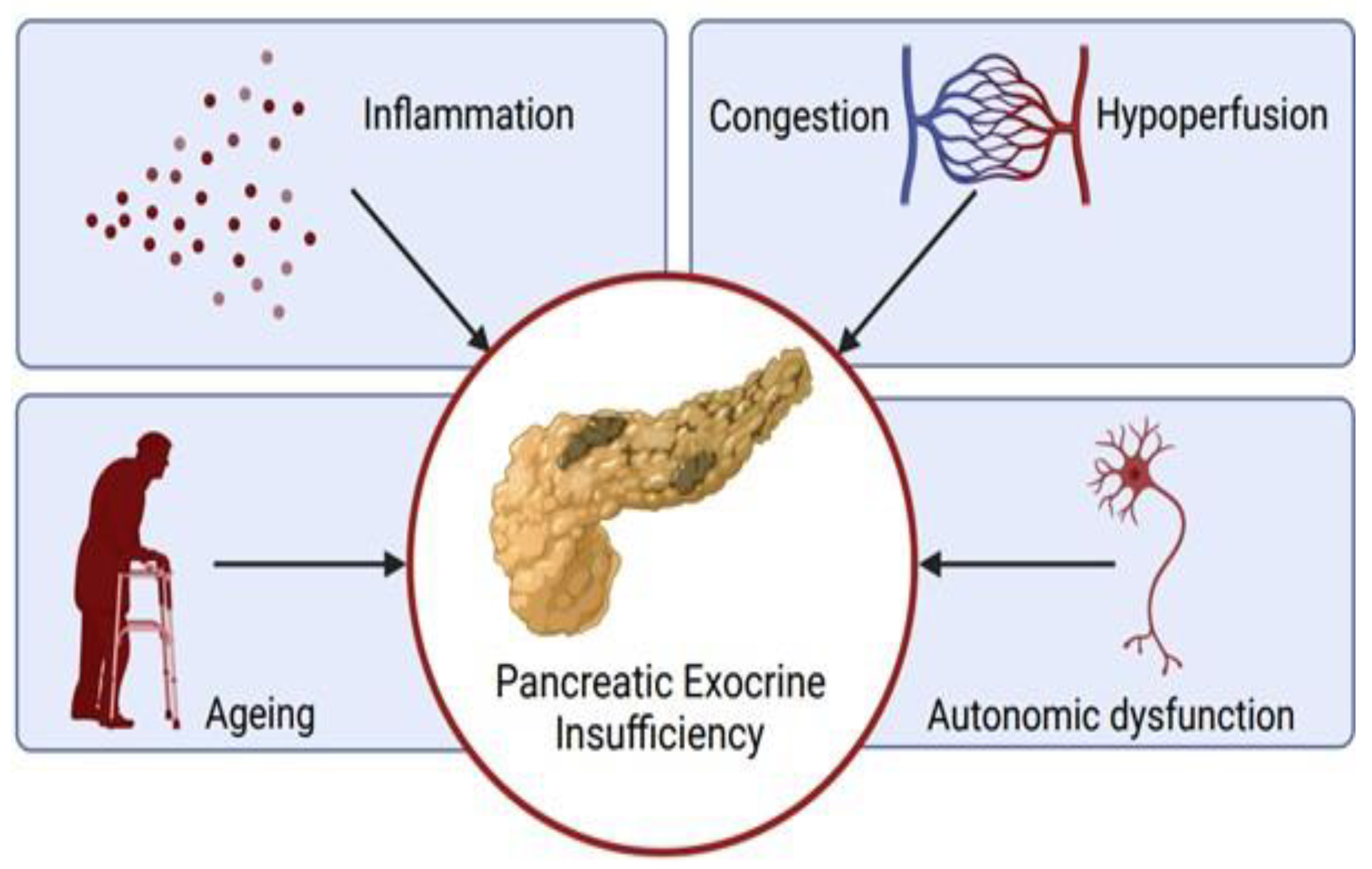
5. Mechanisms of Pancreatic Damage in Heart Failure
5.1. Ageing
5.2. Hemodynamics
5.3. Inflammation
5.4. Autonomic Dysfunction
6. Summary and Clinical Implications
7. Conclusions
Author Contributions
Funding
Institutional Review Board Statement
Informed Consent Statement
Data Availability Statement
Conflicts of Interest
References
- McDonagh, T.A.; Metra, M.; Adamo, M.; Gardner, R.S.; Baumbach, A.; Böhm, M.; Burri, H.; Butler, J.; Čelutkienė, J.; Chioncel, O.; et al. 2021 ESC Guidelines for the diagnosis and treatment of acute and chronic heart failure. Eur. Heart J. 2021, 42, 3599–3726. [Google Scholar] [CrossRef] [PubMed]
- Harjola, V.P.; Mullens, W.; Banaszewski, M.; Bauersachs, J.; Brunner-La Rocca, H.P.; Chioncel, O.; Collins, S.P.; Doehner, W.; Filippatos, G.S.; Flammer, A.J.; et al. Organ dysfunction, injury and failure in acute heart failure, from patho-physiology to diagnosis and management. A review on behalf of the Acute Heart Failure Committee of the Heart Failure As-sociation (HFA) of the European Society of Cardiology (ESC). Eur. J. Heart Fail. 2017, 19, 821–836. [Google Scholar] [CrossRef] [PubMed]
- Damman, K.; van Deursen, V.M.; Navis, G.; Voors, A.A.; van Veldhuisen, D.J.; Hillege, H.L. Increased Central Venous Pres-sure Is Associated with Impaired Renal Function and Mortality in a Broad Spectrum of Patients with Cardiovascular Disease. J. Am. Coll. Cardiol. 2009, 53, 582–588. [Google Scholar] [CrossRef] [PubMed]
- Boorsma, E.M.; ter Maaten, J.M.; Voors, A.A.; van Veldhuisen, D.J. Renal Compression in Heart Failure, The Renal Tam-ponade Hypothesis. J. Am. Coll. Cardiol. 2022, 10, 175–183. [Google Scholar]
- Sundaram, V.; Fang, J.C. Gastrointestinal and Liver Issues in Heart Failure. Circulation 2016, 133, 1696–1703. [Google Scholar] [CrossRef]
- Van Deursen, V.M.; Damman, K.; Hillege, H.L.; van Beek, A.P.; Van Veldhuisen, D.J.; Voors, A.A. Abnormal Liver Function in Relation to Hemodynamic Profile in Heart Failure Patients. J. Card. Fail. 2010, 16, 84–90. [Google Scholar] [CrossRef]
- Valentova, M.; Von Haehling, S.; Bauditz, J.; Doehner, W.; Ebner, N.; Bekfani, T.; Elsner, S.; Sliziuk, V.; Scherbakov, N.; Murín, J.; et al. Intestinal congestion and right ventricular dysfunction: A link with appetite loss, inflammation, and cachexia in chronic heart failure. Eur. Heart J. 2016, 37, 1684–1691. [Google Scholar] [CrossRef]
- Sandek, A.; Bauditz, J.; Swidsinski, A.; Buhner, S.; Weber-Eibel, J.; von Haehling, S.; Schroedl, W.; Karhausen, T.; Doehner, W.; Rauchhaus, M.; et al. Altered Intestinal Function in Patients with Chronic Heart Failure. J. Am. Coll. Cardiol. 2007, 50, 1561–1569. [Google Scholar] [CrossRef]
- Westenbrink, B.D.; Voors, A.A.; De Boer, R.A.; Schuringa, J.J.; Klinkenberg, T.; Van Der Harst, P.; Vellenga, E.; Van Veldhuisen, D.J.; Van Gilst, W.H. Bone marrow dysfunction in chronic heart failure patients. Eur. J. Heart Fail. 2010, 12, 676–684. [Google Scholar] [CrossRef]
- Erkelens, C.D.; van der Wal, H.H.; de Jong, B.M.; Elting, J.W.; Renken, R.; Gerritsen, M.; van Laar, P.J.; van Deursen, V.M.; van der Meer, P.; van Veldhuisen, D.J.; et al. Dynamics of cerebral blood flow in patients with mild non-ischaemic heart failure. Eur. J. Heart Fail. 2017, 19, 261–268. [Google Scholar] [CrossRef]
- Siegmund, A.S.; Pieper, P.G.; Bilardo, C.M.; Gordijn, S.J.; Khong, T.Y.; Gyselaers, W.; van Veldhuisen, D.J.; Dickinson, M.G. Cardiovascular Determinants of Impaired Placental Function in Women with Cardiac Dysfunction. Am. Heart J. 2021. [Google Scholar] [CrossRef] [PubMed]
- Verbrugge, F.H.; Dupont, M.; Steels, P.; Grieten, L.; Malbrain, M.; Tang, W.W.; Mullens, W. Abdominal Contributions to Cardiorenal Dysfunction in Congestive Heart Failure. J. Am. Coll. Cardiol. 2013, 62, 485–495. [Google Scholar] [CrossRef] [PubMed]
- Swan, J.W.; Walton, C.; Godsland, I.F.; Clark, A.L.; Coats, A.J.S.; Oliver, M.F. Insulin resistance in chronic heart failure. Eur. Heart J. 1994, 15, 1528–1532. [Google Scholar] [CrossRef] [PubMed]
- Riehle, C.; Abel, E.D. Insulin Signaling and Heart Failure. Circ. Res. 2016, 118, 1151–1169. [Google Scholar] [CrossRef] [PubMed]
- Melenovsky, V.; Benes, J.; Franekova, J.; Kovar, J.; Borlaug, B.A.; Segetova, M.; Tura, A.; Pelikanova, T. Glucose homeostasis, pancreatic endocrine function, and outcomes in ad-vanced heart failure. J. Am. Heart Assoc. 2017, 6, e005290. [Google Scholar] [CrossRef]
- Isaacson, A.; Spagnoli, F.M. Pancreatic cell fate specification: Insights into developmental mechanisms and their application for lineage reprogramming. Curr. Opin. Genet. Dev. 2021, 70, 32–39. [Google Scholar] [CrossRef]
- Vollmar, B.; Janata, J.; Yamauchi, J.; Wolf, B.; Heuser, M.; Menger, M.D. Exocrine, but not endocrine, tissue is susceptible to microvascular ischemia/reperfusion injury following pancreas transplantation in the rat. Transpl. Int. 1999, 12, 50–55. [Google Scholar] [CrossRef]
- Machens, H.G.; Senninger, N.; Runkel, N.; Frank, G.; Von Kummer, R.; Herfarth, C. Advantages and disadvantages of using the hydrogen clearance technique to measure pancreatic blood flow. Eur. J. Surg. 1992, 158, 113–116. [Google Scholar]
- Lewis, M.P.N.; Reber, H.; Ashley, S. Pancreatic Blood Flow and Its Role in the Pathophysiology of Pancreatitis. J. Surg. Res. 1998, 75, 81–89. [Google Scholar] [CrossRef]
- Parks, D.A.; Jacobson, E.D. Physiology of the Splanchnic Circulation. Arch. Intern. Med. 1985, 145, 1278–1281. [Google Scholar] [CrossRef]
- Kvietys, P.R.; McLendon, J.M.; Bulkley, G.B.; A Perry, M.; Granger, D.N. Pancreatic circulation: Intrinsic regulation. Am. J. Physiol. Content 1982, 242. [Google Scholar] [CrossRef] [PubMed]
- Widdison, A.L.; Karanjia, N.D.; A Reber, H. Pancreatic vascular regulation in chronic pancreatitis in cats. Gut 1995, 36, 133–136. [Google Scholar] [CrossRef] [PubMed][Green Version]
- Hainsworth, R. The Importance of Vascular Capacitance in Cardiovascular Control. Physiology 1990, 5, 250–254. [Google Scholar] [CrossRef]
- Gelman, S.; Warner, D.S.; Warner, M.A. Venous Function and Central Venous Pressure. Anesthesiology 2008, 108, 735–748. [Google Scholar] [CrossRef]
- Fudim, M.; Jones, W.S.; Boortz-Marx, R.L.; Ganesh, A.; Green, C.L.; Hernandez, A.F.; Patel, M.R. Splanchnic Nerve Block for Acute Heart Failure. Circulation 2018, 138, 951–953. [Google Scholar] [CrossRef]
- Fudim, M.; Patel, M.R.; Boortz-Marx, R.; Borlaug, B.A.; DeVore, A.D.; Ganesh, A.; Green, C.L.; Lopes, R.D.; Mentz, R.J.; Patel, C.B.; et al. Splanchnic Nerve Block Mediated Changes in Stressed Blood Volume in Heart Fail. J. Am. Coll. Cardiol. 2021, 9, 293–300. [Google Scholar]
- Birch, D.J.; Turmaine, M.; Boulos, P.B.; Burnstock, G. Sympathetic Innervation of Human Mesenteric Artery and Vein. J. Vasc. Res. 2008, 45, 323–332. [Google Scholar] [CrossRef]
- Khan, D.; Moffet, C.R.; Flatt, P.R.; Kelly, C. Role of islet peptides in beta cell regulation and type 2 diabetes therapy. Peptides 2018, 100, 212–218. [Google Scholar] [CrossRef]
- Zhou, Q.; Melton, D.A. Pancreas regeneration. Nature 2018, 557, 351–358. [Google Scholar] [CrossRef]
- Singh, M.; Webster, P.D. Neurohormonal control of pancreatic secretion. Gastroenterology 1978, 74, 294–309. [Google Scholar] [CrossRef]
- Owyang, C.; Logsdon, C.D. New insights into neurohormonal regulation of pancreatic secretion. Gastroenterology 2004, 127, 957–969. [Google Scholar] [CrossRef] [PubMed]
- Love, J.A.; Yi, E.; Smith, T.G. Autonomic pathways regulating pancreatic exocrine secretion. Auton. Neurosci. 2007, 133, 19–34. [Google Scholar] [CrossRef] [PubMed]
- Konturek, S.J.; Konturek, J.; Lamers, C.B.; Tasler, J.; Bilski, J. Role of secretin and CCK in the stimulation of pancreatic secretion in conscious dogs. Effects of atropine and somatostatin. Int. J. Pancreatol. 1987, 2, 223–235. [Google Scholar] [CrossRef] [PubMed]
- Phillips, M.E.; Hopper, A.D.; Leeds, J.S.; Roberts, K.J.; McGeeney, L.; Duggan, S.N.; Kumar, R. Consensus for the management of pancreatic exocrine insufficiency, UK practi-cal guidelines. BMJ Open Gastroenterol. 2021, 8, e000643. [Google Scholar] [CrossRef]
- Keller, J.; Layer, P. Human pancreatic exocrine response to nutrients in health and disease. Gut 2005, 54, vi1–vi28. [Google Scholar] [CrossRef]
- Sachs, E.F.; Hurwitz, F.J.; Bloch, H.M.; Milne, F.J. Pancreatic exocrine hypofunction in the wasting syndrome of end-stage renal disease. Am. J. Gastroenterol. 1983, 78, 170–173. [Google Scholar]
- Wang, S.; Ma, L.; Zhuang, Y.; Jiang, B.; Zhang, X. Screening and risk factors of exocrine pancreatic insufficiency in critically ill adult patients receiving enteral nutrition. Crit. Care 2013, 17, R171. [Google Scholar] [CrossRef]
- Partelli, S.; Frulloni, L.; Minniti, C.; Bassi, C.; Barugola, G.; D’Onofrio, M.; Crippa, S.; Falconi, M. Faecal elastase-1 is an independent predictor of survival in advanced pancreatic cancer. Dig. Liver Dis. 2012, 44, 945–951. [Google Scholar] [CrossRef]
- Garcia, D.D.L.I.; Vallejo-Senra, N.; Iglesias-Garcia, J.; López-López, A.; Nieto, L.; Domínguez-Muñoz, J.E. Increased Risk of Mortality Associated with Pancreatic Exocrine Insufficiency in Patients with Chronic Pancreatitis. J. Clin. Gastroenterol. 2018, 52, e63–e72. [Google Scholar] [CrossRef]
- Arvanitakis, M.; Ockenga, J.; Bezmarevic, M.; Gianotti, L.; Krznarić, Ž.; Lobo, D.N.; Löser, C.; Madl, C.; Meier, R.; Phillips, M.; et al. ESPEN guideline on clinical nutrition in acute and chronic pancreatitis. Clin. Nutr. 2020, 39, 612–631. [Google Scholar] [CrossRef]
- Vanga, R.R.; Tansel, A.; Sidiq, S.; El-Serag, H.B.; Othman, M.O. Diagnostic Performance of Measurement of Fecal Elastase-1 in Detection of Exocrine Pancreatic Insufficiency: Systematic Review and Meta-analysis. Clin. Gastroenterol. Hepatol. 2018, 16, 1220–1228.e4. [Google Scholar] [CrossRef] [PubMed]
- Safdi, M.; Bekal, P.K.; Martin, S.; Saeed, Z.A.; Burton, F.; Toskes, P.P. The effects of oral pancreatic enzymes (Creon 10 cap-sule) on steatorrhea. A multicenter, placebo-controlled, parallel group trial in subjects with chronic pancreatitis. Pancreas 2006, 33, 156–162. [Google Scholar] [CrossRef] [PubMed]
- Whitcomb, D.C.; A Lehman, G.; Vasileva, G.; Malecka-Panas, E.; Gubergrits, N.; Shen, Y.; Sander-Struckmeier, S.; Caras, S. Pancrelipase Delayed-Release Capsules (CREON) for Exocrine Pancreatic Insufficiency due to Chronic Pancreatitis or Pancreatic Surgery: A Double-Blind Randomized Trial. Am. J. Gastroenterol. 2010, 105, 2276–2286. [Google Scholar] [CrossRef] [PubMed]
- Bruno, M.J.; Haverkort, E.B.; Tijssen, G.P.; Tytgat, G.N.J.; van Leeuwen, D.J. Placebo controlled trial of enteric coated pan-creatin microsphere treatment in patients with unresectable cancer of the pancreatic head region. Gut 1998, 42, 92–96. [Google Scholar] [CrossRef] [PubMed]
- Panebianco, A.C.; Scott, S.M.; Dart, C.H.; Takaro, T.; Echegaray, H.M. Acute Pancreatitis following Extracorporeal Circulation. Ann. Thorac. Surg. 1970, 9, 562–568. [Google Scholar] [CrossRef]
- Rose, D.M.; Ranson, J.H.C.; Cunningham, J.N.; Spencer, F.C. Patterns of Severe Pancreatic Injury Following Cardiopulmonary Bypass. Ann. Surg. 1984, 199, 168–172. [Google Scholar] [CrossRef]
- Castillo, C.F.D.; Harringer, W.; Warshaw, A.L.; Vlahakes, G.J.; Koski, G.; Zaslavsky, A.M.; Rattner, D.W. Risk Factors for Pancreatic Cellular Injury after Cardiopulmonary Bypass. N. Engl. J. Med. 1991, 8, 382–387. [Google Scholar] [CrossRef]
- Rattner, D.W.; Gu, Z.-Y.; Vlahakes, G.J.; Warshaw, A.L. Hyperamylasemia After Cardiac Surgery. Incidence, significance, and management. Ann. Surg. 1989, 209, 279–283. [Google Scholar] [CrossRef]
- Nys, M.; Venneman, I.; Deby-Dupont, G.; Preiser, J.-C.; Vanbelle, S.; Albert, A.; Camus, G.; Damas, P.; Larbuisson, R.; Lamy, M. Pancreatic cellular injury after cardiac surgery with cardiopulmonary bypass. Shock 2007, 27, 474–481. [Google Scholar] [CrossRef]
- Herline, A.J.; Pinson, C.W.; Wright, J.K.; Debelak, J.; Shyr, Y.; Harley, D.; Merrill, W.; Starkey, T.; Pierson, R.; Chapman, W.C. Acute pancreatitis after cardiac transplantation and other cardiac procedures: Case-control analysis in 24,631 patients. Am. Surg. 1999, 65, 819–825. [Google Scholar]
- Haas, G.S.; Warshaw, A.L.; Daggett, W.M.; Aretz, H.T. Acute pancreatitis after cardiopulmonary bypass. Am. J. Surg. 1985, 149, 508–515. [Google Scholar] [CrossRef]
- Dembiński, A.; Warzecha, Z.; Ceranowicz, P.; Stachura, J.; Tomaszewska, R.; Konturek, S.J.; Sendur, R.; Dembiński, M.; Pawlik, W.W. Pancreatic damage and regeneration in the course of ischemia-reperfusion induced pancreatitis in rats. J. Physiol. Pharmacol. 2001, 52, 221–235. [Google Scholar] [PubMed]
- Vollmar, B.; Menger, M.D. Microcirculatory dysfunction in acute pancreatitis, A new concept of pathogenesis involving vas-omotion-associated arteriolar constriction and dilation. Pancreatology 2003, 3, 181–190. [Google Scholar] [CrossRef] [PubMed]
- Kusterer, K.; Enghofer, M.; Zendler, S.; Blochle, C.; Usadel, K.H. Microcirculatory changes in sodium taurocholate-induced pancreatitis in rats. Am. J. Physiol. Liver Physiol. 1991, 260, G346–G351. [Google Scholar] [CrossRef]
- Yoshimura, A.; Ohmori, T.; Yamada, S.; Kawaguchi, T.; Kishimoto, M.; Iwanaga, T.; Miura, N.; Fukushima, R. Comparison of pancreatic and renal blood flow in a canine tachycardia-induced cardiomyopathy model. J. Veter-Med. Sci. 2020, 82, 836–845. [Google Scholar] [CrossRef]
- Warshaw, A.L.; Oʼhara, P.J. Susceptibility of the Pancreas to Ischemic Injury in Shock. Ann. Surg. 1978, 188, 197–201. [Google Scholar] [CrossRef]
- Reilly, P.M.; Toung, T.J.; Miyachi, M.; Schiller, H.J.; Bulkley, G.B. Hemodynamics of pancreatic ischemia in cardiogenic shock in pigs. Gastroenterology 1997, 113, 938–945. [Google Scholar] [CrossRef]
- Yoshimura, A.; Ohmori, T.; Itou, K.; Ishi, R.; Matsumura, Y.; Wada, Y.; Kishimoto, M.; Iwanaga, T.; Miura, N.; Suzuki, K.; et al. Histopathological changes in the pancreas due to decreased pancreatic blood flow in a canine tachycardia-induced cardiomyopathy model. J. Veter-Med. Sci. 2021, 83, 780–783. [Google Scholar] [CrossRef]
- Takahashi, T.; Yaginuma, N. Ischemic Injury of the Human Pancreas. Its Basic Patterns Correlated with the Pancreatic Mi-crovasculature. Pathol. Res. Pract. 1985, 179, 645–651. [Google Scholar] [CrossRef]
- Nagaya, N.; Uematsu, M.; Kojima, M.; Date, Y.; Nakazato, M.; Okumura, H.; Hosoda, H.; Shimizu, W.; Yamagishi, M.; Oya, H.; et al. Elevated Circulating Level of Ghrelin in Cachexia Associated with Chronic Heart Failure, Relationships between ghrelin and anabolic/catabolic factors. Circulation 2001, 104, 2034–2038. [Google Scholar] [CrossRef]
- Schulze, P.C.; Kratzsch, J.; Linke, A.; Schoene, N.; Adams, V.; Gielen, S.; Erbs, S.; Moebius-Winkler, S.; Schuler, G. Elevated serum levels of leptin and soluble leptin receptor in patients with ad-vanced chronic heart failure. Eur. J. Heart Fail. 2003, 5, 33–40. [Google Scholar] [CrossRef]
- Deis, T.; Balling, L.; Rossing, K.; Boesgaard, S.; Kistorp, C.M.; Wolsk, E.; Gøtze, J.P.; Rehfeld, J.F.; Gustafsson, F. Plasma Somatostatin in Advanced Heart Failure, Association with Cardiac Filling Pres-sures and Outcome. Cardiology 2020, 145, 769–778. [Google Scholar] [CrossRef] [PubMed]
- Vujasinovic, M.; Tretjak, M.; Marolt, A.; Tepes, B.; Pusnik, C.S.; Kerbev, M.K. Is exocrine pancreatic insufficiency result of decreased splanchnic circulation in patients with chronic heart failure? JOP 2016, 17, 201–203. [Google Scholar] [CrossRef]
- Özcan, M.; Öztürk, G.Z.; Köse, M.; Emet, S.; Aydın, Ş.; Arslan, K.; Arman, Y.; Akkaya, V.; Tükek, T. Evaluation of malnutrition with blood ghrelin and fecal elastase levels in acute de-compensated heart failure patients. Turk. Kardiyol. Dern. Ars. 2015, 43, 131–137. [Google Scholar]
- Xia, T.; Chai, X.; Shen, J. Pancreatic exocrine insufficiency in patients with chronic heart failure and its possible association with appetite loss. PLoS ONE 2017, 12, e0187804. [Google Scholar] [CrossRef] [PubMed]
- Comfort, M.; Gambill, E.; Baggenstoss, A. Chronic relapsing pancreatitis, a study of twenty-nine cases without associated disease of the biliary or gastro-intestinal tract. Gastroenterology 1946, 6, 376–408. [Google Scholar]
- Takahashi, H.; Okamura, D.; Starr, M.E.; Saito, H.; Evers, B.M. Age-dependent reduction of the PI3K regulatory subunit p85α suppresses pancreatic acinar cell proliferation. Aging Cell 2011, 11, 305–314. [Google Scholar] [CrossRef]
- Von Figura, G.; Wagner, M.; Nalapareddy, K.; Hartmann, D.; Kleger, A.; Guachalla, L.M.; Rolyan, H.; Adler, G.; Rudolph, K.L. Regeneration of the Exocrine Pancreas Is Delayed in Telomere-Dysfunctional Mice. PLoS ONE 2011, 6, e17122. [Google Scholar] [CrossRef]
- Hayashi, K.; Takahashi, T.; Kakita, A.; Yamashina, S. Regional differences in the cellular proliferation activity of the regener-ating rat pancreas after partial pancreatectomy. Arch. Histol. Cytol. 1999, 62, 337–346. [Google Scholar] [CrossRef][Green Version]
- Elsasser, H.P.; Adler, G.; Kern, H.F. Time Course and Cellular Source of Pancreatic Regeneration Following Acute Pancreatitis in the Rat. Pancreas 1986, 1, 421–429. [Google Scholar] [CrossRef]
- Löhr, J.; Panic, N.; Vujasinovic, M.; Verbeke, C. The ageing pancreas: A systematic review of the evidence and analysis of the consequences. J. Intern. Med. 2018, 283, 446–460. [Google Scholar] [CrossRef] [PubMed]
- Wynne, H.A.; Cope, L.H.; Mutch, E.; Rawlins, M.D.; Woodhouse, K.W.; James, O.F.W. The effect of age upon liver volume and apparent liver blood flow in healthy man. Hepatology 1989, 9, 297–301. [Google Scholar] [CrossRef] [PubMed]
- Tsushima, Y.; Kusano, S. Age-Dependent Decline in Parenchymal Perfusion in the Normal Human Pancreas, Measurement by dynamic computed tomography. Pancreas 1998, 17, 148–152. [Google Scholar] [CrossRef] [PubMed]
- Herzig, K.-H.; Purhonen, A.-K.; Räsänen, K.M.; Idziak, J.; Juvonen, P.; Phillps, R.; Walkowiak, J. Fecal pancreatic elastase-1 levels in older individuals without known gastrointestinal diseases or diabetes mellitus. BMC Geriatr. 2011, 11, 4. [Google Scholar] [CrossRef]
- Laugier, R.; Bernard, J.-P.; Berthezene, P.; Dupuy, P. Changes in Pancreatic Exocrine Secretion with Age: Pancreatic Exocrine Secretion Does Decrease in the Elderly. Digestion 1991, 50, 202–211. [Google Scholar] [CrossRef] [PubMed]
- Tal, M.G. Type 2 diabetes, Microvascular ischemia of pancreatic islets? Med. Hypothes. 2009, 73, 357–358. [Google Scholar] [CrossRef] [PubMed]
- Freiburghaus, A.U.; Redha, F.; Ammann, R.W. Does Acute Pancreatitis Progress to Chronic Pancreatitis? A Microvascular Pancreatitis Model in the Rat. Pancreas 1995, 11, 374–381. [Google Scholar] [CrossRef]
- Hackert, T.; Hartwig, W.; Fritz, S.; Schneider, L.; Strobel, O.; Werner, J. Ischemic acute pancreatitis: Clinical features of 11 patients and review of the literature. Am. J. Surg. 2009, 197, 450–454. [Google Scholar] [CrossRef]
- Gullo, L.; Cavicchi, L.; Tomassetti, P.; Spagnolo, C.; Freyrie, A.; D’Addato, M. Effects of ischemia on the human pancreas. Gastroenterology 1996, 111, 1033–1038. [Google Scholar] [CrossRef]
- McNeill, J.; Wilcox, W.C.; Pang, C.C.Y. Vasopressin and angiotensin, reciprocal mechanisms controlling mesenteric conduct-ance. Am. J. Physiol. 1977, 232, H260–H266. [Google Scholar]
- Bailey, R.W.; Bulkley, G.B.; Hamilton, S.R.; Morris, J.B.; Haglund, U.H.; Meilahn, J.E. The fundamental hemodynamic mech-anism underlying gastric “stress ulceration” in cardiogenic shock. Ann. Surg. 1987, 205, 597–612. [Google Scholar] [CrossRef] [PubMed]
- Gunther, S.; A GimbroneJr, M.; Alexander, R.W. Identification and characterization of the high affinity vascular angiotensin II receptor in rat mesenteric artery. Circ. Res. 1980, 47, 278–286. [Google Scholar] [CrossRef] [PubMed]
- Leung, P.S.; Chappell, M.C. A local pancreatic renin-angiotensin system: Endocrine and exocrine roles. Int. J. Biochem. Cell Biol. 2003, 35, 838–846. [Google Scholar] [CrossRef]
- Sakurai, T.; Kudo, M.; Fukuta, N.; Nakatani, T.; Kimura, M.; Park, A.-M.; Munakata, H. Involvement of Angiotensin II and Reactive Oxygen Species in Pancreatic Fibrosis. Pancreatology 2011, 11, 7–13. [Google Scholar] [CrossRef]
- Sherlock, S. The Liver in Heart Failure Relation of Anatomical, Functional, and Circulatory Changes. Heart 1951, 13, 273–293. [Google Scholar] [CrossRef]
- Kuroda, T.; Hirooka, M.; Koizumi, M.; Ochi, H.; Hisano, Y.; Bando, K.; Matsuura, B.; Kumagi, T.; Hiasa, Y. Pancreatic congestion in liver cirrhosis correlates with impaired insulin secretion. J. Gastroenterol. 2014, 50, 683–693. [Google Scholar] [CrossRef]
- Imamura, Y.; Kumagi, T.; Kuroda, T.; Koizumi, M.; Yoshida, O.; Kanemitsu, K.; Tada, F.; Tanaka, Y.; Hirooka, M.; Hiasa, Y. Pancreas stiffness in liver cirrhosis is an indicator of insulin secretion caused by portal hypertension and pancreatic congestion. Hepatol. Res. 2021, 51, 775–785. [Google Scholar] [CrossRef]
- Bhanot, U.K.; Möller, P. Mechanisms of parenchymal injury and signaling pathways in ectatic ducts of chronic pancreatitis, Iimplications for pancreatic carcinogenesis. Lab. Inv. 2009, 89, 489–497. [Google Scholar] [CrossRef]
- Braganza, J.M.; Lee, S.H.; McCloy, R.F.; McMahon, M.J. Chronic pancreatitis. Lancet 2011, 377, 1184–1197. [Google Scholar] [CrossRef]
- Habtezion, A. Inflammation in acute and chronic pancreatitis. Curr. Opin. Gastroenterol. 2015, 31, 395–399. [Google Scholar] [CrossRef]
- Whitcomb, D.C. Clinical practice. Acute pancreatitis. N. Engl. J. Med. 2006, 354, 2142–2150. [Google Scholar] [CrossRef] [PubMed]
- Sah, R.P.; Garg, P.; Saluja, A.K. Pathogenic mechanisms of acute pancreatitis. Curr. Opin. Gastroenterol. 2012, 28, 507–515. [Google Scholar] [CrossRef] [PubMed]
- Gukovskaya, A.S.; Gukovsky, I.; Zaninovic, V.; Song, M.; Sandoval, D.; Pandol, S.J. Pancreatic acinar cells produce, release, and respond to tumor necrosis factor-alpha. Role in regulating cell death and pancreatitis. J. Clin. Investig. 1997, 100, 1853–1862. [Google Scholar] [CrossRef] [PubMed]
- Hoque, R.; Farooq, A.; Mehal, W.Z. Sterile inflammation in the liver and pancreas. J. Gastroenterol. Hepatol. 2013, 28, 61–67. [Google Scholar] [CrossRef]
- Goecke, H.; Forssmann, U.; Uguccioni, M.; Friess, H.; Conejo-Garcia, J.R.; Zimmermann, A.; Baggiolini, M.; Büchler, M.W. Macrophages infiltrating the tissue in chronic pancreatitis express the chem-okine receptor CCR5. Surgery 2000, 128, 806–814. [Google Scholar] [CrossRef]
- Emmrich, J.; Weber, I.; Nausch, M.; Sparmann, G.; Koch, K.; Seyfarth, M.; Löhr, M.; Liebe, S. Immunohistochemical Characterization of the Pancreatic Cellular Infiltrate in Normal Pancreas, Chronic Pancreatitis and Pancreatic Carcinoma. Digestion 1998, 59, 192–198. [Google Scholar] [CrossRef]
- Niederau, C.; Niederau, M.; Lüthen, R.; Strohmeyer, G.; Ferrell, L.D.; Grendell, J.H. Pancreatic exocrine secretion in acute experimental pancreatitis. Gastroenterology 1990, 99, 1120–1127. [Google Scholar] [CrossRef]
- Evander, A.; Hederström, E.; Hultberg, B.; Ihse, I. Exocrine Pancreatic Secretion in Acute Experimental Pancreatitis. Digestion 1982, 24, 159–167. [Google Scholar] [CrossRef]
- Vincent, D.; Myriam, D.; Frederic, C.; Jacques, D.; Dumasy, V.; Delhaye, M.; Cotton, F.; Deviere, J. Fat Malabsorption Screening in Chronic Pancreatitis. Am. J. Gastroenterol. 2004, 99, 1350–1354. [Google Scholar] [CrossRef]
- Boreham, B.; Ammori, B. A prospective evaluation of pancreatic exocrine function in patients with acute pancreatitis: Correlation with extent of necrosis and pancreatic endocrine insufficiency. Pancreatology 2003, 3, 303–308. [Google Scholar] [CrossRef]
- Hollemans, R.A.; Hallensleben, N.D.; Mager, D.J.; Kelder, J.C.; Besselink, M.G.; Bruno, M.J.; Verdonk, R.C.; van Santvoort, H.C. Pancreatic exocrine insufficiency following acute pancreatitis: Systematic review and study level meta-analysis. Pancreatology 2018, 18, 253–262. [Google Scholar] [CrossRef] [PubMed]
- Aukrust, P.; Gullestad, L.; Ueland, T.; Damås, J.K.; Yndestad, A. Inflammatory and anti-inflammatory cytokines in chronic heart failure, Potential therapeutic implications. Ann. Med. 2005, 37, 74–85. [Google Scholar] [CrossRef] [PubMed]
- Levine, B.; Kalman, J.; Mayer, L.; Fillit, H.M.; Packer, M. Elevated Circulating Levels of Tumor Necrosis Factor in Severe Chronic Heart Failure. N. Engl. J. Med. 1990, 323, 236–241. [Google Scholar] [CrossRef] [PubMed]
- Rauchhaus, M.; Doehner, W.; Francis, D.P.; Davos, C.; Kemp, M.; Liebenthal, C.; Niebauer, J.; Hooper, J.; Volk, H.-D.; Coats, A.S.; et al. Plasma Cytokine Parameters and Mortality in Patients with Chronic Heart Failure. Circulation 2000, 102, 3060–3067. [Google Scholar] [CrossRef] [PubMed]
- Smit, M.D.; Maass, A.H.; de Jong, A.M.; Muller Kobold, A.C.; van Veldhuisen, D.J.; van Gelder, I.C. Role of inflammation in early atrial fibrillation recurrence. Europace 2012, 14, 810–817. [Google Scholar] [CrossRef]
- Ukena, C.; Mahfoud, F.; Kindermann, M.; Kindermann, I.; Bals, R.; Voors, A.A.; van Veldhuisen, D.J.; Böhm, M. The cardiopulmonary continuum systemic inflammation as ‘common soil’ of heart and lung disease. Int. J. Cardiol. 2010, 145, 172–176. [Google Scholar] [CrossRef]
- Markousis-Mavrogenis, G.; Tromp, J.; Ouwerkerk, W.; Devalaraja, M.; Anker, S.D.; Cleland, J.G.; Dickstein, K.; Filippatos, G.S.; van der Harst, P.; Lang, C.C.; et al. The clinical significance of interleukin-6 in heart failure, results from the BIOSTAT-CHF study. Eur. J. Heart Fail. 2019, 21, 965–973. [Google Scholar] [CrossRef]
- Jahng, W.S.; Song, E.; Sweeney, G. Crosstalk between the heart and peripheral organs in heart failure. Exp. Mol. Med. 2016, 48, e217. [Google Scholar] [CrossRef]
- Nijst, P.; Verbrugge, F.H.; Grieten, L.; Dupont, M.; Steels, P.; Tang, W.W.; Mullens, W. The Pathophysiological Role of Interstitial Sodium in Heart Failure. J. Am. Coll. Cardiol. 2015, 65, 378–388. [Google Scholar] [CrossRef]
- Eckberg, D.L.; Drabinsky, M.; Braunwald, E. Defective Cardiac Parasympathetic Control in Patients with Heart Disease. N. Engl. J. Med. 1971, 285, 877–883. [Google Scholar] [CrossRef]
- Brouwer, J.; Van Veldhuisen, D.J.; Man In’t Veld, A.J.; Dunselman, P.H.; Boomsma, F.; Haaksma, J.; Lie, K.I.; Dutch Ibopamine Multicenter Trial (DIMT) Study Group. Heart rate variability in patients with mild to moderate heart failure, Effects of neurohormonal modulation by digoxin and ibopamine. J. Am. Coll. Cardiol. 1995, 26, 983–990. [Google Scholar] [CrossRef]
- Tuininga, Y.; Van Veldhuisen, D.J.; Brouwer, J.; Haaksma, J.; Crijns, H.J.; Veld, A.J.M.I.; Lie, K.I. Heart rate variability in left ventricular dysfunction and heart failure: Effects and implications of drug treatment. Heart 1994, 72, 509–513. [Google Scholar] [CrossRef] [PubMed]
- van den Berg, M.P.; Tjeerdsma, G.; Lefrandt, J.D.; Smit, A.J.; van den Meiracker, A.H.; van Roon, A.M.; Boomsma, F.; van Veldhuisen, D.J. Effect of lower body negative pressure in patients with mild conges-tive heart failure. Am. J. Cardiol. 2003, 91, 759–762. [Google Scholar] [CrossRef]
- Newihi H el Dooley, C.P.; Saad, C.; Staples, J.; Zeidler, A.; Valenzuela, J.E. Impaired exocrine pancreatic function in diabetics with diarrhea and peripheral neuropathy. Dig. Dis. Sci. 1988, 33, 705–710. [Google Scholar] [CrossRef] [PubMed]
- Sangnes, D.A.; Bergmann, E.S.; Moss, R.M.; Engjom, T.; Søfteland, E. Pancreatic exocrine insufficiency in diabetes is associated with autonomic dysfunction. Scand. J. Gastroenterol. 2021, 56, 1222–1228. [Google Scholar] [CrossRef]
- Von Haehling, S.; Doehner, W.; Anker, S.D. Nutrition, metabolism, and the complex pathophysiology of cachexia in chronic heart failure. Cardiovasc. Res. 2007, 73, 298–309. [Google Scholar] [CrossRef]
- Molfino, A.; Papa, A.; Gasperini-Zacco, M.L.; Muscaritoli, M.; Amoroso, A.; Cascino, A.; Catalano, C.; Albanese, C.V.; Laviano, A. Left ventricular mass correlates with lean body mass in patients with disease-associated wasting. J. Cachexia Sarcopenia Muscle 2014, 5, 251–252. [Google Scholar] [CrossRef]
- Berry, C.; Clark, A.L. Catabolism in chronic heart failure. Eur. Heart J. 2000, 21, 521–532. [Google Scholar] [CrossRef]
- Drott, C.; Persson, H.; Lundholm, K. Cardiovascular and metabolic response to adrenaline infusion in weight-losing patients with and without cancer. Clin. Physiol. 1989, 9, 427–439. [Google Scholar] [CrossRef]
- Streng, K.W.; Hillege, H.L.; ter Maaten, J.M.; van Veldhuisen, D.J.; Dickstein, K.; Ng, L.L.; Samani, N.J.; Metra, M.; Ponikowski, P.; Cleland, J.G.; et al. Clinical implications of low estimated protein intake in patients with heart failure. J. Cachexia Sarcopenia Muscle 2022, 13, 1762–1770. [Google Scholar] [CrossRef]
- de Rijk, F.E.; van Veldhuisen, C.L.; Besselink, M.G.; van Hooft, J.E.; van Santvoort, H.C.; van Geenen, E.J.; Hegyi, P.; Löhr, J.-M.; Dominguez-Munoz, J.E.; de Jonge, P.J.F.; et al. Diagnosis and treatment of exocrine pancreatic insufficiency in chronic pancreatitis: An international expert survey and case vignette study. Pancreatology 2022, 22, 457–465. [Google Scholar] [CrossRef] [PubMed]
- Bomer, N.; Pavez-Giani, M.G.; Beverborg, N.G.; Cleland, J.G.F.; van Veldhuisen, D.J.; van der Meer, P. Micronutrient deficiencies in heart failure: Mitochondrial dysfunction as a common pathophysiological mechanism? J. Intern. Med. 2022, 291, 713–731. [Google Scholar] [CrossRef] [PubMed]
- Schöttker, B.; Haug, U.; Schomburg, L.; Köhrle, J.; Perna, L.; Müller, H.; Holleczek, B.; Brenner, H. Strong associations of 25-hydroxyvitamin D concentrations with all-cause, cardiovascular, cancer, and respiratory disease mortality in a large cohort study. Am. J. Clin. Nutr. 2013, 97, 782–793. [Google Scholar] [CrossRef]
- Perna, L.; Schöttker, B.; Holleczek, B.; Brenner, H. Serum 25-Hydroxyvitamin D and Incidence of Fatal and Nonfatal Cardi-ovascular Events, A Prospective Study with Repeated Measurements. J. Clin. Endocrinol. Metab. 2013, 98, 4908–4915. [Google Scholar] [CrossRef]
- Meems, L.M.; Van Der Harst, P.; Van Gilst, W.H.; De Boer, R.A. Vitamin D biology in heart failure: Molecular mechanisms and systematic review. Curr. Drug Targets 2011, 12, 29–41. [Google Scholar] [CrossRef] [PubMed]
- Meems, L.M.G.; De Borst, M.H.; Postma, D.S.; Vonk, J.M.; Kremer, H.P.H.; Schuttelaar, M.-L.A.; Rosmalen, J.; Weersma, R.K.; Wolffenbuttel, B.H.R.; Scholtens, S.; et al. Low levels of vitamin D are associated with multimorbidity: Results from the LifeLines Cohort Study. Ann. Med. 2015, 47, 474–481. [Google Scholar] [CrossRef]
- Liu, L.C.; Voors, A.A.; Van Veldhuisen, D.J.; van der Veer, E.; Belonje, A.M.; Szymanski, M.K.; Silljé, H.H.; van Gilst, W.; Jaarsma, T.; De Boer, R.A. Vitamin D status and outcomes in heart failure patients. Eur. J. Heart Fail. 2011, 13, 619–625. [Google Scholar] [CrossRef]
- Schroten, N.F.; Ruifrok, W.P.; Kleijn, L.; Dokter, M.M.; Silljé, H.H.; Heerspink, H.J.L.; Bakker, S.J.; Kema, I.P.; van Gilst, W.H.; van Veldhuisen, D.J.; et al. Short-term vitamin D3 supplementation lowers plasma renin activity in pa-tients with stable chronic heart failure, an open-label, blinded end point, randomized prospective trial (VitD-CHF trial). Am. Heart J. 2013, 166, 357–364.e2. [Google Scholar] [CrossRef]
- Zittermann, A.; Ernst, J.B.; Prokop, S.; Fuchs, U.; Dreier, J.; Kuhn, J.; Knabbe, C.; Birschmann, I.; Schulz, U.; Berthold, H.K.; et al. Effect of Vitamin D on all-causemortality in heart failure (EVITA), A 3-year ran-domized clinical trial with 4000 IU Vitamin D daily. Eur. Heart J. 2017, 38, 2279–2286. [Google Scholar] [CrossRef]
- de Boer, R.A.; Meems, L.M.G.; van Veldhuisen, D.J. Vitamin D supplementation in heart failure, Case closed? Eur. Heart J. 2017, 38, 2287–2289. [Google Scholar] [CrossRef]
- Zittermann, A.; Ernst, J.B.; Prokop, S.; Fuchs, U.; Dreier, J.; Kuhn, J.; Berthold, H.K.; Pilz, S.; Gouni-Berthold, I.; Gummert, J.F. Vitamin D supplementation and bone turnover in advanced heart failure: The EVITA trial. Osteoporos. Int. 2017, 29, 579–586. [Google Scholar] [CrossRef] [PubMed]


| Test | Advantages | Disadvantages |
|---|---|---|
| Non-invasive | ||
| Fecal elastase-1 (FE-1) single stool measurement |
|
|
| Coefficient of fat absorption (CFA) 3-day stool collection |
|
|
| 13C-mixed triglyceride breath test |
|
|
| Trial of PERT Pancreatic Enzyme Replacement Therapy |
|
|
| Invasive | ||
| CCK-stimulated pancreatic function test measuring enzyme secretion acinar cells in duodenum |
|
|
| Secretin stimulated pancreatic function test measuring HCO3− and fluid secretion ductal cells in duodenum |
|
|
| Secretin—CCK test, combination of the above | ||
| Imaging | ||
|
|
|
Publisher’s Note: MDPI stays neutral with regard to jurisdictional claims in published maps and institutional affiliations. |
© 2022 by the authors. Licensee MDPI, Basel, Switzerland. This article is an open access article distributed under the terms and conditions of the Creative Commons Attribution (CC BY) license (https://creativecommons.org/licenses/by/4.0/).
Share and Cite
Dams, O.C.; Vijver, M.A.T.; van Veldhuisen, C.L.; Verdonk, R.C.; Besselink, M.G.; van Veldhuisen, D.J. Heart Failure and Pancreas Exocrine Insufficiency: Pathophysiological Mechanisms and Clinical Point of View. J. Clin. Med. 2022, 11, 4128. https://doi.org/10.3390/jcm11144128
Dams OC, Vijver MAT, van Veldhuisen CL, Verdonk RC, Besselink MG, van Veldhuisen DJ. Heart Failure and Pancreas Exocrine Insufficiency: Pathophysiological Mechanisms and Clinical Point of View. Journal of Clinical Medicine. 2022; 11(14):4128. https://doi.org/10.3390/jcm11144128
Chicago/Turabian StyleDams, Olivier C., Marlene A. T. Vijver, Charlotte L. van Veldhuisen, Robert C. Verdonk, Marc G. Besselink, and Dirk J. van Veldhuisen. 2022. "Heart Failure and Pancreas Exocrine Insufficiency: Pathophysiological Mechanisms and Clinical Point of View" Journal of Clinical Medicine 11, no. 14: 4128. https://doi.org/10.3390/jcm11144128
APA StyleDams, O. C., Vijver, M. A. T., van Veldhuisen, C. L., Verdonk, R. C., Besselink, M. G., & van Veldhuisen, D. J. (2022). Heart Failure and Pancreas Exocrine Insufficiency: Pathophysiological Mechanisms and Clinical Point of View. Journal of Clinical Medicine, 11(14), 4128. https://doi.org/10.3390/jcm11144128