Review

Fusarium Dry Rot of Garlic Bulbs Caused by Fusarium proliferatum: A Review

Department of Producción Agraria, School of Agricultural, Food and Biosystems Engineering, Universidad Politécnica de Madrid, Avenida Puerta de Hierro, 4, 28040 Madrid, Spain
*
Author to whom correspondence should be addressed.
Horticulturae 2022, 8(7), 628; https://doi.org/10.3390/horticulturae8070628
Submission received: 30 May 2022 / Revised: 7 July 2022 / Accepted: 8 July 2022 / Published: 12 July 2022
(This article belongs to the Section Plant Pathology and Disease Management (PPDM))

Abstract

:
Fusarium dry rot (FDR) is a postharvest disease of garlic crops causing yield losses worldwide. Fusarium proliferatum has been identified as the main species causing the disease. Symptoms begin as small brown lesions with a dehydrated appearance that can progress to cover the entire clove during the storage period. Symptoms on growing plants cause brown lesions on the basal plates and roots, and sometimes damping-off is observed. F. proliferatum is a polyphagous pathogen with a wide range of hosts. This pathogen colonizes garlic roots, remaining as a latent pathogen, and develops rot during storage. The pathogen can overwinter in the soil, infested crop residues, and weeds. The fungus can also persist on garlic cloves, acting as primary inoculum in the field and contributing to the long-distance spread. Using healthy plant material, rotating crops, burying crop residues, avoiding bulb injury during harvest and subsequent handling, and providing appropriate postharvest environmental conditions are crucial factors that greatly influence the disease severity. Choosing a suitable non-host crop to achieve truly effective rotation is sometimes difficult. Chemical control in the form of seed treatments or field spraying of the crop has a limited effect on controlling FDR. Field applications of biological control agents have shown some efficacy, but conditions to optimize their activity must be determined. Moreover, different soil management strategies to reduce soil inoculum must be also studied.

Graphical Abstract

1. Introduction

Garlic (Allium sativum) is a horticultural crop of great importance throughout the world and is highly valued for its culinary and medicinal properties. Its secondary metabolites have shown excellent health-promoting and disease-preventing effects through its antioxidant, anti-inflammatory, and lipid-lowering properties [1]. The worldwide production of garlic has been steadily increasing over the last 20 years. In 2020, more than 28 million tons were produced on more than 1.6 million ha, mostly located in tropical and temperate regions [2,3]. Garlic is a strategic crop for many regions from not only the economic point of view, but also the social [4]. In Europe, the cultivated area amounts to 102,824 hectares, of which more than half is located in three countries: Spain (27,940 ha), Ukraine (23,800 ha), and Russian Federation (20,619 ha). In the EU, the production of the four main producing countries (Spain, Italy, Romania, and France) adds up to 40,120 tonnes per year, almost half of the total production of the European continent [3].
In EU countries, garlic is usually cultivated with high quality standards. It is sown in late autumn. The ripening period starts in April and the garlic bulbs are harvested in summer [5]. After harvest, bulbs are spread on the ground and sun-dried for 10–30 days, until the moisture content reaches 70–75%. Sometimes bulbs are placed in containers through which hot air passes to reduce humidity before they enter the storage chamber. Then, the product is delivered to storage houses, and, depending on the zone, whole garlic plants, including roots and leaves (in northern Italy) or cleaned bulbs (in Spain) are stored in cold chambers (−4 °C) for 6–8 months [6].
There are several diseases that affect the crop, some of them easy to control chemically, as in the case of rust [7], and others perhaps more complicated to control because of the biology of the pathogen itself, such as Stemphylium leaf blight [8]. Among the key diseases of the crop, different postharvest diseases and disorders lead to great economic losses. Postharvest rot caused by fungi is one of the main causes of garlic bulb loss during storage. The postharvest handling processes, including drying, storage, transportation, and marketing of the bulbs, influence the incidence and the severity of the diseases. Some pathogens initiate their infection at the beginning of the garlic cultivation cycle, and the disease then progresses during the storage period. Some microorganisms may remain dormant and then develop during the postharvest period, causing losses of economic importance or acting as inoculum reservoirs in cloves when they are used for sowing. The most important fungal diseases on the bulb are white rot, green rot, and dry rot caused by Sclerotium cepivorum, Penicillium spp., and Fusarium spp. Other fungal diseases are skin blotch and black mold on garlic bulbs caused by Embelllisia allii, Botrytis porri, and Aspergillus niger [9]. However, the most recently reported disease is the Fusarium dry rot (FDR) cited by the main producing countries, causing up to 30% of postharvest yield losses of garlic [10]. Most literature on FDR relates to the fungus Fusarium proliferatum as the main causal agent of the disease. Moreover, F. proliferatum is capable of producing a broad range of toxins, such as fumonisins (FB1, FB2, and FB3), moniliformin (MON), beauvericin (BEA), fusaric acid (FA), and fusaproliferin (FUP), which can be toxic to humans and animals [11]. Contamination with fumonisins has been reported on garlic [12,13,14,15,16]. The ability of F. proliferatum isolated from garlic to produce fumonisin, BEA, FA, FUP, and MON has been proven [17,18,19,20]. This discovery adds to the importance of controlling this pathogen during the postharvest of garlic.
Many published papers have described the first detection of the disease in different countries, but few studies are related to disease epidemiology or crop management. The characteristics of the crop cycle make the control of these postharvest diseases a difficult task. On the one hand, the plants dry up at the end of their production period, preventing or greatly hindering the translocation of the fungicides applied to the bulbs. The safety periods set for the different active ingredients, together with the long post-harvest periods in the field, chamber, and warehouse, demonstrate low effectiveness in the control of postharvest diseases. On the other hand, the distribution of the cloves inside the bulbs, covered by different cataphylls that maintain humidity to a great extent, creates favorable conditions for the establishment of the fungus and subsequent development of the disease. This article aims to review the state-of-the-art research on postharvest garlic dry rot, with special emphasis on the different studies carried out on the aetiology and epidemiology of the disease, and the different techniques used for its control.

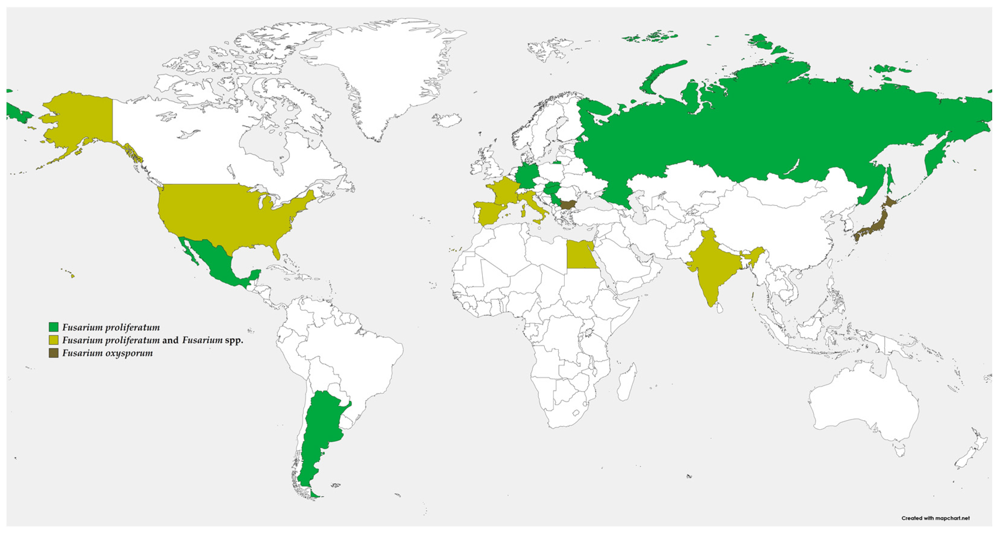
2. Distribution and Importance
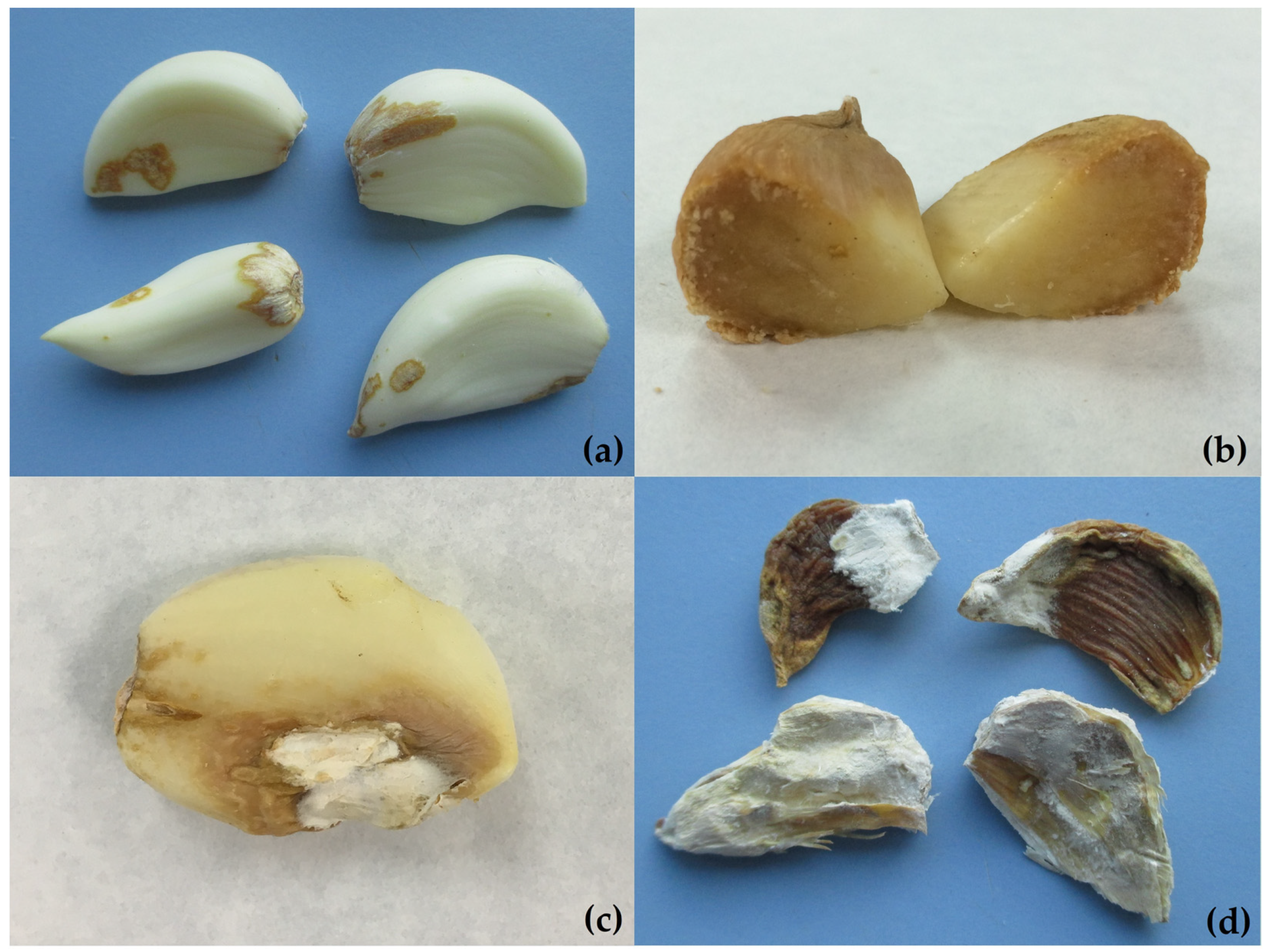
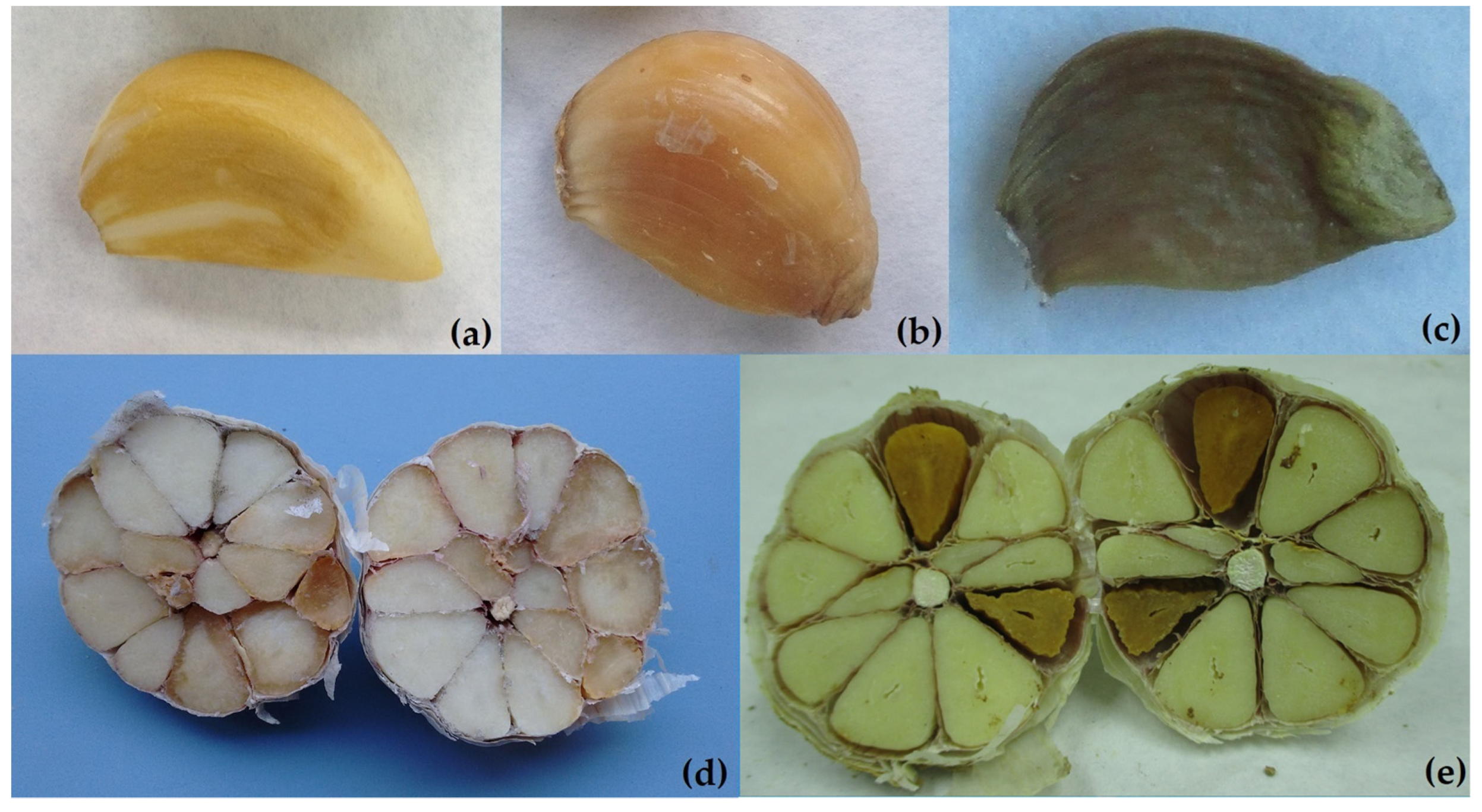
FDR was first described in Japan [21] in 1986, as caused by F. oxysporum, but 4 years later in Hungary, Simey [22] identified F. proliferatum var. minus as the causal agent of garlic rot. Almost 20 years later, it was reported in Bulgaria [23], again associated with F. oxysporum and other Fusarium spp. However, in the last 20 years, postharvest dry rot has been reported in America (United States [24], Argentina [25], and México [26]), Europe (Germany [13], Spain [27], Italy [10], France [28], Serbia [29], and Slovakia [30]), Africa (Egypt [31,32]), and Asia (Russia [33] and India [34]), where F. proliferatum has been identified as the main cause of dry rot in harvested garlic bulbs (Figure 1). F. proliferatum is the most frequently isolated species from symptomatic cloves. Moreover, F. oxysporum [2,16,32,35] and F. solani [9,31] have also been reported. Fusarium culmorum [23] and F. verticillioides [35] have rarely been isolated [16]. In Spain, Gálvez and Palmero [9] reported isolation percentages of F. proliferatum over 80%, agreeing with Chrétien et al. [2] in France. In all cases, F. proliferatum has been the predominant Fusarium species associated with dry rot.
FDR is the most frequently detected postharvest disease in Spain, found in up to 50% of the samples analyzed [9], coinciding with the results of Chrétien et al. [2] in France, who observed 55% of the peeled cloves with typical Fusarium symptoms. In Italy, FDR was observed in 71.4% at harvest and 82.1% during postharvest storage [16]. Tonti et al. [10] observed that the disease developed during the drying process and stated that it could affect almost 30% of the stored bulbs. Moharam et al. [31] detected Fusarium spp. in up to 66.7% of the samples analyzed in Egypt. In India, approximately 60% of the garlic bulbs were affected by FDR [34].

3. Symptomatology

The appearance of the disease caused by Fusarium spp. on postharvest garlic bulbs differs depending upon time and environmental conditions. At the initial phase, brown lesions with a dehydrated appearance may emerge in different zones of the garlic cloves (Figure 2a). These lesions are superficial or penetrate several millimeters and can progress (Figure 2b) to cover the entire clove during the storage period. Sometimes, infected garlic cloves appear with softened water-soaked lesions that progress to tan-colored lesions and are covered with white/pink mycelia (Figure 2c). Finally, infected cloves dry out and crinkle (Figure 2d). These symptoms are described consistently by different authors [9,10,16,24,28].
FDR can be confused with the damage caused by eriophyid mites, which produce a superficially dehydrated appearance that subsequently darkens. Symptoms are mainly observed in the apical area of the clove (Figure 3a). It is easy to distinguish by observing the presence of mites under the binocular loupe. Cloves affected by physiological disorders such as waxy breakdown turn deep yellow/amber at 20 or 25 days after harvest [36] and subsequently darken (Figure 3b). They are also easily confused with FDR, but these soft cloves are completely affected and emit a characteristic odor. Moreover, this physiological disorder can be found together with FDR after long periods of storage (Figure 3c). The flesh of cloves affected with waxy breakdown are entirely affected after 40–45 days; conversely, FDR does not always affect the entire clove (Figure 3d,e).
At harvest, the bulbs affected with FDR are externally symptomless. When peeled, however, cloves appear with small brown spots with or without mycelia. Dry rot symptoms on garlic cloves are mainly observed during the dry storage period [28], even in 4 °C cold chambers [16]. The first symptoms generally appear 2 months after harvest [28].
Fusarium spp. also produce symptoms on the plants during the crop, causing the disease known as Fusarium basal rot (FBR) on different Allium species [37]. FBR is also known as Fusarium rot or Fusarium wilt or basal plate rot and sometimes referred to as “damping-off” or “dieback” disease of the seedlings. The symptoms of affected plants are yellowing of leaves, followed by withered and curly leaves. Moreover, the disease symptoms can be rotting of the entire bulb plates, and white mycelium can be observed easily. Sometimes, symptoms may not be apparent at harvest and advance during storage. These symptoms have been described on Allium spp. crops such as onion (Allium cepa) and leeks (A. porrum) [37,38], and Stankovic et al. [17] reported the golden to yellowish-brown lesions on garlic roots, becoming dark brown and necrotic with time. Brown to tan spots were first observed at early growth stages, almost exclusively on the basal plate [39]. Chrétien et al. [40] reported that during the storage of garlic bulbs, the browning of cloves starts from the basal plates and extends to the tops of the cloves, indicating that F. proliferatum rapidly infects garlic root tissues before infecting clove tissues but does not penetrate healthy cells. Nevertheless, it is possible to observe brown lesions distributed throughout the garlic clove and the bulb. Moharam et al. [31] reported differences in the pathogenicity of the different Fusarium species according to the vegetative state of the garlic plants. For example, F. oxysporum highly reduced clove germination, produced extensive seedling damping-off, and caused high disease severity of rotted roots/cloves followed by F. solani, and F. proliferatum infections, which greatly reduced clove germination and caused the cloves of stored bulbs to rot during the harvesting (Table 1). Most researchers agree that the economic losses caused by F. proliferatum begin with maturation in the field and therefore depend on the initial inoculum and environmental conditions from harvest time and later on storage conditions.

4. Taxonomy and Identification
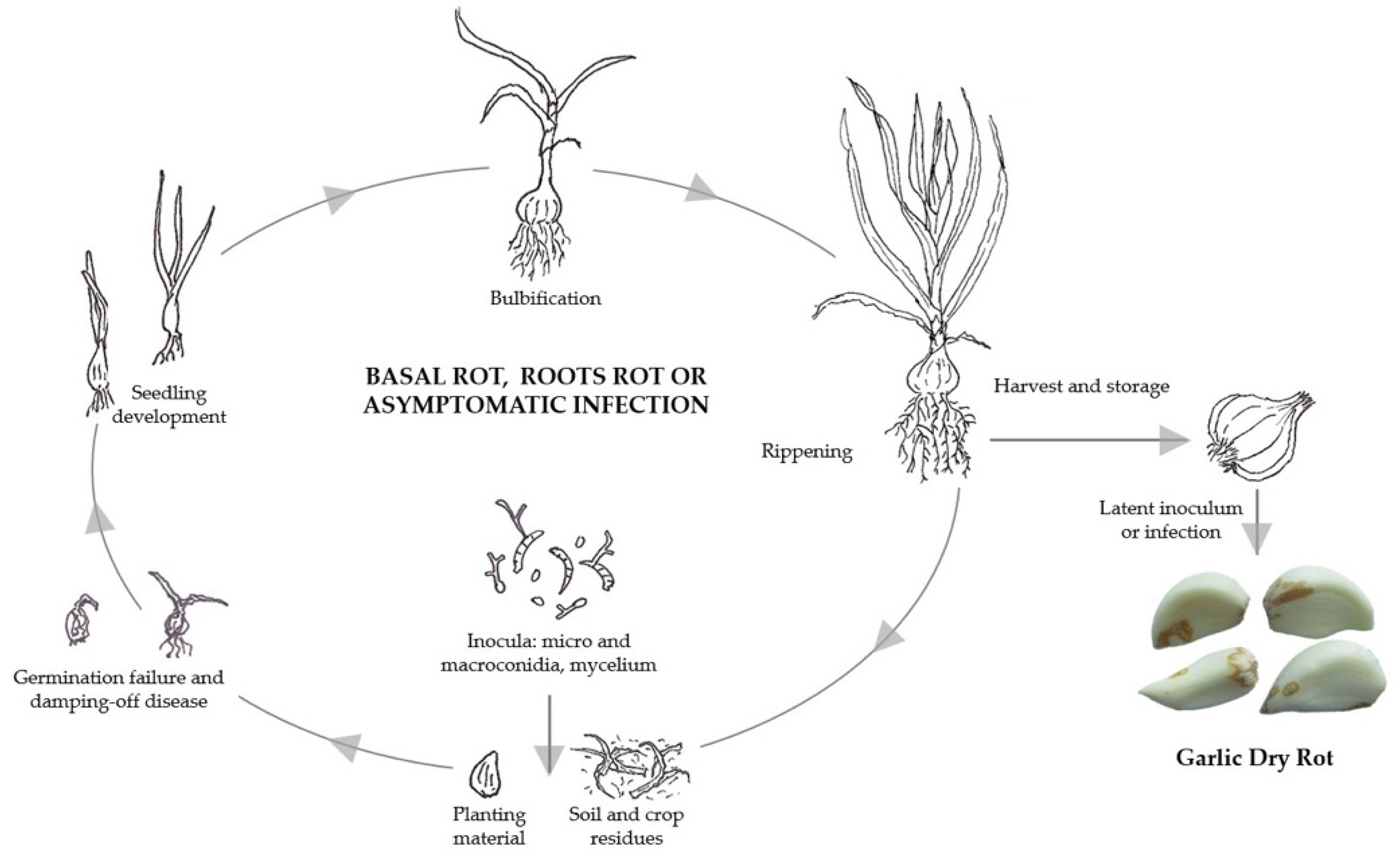
Fusarium proliferatum belongs to the Liseola section of the Fusarium genus [43], and its teleomorph, Gibberella intermedia, belongs to the Fusarium fujikuroi species complex (FSSC) [44,45], which consists of at least 13 reproductively different biological species (mating populations) [46]. For decades, this fungus was known as Fusarium moniliforme, but as information on host range and morphological variation has accumulated, F. moniliforme has been resolved into an increasing number of distinct species within the FFSC [47], including F. proliferatum and F. verticillioides. The community working with Fusarium by broad consensus recommended that F. moniliforme should not be used [48]. In fact, these fungal species share many morphological and biochemical characteristics, and misidentification can occur [49].
F. proliferatum is a heterothallic species with two different mating types (MAT-1 and MAT-2 idiomorphs). The formation of the fruiting bodies with ascospores can be observed by sexual crossing using tester strains. However, the cloning and sequencing of different mating type genes by PCR assays have allowed the identification of MAT gene idiomorphs in F. proliferatum [50]. Gálvez et al. [51] reported the presence of the two types of mating alleles and the possible (even if limited) occurrence of sexual reproduction of the F. proliferatum isolates in garlic fields. Sexual reproduction could enhance the adaptation to environmental changes by conferring genetic variability onto the population. This could influence their response to the application of fungicides, biocontrol agents, or even to climate change. In fact, a high variability in the F. proliferatum garlic population was demonstrated by the phylogenetic analysis inferred from TEF1 (translation elongation factor) and FUM1 (fumonisin biosynthesis) genes, the mycotoxigenic profile (fumonisin B1, B2 and B3, beauvericin and moniliformin), and the mycelial growth rate. The worldwide distribution of the garlic crop, the interchange of plant material for sowing, and the colonization of the plant by the pathogen without visible infection symptoms [19,52] might explain this high intraspecific variability. It has already been observed in F. verticillioides [53].
F. proliferatum colonies on Potato-Dextrose-Agar grow quickly with white to purple mycelia and catenate microconidia (club-shaped with a flattened base, aseptate) and produce mono- and polyphialides. The curved macroconidia usually are 3–5 septate. Chlamydospores are not produced. Fusarium proliferatum is morphologically indistinguishable from F. fujikuroi and can only be distinguished by testing sexual cross-fertility or DNA sequencing [49]. Sequencing the TEF1 gene has been identified as a good single-locus identification tool in Fusarium because it shows high sequence polymorphism among closely related species, and non-orthologous copies of the gene have not been detected in the genus [54]. Moreover, there are specific primer pairs for PCR assays for F. proliferatum based on calmodulin gene sequences [55,56] and on the IGS region of the rDNA gene [57]. Galvez et al. [58] reported that specific IGS primers did not result in an amplification product for all garlic isolates. The IGS region may be too variable to be a reliable target for the identification of this fungus. Thus, DNA sequencing is the best option for the identification to species level using the TEF1 gene and currently, it is complemented with multilocus sequence typing (MLST) through the addition of two partial sequences from the two largest DNA-directed RNA polymerase subunits (RPB1 and RPB2) [59,60]. In fact, in most studies on garlic, F. proliferatum has been identified by sequencing of the TEF1 gene [2,9,10,28,29,30]. Torres-Cruz et al. [61] discussed using portions of three phylogenetically informative genes (i.e., TEF1, RPB1, and RPB2) for resolving at or near the species level in every Fusarium species. Recently, Anisimova et al. [33] used these same informative genes for the identification of F. proliferatum on garlic cloves.

5. Life Cycle

Fusarium proliferatum is characterized by not forming chlamydospores (thick-walled resting spores); however, it is capable of persisting in the form of mycelia and micro- and macroconidia. Sclerotia may develop in some isolates, but they are not taxonomic criteria, although they may be indicative of a high level of sexual female fertility [49]. The hyphae of F. proliferatum have the ability to thicken in response to stresses such as low temperatures [62]. In addition, it has a marked competitive saprophytic ability, an intrinsic characteristic that allows it to survive in the absence of a host.
The pathogen is mainly soilborne where it survives in dormancy, but the effect of the amount of soil inocula on the subsequent development of garlic dry rot is not well established. Based on previous studies, the inoculum in the soil seems to have little relevance, and the dry rot incidence of F. proliferatum and F. oxysporum may depend to a large extent on the growing year [16]. There was no correlation between the density of propagules in the soil at the beginning of the garlic crop and the disease severity on the bulbs. The main source of the primary inoculum of F. proliferatum is soil, although it can also be found in the planting material itself, crop residues, weeds [63,64], or neighboring crops. Propagules of this pathogen can also be found in the air [65,66], irrigation water, rainwater and even atmospheric dust [67]. Gil-Serna et al. [68] evaluated rainwater as a source of the inoculum that produces garlic rot and the results revealed that, although the pathogenic ability of isolates from rainwater was lower than that of isolates obtained from rooted garlic, all were pathogenic to the three garlic varieties tested (Purple, White, and Purple Chinese).
The infected plant material can also contribute to the subsequent development of the disease by acting as primary inoculum in the field [66,69,70], as well as to the long-distance spread of disease linked to the trade in seed cloves. Fusarium proliferatum has been shown to persist quiescently in garlic cloves [41,71]. There are no studies available that explain the role played by garlic harvest debris as a source of the primary inoculum of F. proliferatum in the field, and its relationship to dry rot of the bulbs needs to be clarified. Previous works have indicated that crop residues of corn [69], wheat [72], and asparagus [73] are the main sources of the primary inoculum of F. proliferatum in those crops.
Primary infection occurs when the fungus germination is induced by garlic root exudates and, as occurs in onions [74], it penetrates directly into roots, possibly at points of active root growth in the early stages of germination. Nevertheless, Chrétien et al. [40] reported that F. proliferatum rapidly infects clove tissues at the outline/contour of the basal plates when the roots emerge, without penetrating healthy cells. Many species of Fusarium are viewed as opportunistic or weak pathogens that are capable of attacking only plants that were weakened previously by some other stress [69].
The infection of Fusarium results in a certain delay of garlic seedling emergence. Pre-emergence damping-off is observed only with high concentrations of the pathogen or after planting cloves with very advanced symptoms of the disease. However, Mondani et al. [39] did not find a direct correlation between the incidence of basal rot in the field and the severity of dry rot at the harvest of bulbs; this could be explained by the latent presence of the fungus, which penetrates through the wounds without generating visible symptoms [40]. Fusarium proliferatum has been cited as an endophyte in different plants without showing visible symptoms [19,49,70,75]. This fungus is widely distributed in the host tissue of corn, sorghum, and soybean under field conditions, and it takes advantage of plant stress conditions for growing and causing the disease. Although it can penetrate effectively owing to the production of enzymes and toxins that destroy host tissues prior to colonization [39], the proliferation of the fungus in garlic tissue was reduced in the presence of thick-walled cells, and was directed toward the parenchyma. Chrétien et al. [40] described that F. proliferatum on garlic cloves does not invade host conductive tissues and progresses from the points of infection in all directions.
Most infected garlic seedlings can still survive, perhaps with some developmental delay. Fusarium infections are first observed [39] in early growth stages (BBCH 15 crop phenology of López-Bellido et al. [5]), although in most cases the infection is difficult to observe in the field. Symptoms become more noticeable during crop maturity or after harvest [17,76]. Destroyed tissues were observed beyond the margins where fungal growth occurred, indicating that the fungus deployed strategies such as enzyme and toxin production to destroy host tissues prior to colonization. This could explain why, during storage, a greater presence of rotten bulbs is detected; owing to the production of toxins and enzymes from the latent inoculum of the tunics or latent infections, they develop rot during storage [17] (Figure 4).

6. Host Range and Varietal Response

Fusarium proliferatum has been reported as a cosmopolite pathogen in many important crops including maize [77], wheat [78], rice [79], sorghum [80], asparagus [81], date palm [82], onion [83], chive [84], potato [85], sunflower [86], soybean [87], cowpea [88], oat [89] banana [90], blueberry [91], alfalfa [92], strawberry [93], grape [94], cotton [95], melon [96], and cauliflower [97], among others, inducing damage and symptoms including root rot, dry rot, crown rot, stem rot, trunk canker, and vascular wilt, depending on the host [98,99,100].
Although in recent years citations of the pathogenicity of F. proliferatum have multiplied, including various crops in very different climatic zones, a clear parasitic specificity has not been identified, such as occurs in the special forms of other species of the same genus such as F. oxysporum [101]. In the same way, members of the FSSC collectively have a very broad host range and were subdivided into formae speciales. Recent phylogenetic analysis has revealed that these correspond to biologically and phylogenetically distinct species [102].
Okello et al. [103] studied the cross-pathogenicity of several Fusarium species, demonstrating that F. proliferatum from either soybean or corn was pathogenic to both crops. Many other studies have included inoculations of the recovered isolates on cultivars of the same species to confirm their pathogenicity, but on very few occasions have cross-inoculations been carried out on other hosts to identify the degree of specificity.
Based on the bibliography consulted, it seems that F. proliferatum has a very broad genetic variability but does not present host specificity, although a certain consistency can be identified in the association of the pathogen with different species of the Allium genus. Fusarium proliferatum has been reported on Alliaceae in many production areas [17,84,104,105]. The inoculation of F. proliferatum isolates from diseased garlic on other Allium species provided information on its pathogenicity and its host range within Allium spp.
There are few works where cross-inoculations have been conducted. Stankovic et al. [17] reported pathogenicity after inoculations on garlic and onion using isolates obtained from diseased plants in the field, although only a single cultivar of each crop species was inoculated. Galván et al. [106] inoculated 12 plants per cultivar from seven Allium species screening for resistance, but only one isolate of F. proliferatum was used in that study. High levels of resistance to each isolate were found in scallion (A. fistulosum) and chives (A. schoenoprasum) accessions, whereas A. cepa, A. pskemense, A. roylei, and A. galanthum showed intermediate levels of resistance.
Palmero et al. [18] revealed a clear susceptibility to six isolates of F. proliferatum originating from diseased garlic in onion, leek, chives, and scallion. Germination and seedling emergence were also seriously affected in onion and leek after inoculation of un-germinated and pregerminated seeds with F. proliferatum. In general, chives and scallion cultivars had the lowest disease severity index (DSI) values; in contrast, onion had the highest DSI scores.
Palmero et al. [18] reported differential varietal responses to the pathogen depending on the cultivar. The two different cultivars tested showed significant differences for leek (p < 0.05), garlic (p < 0.001), and onion (p < 0.001). A greater number of varieties of all varietal types marketed in Europe were evaluated by Palmero et al. [107], and F. proliferatum was pathogenic in all 17 commercial varieties tested. Furthermore, a greater degree of varietal susceptibility was confirmed in white and Chinese cultivars (81.84 ± 16.44 and 87.5 ± 23.19% symptomatic cloves, respectively) versus purple cultivars (49.06 ±13.42% symptomatic cloves).
Regarding the current feasibility of gene introgression, Galván et al. [106] identified A. fistulosum, A. roylei, and A. galanthum as potential sources for the transfer of resistance to Fusarium into onion. In an apomictic species such as garlic, propagated vegetatively and where the clove is sown (a clone of the mother plant), genetic improvement through sexual crossing was ruled out. Nor does there seem to be resistant plant material among the available cultivars, although tolerance ranges were observed, possibly owing to the morphology of the bulb itself (more compact bulbs seem more tolerant).
Disease control of FDR in stored garlic is not well established. Although no resistant varieties are available, this information will allow a certain management of varieties in areas of high pathogen pressure; together with cultural and postharvest control, it is essential to adjust growing cycles and select appropriate plant material to reduce disease incidence.

7. Cultural Control and Storage Conditions

Cultural practices along with good storage conditions for harvested garlic bulbs are crucial factors that greatly influence the incidence and severity of postharvest diseases. Among them, using healthy plant material, avoiding bulb injury during harvest and subsequent handling, and providing appropriate postharvest conditions are key factors for disease control.
Producers generally conduct the disinfection of planting cloves by applying fungicides in pre-planting. In the past few years, the availability of authorized products in garlic cultivation has decreased, for example, in Spain different commercial products with prochloraz as an active ingredient, which were routinely applied in pre-sowing treatments against different diseases of sprouting germination such as green rot (Penicillium allii) or brown rot (Sclerotium cepivorum), have been prohibited.
In Spain, the big garlic producers use techniques such as thermotherapy to disinfect the material or the in vitro culture of meristems for cultivating pathogen-free plants. These techniques are used mainly for the elimination of viruses or other pathogens, such as S. cepivorum or nematodes, either alone or in combination [108,109], although they have a similar effect against F. proliferatum propagules. Palmero et al. [107] studied the effect of thermotherapy and demonstrated in vitro substantial decreases in the conidial viability of F. proliferatum at 50 °C. Sodium hypochlorite and hydrocyanic acid have also been used to disinfect garlic cloves [110,111], although in this case, no studies are available on the effectiveness of these techniques against Fusarium.
As an alternative way to reduce the inoculum on the seed cloves, Dugan et al. [112] reported that the planting of aerial bulbils (borne in umbels at the apex of stalks known as scapes) can strongly reduce the infection of propagation material by F. proliferatum (compared with the seed cloves).
Regarding the reduction of the soil inoculum of Fusarium spp., the most economically and environmentally viable technique is crop rotation. However, in garlic cultivation the effect of rotations on the management of dry rot caused by F. proliferatum is still not well known. Molinero-Ruiz et al. [113] discovered that the root tissues of asymptomatic species (maize, wheat, potato, and sunflower) can serve as carriers and sources of the inoculum of Fusarium spp. until susceptible hosts such as garlic are available for infection, and severe outbreaks of Fusarium diseases can be expected with these particular crop rotations.
A crop rotation of 4 years with a non-susceptible host is recommended before planting another onion crop in a field affected with Fusarium basal rot [74]. As indicated before, F. proliferatum is a polyphagous pathogen with a wide range of hosts, affecting them to greater or lesser extents, depending on the host, and the choice of a suitable non-host crop is sometimes difficult for rotation to be truly effective.
Inoculation experiments with F. proliferatum in substrate and crop debris conducted in Madrid (Spain) suggest that this fungus can survive up to 2 years under field conditions. However, it appears that there was a drastic loss of viability of the propagules (almost 90%) during the first year of sampling (Figure 5, unpublished data [114]).
At the end of the sampling period (after 2 years of field analysis) the reduction in inoculum density was 95%. The experimental results indicated that F. proliferatum survives seven times higher in the garlic crop residues than in the inoculated substrate samples. Therefore, cultural practices related to the management of residues after harvesting garlic also deserve attention. A priori, the condition of soilborne disease seems to discourage the burial of crop residues; however, if the fields are plowed and these residues are buried (20 cm deep), fewer propagules should survive (Figure 5).
This study suggests that garlic crop residues should be incorporated into the soil soon after harvest. These results agreed with those reported by Cotten and Munkvold [115] in their study of F. proliferatum on buried maize residues.
The above results suggested also that a 1-year rotation with a non-susceptible host would be sufficient to substantially reduce the inoculum. However, Nivall and Kommedah [62] reported that this species (referred to in the work as F. moniliforme) is capable of thickening the hyphae to withstand low temperatures, and Manzo and Claflin [116] did not observe loss of viability of the conidia and hyphae of F. moniliforme when incubated at −16 °C for 6 months. Gradual decomposition of materials in the soil by microorganisms, availability of nutrients in the samples, moisture content, pH, retention capacity of water, or absence of chlamydospores could be the influencing factors for the low survival of the fungus in the field [117,118,119,120,121,122,123,124,125].
Cultural practices have been developed for different horticultural crops to cope with the withdrawal of the most effective but controversial chemical soil fumigants. Solarization (alone or in combination with organic matter amendments) is a soil disinfection practice used in southern European countries [126]. Although there is no literature on the use of these techniques in garlic crops, the previous results of Carrieri et al. [127] on onion and Borrego-Benjumea et al. [128] on asparagus indicated that they reduced the inoculum levels of the pathogen in these crops. These techniques increase soil temperature and produce changes in the microbial soil community as well as in the chemical and physical properties of the soil but require covering the soil with a plastic film for 4–6 weeks during the part of the year with the highest solar radiation and temperatures. Although this might be an alternative for small family farms, it is not a viable alternative for large extensions of land.
Until now, the most effective way to control the progress of the disease has been by managing the temperature and humidity conditions after harvest. Clove rot incidence and severity increased progressively with the increase in storage time at room temperature (20 °C and 25 °C) over several months. When garlic bulbs were stored in a refrigerator (4–5 °C) there was a much slower progression of clove rot incidence and severity compared to that recorded during the same period at room temperature [32,107]. Conversely, Mondani et al. [16] observed a 26% higher disease severity in freshly harvested bulbs than those analyzed after 6 months of storage in a cold room (−4 °C).
Palmero et al. [107] demonstrated that low temperature slows the activity of F. proliferatum but does not affect its vitality, and the growth and metabolism of the fungus restarts rapidly once the temperature increases. Relative humidity (RH) of 60–70% is considered optimal for garlic storage, as higher RH favors fungal growth and lower RH causes excessive moisture loss from the bulbs [129]. Water activity values ranging from 0.85 up to 0.95 aw were found for garlic bulbs during storage in large storehouses [16], and resulted positively correlated with the incidence of F. proliferatum on basal plates during 6 months storage. A value of aw above 0.90 is useful for Fusarium, and metabolic activities occur under this condition, even if at a very slow rate because of the low temperature (−4 °C ) in the cold storage room [130,131].

8. Chemical and Biological Control

Few studies are available regarding the effectiveness of chemical treatments in reducing F. proliferatum occurrence in garlic. Most of them are in vitro studies [32,132], although there are some trials available where researchers have scaled up the application of fungicides as either clove preplant treatments [6,35,133] or through field spray application [134].
When comparing groups of fungicides, previous in vitro studies have indicated the greater efficacy of demethylation inhibitor fungicides (DMI), which inhibit the demethylation step in sterol biosynthesis that is needed in fungal cell walls. Within this group, the triazole chemical group (e.g., prothioconazole, cyproconazole, and tebuconazole) [135] and azoles (e.g., prochloraz, difenoconazole, and fluquinconazole) [136] have proved more effective against Fusarium spp. than strobilurin fungicides (e.g., azoxystrobin and kresoxim-methyl) based on EC50 and EC90 values [134]. Dubos et al. [137,138] and Pasquali et al. [139] described the Fusarium species as intrinsically resistant to complex III respiration inhibitors (QoI) such as trifloxystrobin and the succinate dehydrogenase inhibitor (SDHI) group.
Mixtures of different active ingredients within different chemical groups reduce the risk of fungicide resistance [140]. In several of the reviewed works [133,134], the active ingredient tebuconazole was evaluated together with another active ingredient (fluopyram, trifloxystrobin, or azoxystrobin). All are included in the biochemical mode of action C (respiration) by the FRAC. Fluopyram belongs to the SDHI group and was reported to be effective in controlling different plant pathogens [141,142,143]. The other two belong to the QoI group. Fungicides belonging to these groups have been tested on different Fusarium spp. Pasquali et al. [139] demonstrated that the complex II and complex III respiration inhibitors isopyrazam (SDHI) and trifloxystrobin (QoI) were unable to inhibit F. culmorum up to a concentration of 1 mM. Trifloxystrobin concentrations ≤0.0003 mM did not significantly inhibit F. graminearum [137]. Maitlo et al. [144] also evaluated the active ingredient dimethomorph, obtaining low percentages of inhibition (41.75% at 1000 ppm). Chen et al. [145] evaluated the in vitro activity of pyraclostrobin in inhibiting the mycelial growth of F. asiaticum and F. graminearum isolates reporting EC50 in the range of 0.010–0.135 μg mL−1. However, Galvez et al. [134] reported that EC50 values were always higher (3.59 ppm) when analyzing F. proliferatum causing FDR on garlic.
Among the few specific studies on F. proliferatum in garlic, Dugan et al. [35] reported the effectiveness of benomyl (methyl benzimidazole carbamate affecting cyto-skeleton and motor protein) in preventing fungal rot in superficially wounded bulbs. However, this fungicide is currently forbidden in the EU (Directive 2009/128/EC). In in vitro assays carbendazim also had a strong inhibition effect on fungal growth followed by methalaxyl 8% + Mancozeb 64% and thiophanate methyl [32].
Palmero et al. [132] evaluated the efficacy of different chemical groups of fungicides in reducing F. proliferatum mycelial growth. In vitro test results on seven strains of F. proliferatum isolated from rotten bulbs of garlic demonstrated that tebuconazole was the most effective in inhibiting mycelial growth of isolates in diseased garlic. These results were consistent with previous studies on F. proliferatum isolated from other crop species such as wheat [146] as well as on different Fusarium species [135,139,147].
Mondani et al. [6,133] tested in vitro six commercial chemical fungicides (all DMI fungicides alone or mixed with other modes of action). In vivo efficacy was also assessed by dipping before sowing and applying as spray treatment at planting. The mixture of propiconazole + prochloraz showed the greatest growth inhibition of F. proliferatum (around 100%). The researchers reported that the chemical worked optimally at 10 °C, and efficacy decreased with the temperature increase. The results, however, cannot be extrapolated to assess the efficacy in garlic against the two species used in the study since the work was carried out with a single isolate of F. oxysporum and only one of the two F. proliferatum isolates came from diseased garlic. Among the six commercial chemical fungicides used in the in vitro test, two were also evaluated in the in vivo test. In this case both DMI treatments again significantly reduced disease severity compared to the inoculated control (11.1–24.8% versus 65.6%), although the experiment was stopped at the plant growth stage BBCH 14 (four leaves visible). This was remarkable because the postharvest dry rot disease develops mainly during the storage period. In a subsequent study, Italian researchers carried out a much more exhaustive monitoring of the disease during the cropping after applying the fungicides by wetting the cloves, spraying the products into the planting furrows, and analyzing them for 2 consecutive years, not merely for the three crop growth stages of BBCH 15 (5th leaf folded clearly visible (>3 cm); 1st leaf unfolded), 45 (50% of expected bulbs diameter reached) and 49 (100% of expected bulbs diameter reached; plant still erect and several leaves (3–5) green), but also for the 3-, 6-, and 9-month storage periods at −4 °C and 15 days after the bulbs were transferred to room temperature. In neither of the two cases significant differences were found between the fungicidal treatments and the untreated control, nor between the 2 years analyzed, and the time of greatest incidence of the disease was determined to be after 15 days at room temperature. Experimental results revealed that all the fungicidal treatments tested failed to control garlic bulb rot during and after storage. Dugan et al. [35] found that although the results of miscellaneous fungicide trials sometimes attained significant control (e.g., benomyl, fludioxonil, and thiophanate methyl), some experiments failed to demonstrate any effect on bulb rot control.
Some authors have shown a clear correlation between the trial year and the severity of garlic clove rot [6,35,134]. This suggests that there are factors other than fungicide effectiveness or the amount of initial inoculum in fields contributing to the disease occurrence. Specifically, the environmental conditions of relative humidity and temperature during the process of drying bulbs [107], could greatly influence the disease development.
Among the factors to consider for adequate control of soil diseases in the field are the date of fungicide application, the number of applications, environmental conditions, the pressure of inoculum in soil, and the nature and concentration of fungicides applied [41,146,148,149].
On the other hand, although there are significant differences in the activity spectra of DMI fungicides, and they show no cross-resistance with other classes of Sterol Biosynthesis Inhibitors (SBI), it is generally wise to accept that cross-resistance is present between DMI fungicides active against the same fungus.
The slow translocation that takes place in the last days before harvest, together with the period of drying in the field after harvest (about 7 days) and the long period of storage before sale can explain the limited effectiveness of systemic fungicides sprayed in the field in controlling postharvest rot.
As the disease is detected in the early crop stages in the field [133], the timely application of biological control agents (BCAs) can help protect the crop, although few studies have tested their application in garlic crops. Elshahawy et al. [32] reported the effectiveness of Trichoderma harzianum, T. viride, T. koningii, T. virens, and T. album in vitro against F. proliferatum isolated from garlic. Bjelic et al. [150] tested the effectiveness of selected Bacillus subtilis isolates from the soil as biocontrol agents, describing high antifungal activity (up to 71% reduction of fungal growth) and significant reduction of garlic clove infection (up to 58% reduction in rot symptoms) in situ. Recently, Guo et al. [151] reported the in vitro inhibitory effect of Serrata marcescens isolated from the cuticle of earthworms on F. proliferatum growth and conidial germination, and Ahmed et al. [152] reported that silver nanoparticles synthesized by Pleurotus ostreatus had a great effect on mycelial growth reduction.
Mondani et al. [133] studied the effect of four selected BCAs including B. subtilis, F. oxysporum, Streptomyces grioseoviridis, and the mix T. harzianum + T. gamsii based on their different modes of action. In all cases, they observed decreased in vitro growth of the garlic F. proliferatum isolate, with an average growth inhibition of 64.9%, although the BCAs were generally more effective in controlling F. proliferatum from garlic compared to F. proliferatum from maize. Decoupled data by fungal species are not provided, although the authors indicated that the most effective compound was based on Trichoderma harzianum + T. gamsii. In the field trials to evaluate the efficacy of coating products, the fungicides always showed lower disease severity than the tested BCA, although with no significant difference between the treatments of B. subtilis and S. griseoviridis. The activity of the different BCAs was closely linked to the conditions of water activity and temperature, and choosing the microorganism adapted to the environmental conditions at the time of its implantation (at the early crop stages) will be crucial to the success of the treatment. In a recent study, Mondani et al. [6] tested the efficacy of seed clove treatment on disease severity during garlic storage. Among the BCAs tested, S. griseoviridis controlled dry rot in the garlic bulbs. Bacillus subtilis and Trichoderma spp. were less effective in reducing visible symptoms. Bacillus subtilis was the BCA that reduced the percentage of incidence of F. proliferatum to a greater extent, although without significant differences compared to the untreated control.
Again, as was the case with chemical fungicides, BCA treatments have had a very limited postharvest impact, especially when the bulbs were stored at room temperature. Although BCAs have shown efficacy in some cases and the absence of a safety period could open possible BCA applications close to the harvest dates (when chemical control is banned), the disease has not yet been controlled satisfactorily, and it is necessary to continue investigating alternatives

9. Conclusions and Future Prospects

Experimental results have indicated that FDR disease control does not exclusively involve the use of fungicide treatments on pre-planting but also that techniques of integrated disease management can be effective. The latter includes the use of disease-free seed (cloves from in vitro culture), thermotherapy (hot water treatment), rotations with non-susceptible crops, and soil desinfestation by solarization (alone or conbined with organic matter incorporation).
Tissue culture allows us to obtain germplasm free of fungi and viruses, and this is routinely used by garlic producers in Europe. Thermotherapy has also been used during pre-sowing to treat seed cloves against fungal, nematode, and mite pests, and it has been demonstrated to achieve substantial decreases in vitro in the conidial viability of F. proliferatum.
The presence of inoculum in planting cloves has been described, and it may be important in the subsequent development of the disease. It seems that the soil inoculum present in cultivated fields is sufficient to constitute the initial source of inoculum. On the other hand, several studies have determined F. proliferatum to be associated with garlic plants during the cropping season, opening the door to addressing preventive control of the disease during cultivation.
Unfortunately, plant material resistant to the disease is not available, and even with all the above measures, the disease has not yet been controlled in a totally effective way. Therefore, it is necessary to explore other control measures for this pathogen. Undoubtedly, future measures will include the use of biological control agents that are not subject to the pre-harvest interval (PHI), do not generate resistance or cross-resistance, and are capable of acting even when the crop has been harvested. The variability among control agents is enormous. Future studies will enable us to determine not only the most suitable treatment for disease control, but also the best application time during or before crop planting.
Among other sustainable techniques, biofumigation could be an interesting option to investigate. This technique consists of the use of green manure of crops such as Brassicaceae, that release biocidal molecules after their incorporation into the soil, enhancing soil health for the next crop by reducing the level of harmful soilborne pests. Such toxic compounds derive from the enzymatic breakdown of glucosinolates contained in brassica cells. Again, no information is available on the effect of this cultural practice on F. proliferatum, but it would be worth testing this method for garlic crops. It is a promising option in places where crop rotation with garlic allows it.
Strict control of environmental conditions during postharvest storage is a key factor in avoiding the garlic dry rot disease caused by F. proliferatum. However, it is not sufficient to completely control the disease. It is necessary to implement strategies for integrated control of the pathogen that extend from the disinfection of planting cloves to control strategies during the development of garlic plants in the field.

Author Contributions

Conceptualization, D.P. and L.G.; methodology, L.G.; software, L.G.; validation, D.P.; formal analysis, L.G.; investigation, D.P.; resources, D.P.; data curation, L.G.; writing—original draft preparation, L.G.; writing—review and editing, L.G. and D.P.; visualization, L.G.; supervision, D.P.; project administration, D.P.; funding acquisition, D.P. All authors have read and agreed to the published version of the manuscript.

Funding

This research received no external funding.

Data Availability Statement

Not applicable.

Conflicts of Interest

The authors declare no conflict of interest.

References

  1. Ansary, J.; Forbes-Hernández, T.Y.; Gil, E.; Cianciosi, D.; Zhang, J.; Elexpuru-Zabaleta, M.; Simal-Gandara, J.; Giampieri, F.; Battino, M. Potential health benefit of garlic based on human intervention studies: A brief overview. Antioxidants 2020, 9, 619. [Google Scholar] [CrossRef] [PubMed]
  2. Chretien, P.L.; Morris, C.E.; Duffaud, M.; Leyronas, C. Aetiology of garlic rot, an emerging disease in France. Plant Pathol. 2021, 70, 1276–1291. [Google Scholar] [CrossRef]
  3. FAOSTAT. FAO Statistical Division 2020. Available online: https://www.fao.org/faostat/es/#data/QC/visualiz (accessed on 25 April 2022).
  4. Spagnoli, S. La qualità carta vincente contro l’import a basso prezzo. Agricoltura 2014, 6, 36–37. [Google Scholar]
  5. Lopez-Bellido, F.J.; Lopez-Bellido, R.J.; Muñoz-Romero, V.; Fernandez-Garcia, P.; Lopez-Bellido, L. New phenological growth stages of garlic (Allium sativum). Ann. Appl. Biol. 2016, 169, 423–439. [Google Scholar] [CrossRef]
  6. Mondani, L.; Chiusa, G.; Battilani, P. Efficacy of chemical and biological spray seed treatments in preventing garlic dry rot. Phytopathol. Mediterr. 2022, 61, 27–37. [Google Scholar] [CrossRef]
  7. Palmero, D.; Gálvez, L.; Arias, M.; González, C. La roya del ajo (Puccinia allii). Terralia 2016, 108, 56–60. [Google Scholar]
  8. Gálvez, L.; Gil-Serna, J.; García, M.; Iglesias, C.; Palmero, D. Stemphylium leaf blight of garlic (Allium sativum) in Spain: Taxonomy and in vitro fungicide response. Plant Pathol. J. 2016, 32, 388–395. [Google Scholar] [CrossRef]
  9. Gálvez, L.; Palmero, D. Incidence and etiology of postharvest fungal diseases associated with bulb rot in garlic (Alllium sativum) in Spain. Foods 2021, 10, 1063. [Google Scholar] [CrossRef]
  10. Tonti, S.; Prà, M.D.; Nipoti, P.; Prodi, A.; Alberti, I. First report of Fusarium proliferatum causing rot of stored garlic bulbs (Allium sativum L.) in Italy. J. Phytopathol. 2012, 160, 761–763. [Google Scholar] [CrossRef]
  11. Desjardins, A.E. Fusarium Mycotoxins: Chemistry, Genetics, and Biology; APS Press: St. Paul, MN, USA, 2006. [Google Scholar]
  12. Boonzaaijer, G.; van Osenbruggen, W.; Kleinnijenhuis, A.; van Dongen, W. An exploratory investigation of several mycotoxins and their natural occurrence in flavour ingredients and spices, using a multi-mycotoxin LC-MS/MS method. World Mycotoxin J. 2008, 1, 167–174. [Google Scholar] [CrossRef]
  13. Seefelder, W.; Gossmann, M.; Humpf, H.-U. Analysis of fumonisin B1 in Fusarium proliferatum-infected asparagus spears and garlic bulbs from germany by liquid chromatography-electrospray ionization mass spectrometry. J. Agric. Food Chem. 2002, 50, 2778–2781. [Google Scholar] [CrossRef] [PubMed]
  14. Seefelder, W.; Knecht, A.; Goβmann, M.; Kleta, S.; Büttner, C.; Humpf, H. Occurrence of fumonisins in asparagus (Asparagus officinalis L.) and garlic (Allium sativum L.) from Germany. Mycotoxin Res. 2004, 20, 29–30. [Google Scholar] [CrossRef] [PubMed]
  15. Tonti, S.; Mandrioli, M.; Nipoti, P.; Pisi, A.; Toschi, T.G.; Prodi, A. Detection of fumonisins in fresh and dehydrated commercial garlic. J. Agric. Food Chem. 2017, 65, 7000–7005. [Google Scholar] [CrossRef]
  16. Mondani, L.; Chiusa, G.; Pietri, A.; Battilani, P. Monitoring the incidence of dry rot caused by Fusarium proliferatum in garlic at harvest and during storage. Postharvest Biol. Technol. 2021, 173, 111407. [Google Scholar] [CrossRef]
  17. Stankovic, S.; Levic, J.; Petrovic, T.; Logrieco, A.; Moretti, A. Pathogenicity and mycotoxin production by Fusarium proliferatum isolated from onion and garlic in Serbia. Eur. J. Plant Pathol. 2007, 118, 165–172. [Google Scholar] [CrossRef]
  18. Palmero, D.; De Cara, M.; Nosir, W.; Gálvez, L.; Cruz, A.; WoodWard, S.; González-Jaén, M.T.; Tello, J. Fusarium proliferatum isolated from garlic in Spain: Identification, toxigenic potential and pathogenicity on related Allium species. Phytopathol. Mediterr. 2012, 51, 207–218. [Google Scholar]
  19. Stępień, Ł.; Koczyk, G.; Waśkiewicz, A. Genetic and phenotypic variation of Fusarium proliferatum isolates from different host species. J. Appl. Genet. 2011, 52, 487–496. [Google Scholar] [CrossRef] [Green Version]
  20. Waśkiewicz, A.; Stępień, Ł.; Wilman, K.; Kachlicki, P. Diversity of pea-associated F. proliferatum and F. verticillioides populations revealed by FUM1 sequence analysis and fumonisin biosynthesis. Toxins 2013, 5, 488–503. [Google Scholar] [CrossRef] [Green Version]
  21. Matuo, T.; Miyagawa, M.; Saito, H. Fusarium oxysporum f. sp. garlic nf sp. causing basal rot of garlic. Jpn. J. Phytopathol. 1986, 52, 860–864. [Google Scholar]
  22. Simey, E.I. Garlic rot caused by Fusarium proliferatum (Matsushima) Nirenferg var. minus Nirenferg in Hungary. Növényvédelem 1990, 26, 397–399. [Google Scholar]
  23. Koleva, K. Variety of species and spread of fungi of genus Fusarium related to rotting of garlic. Bulg. J. Agric. Sci. 2004, 10, 177–180. [Google Scholar]
  24. Dugan, F.; Hellier, B.; Lupien, S. First report of Fusarium proliferatum causing rot of garlic bulbs in North America. Plant Pathol. 2003, 52, 426. [Google Scholar] [CrossRef]
  25. Salvalaggio, A.; Ridao, A.d.C. First report of Fusarium proliferatum causing rot on garlic and onion in Argentina. Plant Dis. 2013, 97, 556. [Google Scholar] [CrossRef] [PubMed]
  26. Fuentes, Y.M.O.; Ortiz, J.C.D.; Chávez, E.C.; Castillo, F.D.H.; Olivas, A.F.; Morales, G.G.; Martínez, O.V.; Guerra, R.R. The first report of Fusarium proliferatum causing garlic bulb rots in Mexico. Afr. J. Agric. Res. 2013, 8, 570–573. [Google Scholar]
  27. Palmero, D.; De Cara, M.; Iglesias, C.; Moreno, M.; Gonzalez, N.; Tello, J. First report of Fusarium proliferatum causing rot of garlic bulbs in Spain. Plant Dis. 2010, 94, 277. [Google Scholar] [CrossRef]
  28. Leyronas, C.; Chrétien, P.L.; Troulet, C.; Duffaud, M.; Villeneuve, F.; Morris, C.; Hunyadi, H. First report of Fusarium proliferatum causing garlic clove rot in France. Plant Dis. 2018, 102, 2658. [Google Scholar] [CrossRef]
  29. Ignjatov, M.; Milosević, D.; Ivanović, Ž.; Karaman, M.; Vlajić, S.; Nikolić, Z.; Gvozdanović-Varga, J. Morphological and pathogenic properties of Fusarium proliferatum isolates: The causal agent of garlic (Allium sativum L.): Rot in Serbia. Ratar. Povrt. 2018, 55, 125–129. [Google Scholar] [CrossRef] [Green Version]
  30. Horáková, M.K.; Tancik, J.; Barta, M. Fusarium proliferatum causing dry rot of stored garlic in Slovakia. J. Plant Pathol. 2021, 103, 997–1002. [Google Scholar] [CrossRef]
  31. Moharam, M.H.A.; Farrag, E.S.H.; Mohamed, M.D.A. Pathogenic fungi in garlic seed cloves and first report of Fusarium proliferatum causing cloves rot of stored bulbs in upper Egypt. Arch. Phytopathol. Plant Prot. 2013, 46, 2096–2103. [Google Scholar] [CrossRef]
  32. Elshahawy, I.; Saied, N.; Morsy, A. Fusarium proliferatum, the main cause of clove rot during storage, reduces clove germination and causes wilt of established garlic plants. J. Plant Pathol. 2017, 99, 85–93. [Google Scholar]
  33. Anisimova, O.K.; Seredin, T.M.; Danilova, O.A.; Filyushin, M. first report of Fusarium proliferatum causing garlic clove rot in Russian Federation. Plant Dis. 2021, 105, 3308. [Google Scholar] [CrossRef] [PubMed]
  34. Sankar, N.R.; Babu, G.P. First report of Fusarium proliferatum causing rot of garlic bulbs (Allium sativum) in India. Arch. Phytopathol. Plant Prot. 2012, 1, 8. [Google Scholar] [CrossRef] [PubMed]
  35. Dugan, F.M.; Hellier, B.C.; Lupien, S.L. Pathogenic fungi in garlic seed cloves from the United States and China, and efficacy of fungicides against pathogens in garlic germplasm in Washington State. J. Phytopathol. 2007, 155, 437–445. [Google Scholar] [CrossRef]
  36. Cirrincione, M.A.; Guiñazú, M.E. Paralisis Cerosa (Waxy Breakdowm), en Bulbos de Ajo. Available online: http://anapa.com.br/wp-content/uploads/2017/07/paralisis-cerosa.pdf (accessed on 19 April 2022).
  37. Le, D.; Audenaert, K.; Haesaert, G. Fusarium basal rot: Profile of an increasingly important disease in Allium spp. Trop. Plant Pathol. 2021, 46, 241–253. [Google Scholar] [CrossRef]
  38. Armengol, J.; Moretti, A.; Perrone, G.; Vicent, A.; Bengoechea, J.; García-Jiménez, J. Identification, incidence and characterization of Fusarium proliferatumon ornamental palms in Spain. Eur. J. Plant Pathol. 2005, 112, 123–131. [Google Scholar] [CrossRef]
  39. Mondani, L.; Chiusa, G.; Battilani, P. Fungi associated with garlic during the cropping season, with focus on Fusarium proliferatum and F. oxysporum. Plant Health Prog. 2021, 22, 37–46. [Google Scholar] [CrossRef]
  40. Chretien, P.L.; Laurent, S.; Bornard, I.; Troulet, C.; El Maâtaoui, M.; Leyronas, C. Unraveling the infection process of garlic by Fusarium proliferatum, the causal agent of root rot. Phytopathol. Mediterr. 2020, 59, 285–293. [Google Scholar]
  41. Schwartz, H.F.; Mohan, S.K. Compendium of Onion and Garlic Diseases and Pests; APS Press: St. Paul, MN, USA, 2008. [Google Scholar]
  42. Delgado-Ortiz, J.C.; Ochoa-Fuentes, Y.M.; Cerna-Chávez, E.; Beltrán-Beache, M.; Rodríguez-Guerra, R.; Aguirre-Uribe, L.A.; Vázquez-Martínez, O. Patogenicidad de especies de Fusarium asociadas a la pudrición basal del ajo en el centro norte de México. Rev. Argent. Microbiol. 2016, 48, 222–228. [Google Scholar] [CrossRef] [Green Version]
  43. Nelson, P.E.; Toussoun, T.A.; Marasas, W. Fusarium Species: An Illustrated Manual for Identification; Pennsylvania State University Press: University Park, PA, USA, 1983. [Google Scholar]
  44. Geiser, D.M.; Aoki, T.; Bacon, C.W.; Baker, S.E.; Bhattacharyya, M.K.; Brandt, M.E.; Brown, D.W.; Burgess, L.W.; Chulze, S.; Coleman, J.J. One fungus, one name: Defining the genus Fusarium in a scientifically robust way that preserves longstanding use. Phytopathology 2013, 103, 400–408. [Google Scholar] [CrossRef] [Green Version]
  45. O’Donnell, K.; Cigelnik, E.; Nirenberg, H.I. Molecular systematics and phylogeography of the Gibberella fujikuroi species complex. Mycologia 1998, 90, 465–493. [Google Scholar] [CrossRef]
  46. Lima, C.S.; Pfenning, L.H.; Costa, S.S.; Abreu, L.M.; Leslie, J.F. Fusarium tupiense sp. nov., a member of the Gibberella fujikuroi complex that causes mango malformation in Brazil. Mycologia 2012, 104, 1408–1419. [Google Scholar] [CrossRef] [PubMed]
  47. Proctor, R.H.; Desjardins, A.E.; Moretti, A. Biological and chemical complexity of Fusarium proliferatum. In The Role of Plant Pathology in Food Safety and Food Security; Strange, R.N., Gullino, M.L., Eds.; Springer: Dordrecht, The Netherlands, 2010; pp. 97–111. [Google Scholar]
  48. Seifert, K.A.; Aoki, T.; Baayen, R.P.; Brayford, D.; Burgess, L.W.; Chulze, S.; Gams, W.; Geiser, D.; De Gruyter, J.; Leslie, J.F. The name Fusarium moniliforme should no longer be used. Mycol. Res. 2003, 107, 643–644. [Google Scholar] [CrossRef]
  49. Leslie, J.F.; Summerell, B.A. The Fusarium Laboratory Manual; Blackwell Publishing: Ames, IA, USA, 2006. [Google Scholar]
  50. Kenényi, Z.; Mulé, G.; Moretti, A.; Waalwijk, C.; Hornok, L. Fertility and mating type assessment within Fusarium proliferatum isolates from different host plants. In Proceedings of the the Seventh International Symposium, Poznán, Poland, 4–7 September 2002; pp. 55–68. [Google Scholar]
  51. Gálvez, L.; Urbaniak, M.; Waśkiewicz, A.; Stępień, Ł.; Palmero, D. Fusarium proliferatum–Causal agent of garlic bulb rot in Spain: Genetic variability and mycotoxin production. Food Microbiol. 2017, 67, 41–48. [Google Scholar] [CrossRef] [PubMed]
  52. Jurado, M.; Marín, P.; Callejas, C.; Moretti, A.; Vázquez, C.; González-Jaén, M.T. Genetic variability and fumonisin production by Fusarium proliferatum. Food Microbiol. 2010, 27, 50–57. [Google Scholar] [CrossRef] [PubMed]
  53. Rocha, L.D.O.; Reis, G.M.; Da Silva, V.N.; Braghini, R.; Teixeira, M.M.G.; Correa, B. Molecular characterization and fumonisin production by Fusarium verticillioides isolated from corn grains of different geographic origins in Brazil. Int. J. Food Microbiol. 2011, 145, 9–21. [Google Scholar] [CrossRef]
  54. Geiser, D.M.; del Mar Jiménez-Gasco, M.; Kang, S.; Makalowska, I.; Veeraraghavan, N.; Ward, T.J.; Zhang, N.; Kuldau, G.A.; O’Donnell, K. FUSARIUM-ID v. 1.0: A DNA sequence database for identifying Fusarium. Eur. J. Plant Pathol. 2004, 110, 473–479. [Google Scholar] [CrossRef]
  55. Mulè, G.; Susca, A.; Stea, G.; Moretti, A. A species-specific PCR assay based on the calmodulin partial gene for identification of Fusarium verticillioides, F. proliferatum and F. subglutinans. Eur. J. Plant Pathol. 2004, 110, 495–502. [Google Scholar] [CrossRef]
  56. Mulè, G.; Susca, A.; Stea, G.; Moretti, A. Specific detection of the toxigenic species Fusarium proliferatum and F. oxysporum from asparagus plants using primers based on calmodulin gene sequences. FEMS Microbiol. Lett. 2004, 230, 235–240. [Google Scholar] [CrossRef] [Green Version]
  57. Jurado, M.; Vázquez, C.; Marín, S.; Sanchis, V.; González-Jaén, M.T. PCR-based strategy to detect contamination with mycotoxigenic Fusarium species in maize. Syst. Appl. Microbiol. 2006, 29, 681–689. [Google Scholar] [CrossRef]
  58. Gálvez, L.; Clarkson, J.P.; Palmero, D. IGS region polymorphisms are responsible for failure of commonly used species-specific primers in Fusarium proliferatum isolates from diseased garlic. Plant Pathol. 2020, 69, 713–722. [Google Scholar] [CrossRef]
  59. O’Donnell, K.; Ward, T.J.; Robert, V.A.; Crous, P.W.; Geiser, D.M.; Kang, S. DNA sequence-based identification of Fusarium: Current status and future directions. Phytoparasitica 2015, 43, 583–595. [Google Scholar] [CrossRef] [Green Version]
  60. O’Donnell, K.; Whitaker, B.K.; Laraba, I.; Proctor, R.H.; Brown, D.W.; Broders, K.; Kim, H.-S.; McCormick, S.P.; Busman, M.; Aoki, T.; et al. DNA sequence-based identification of Fusarium: A work in progress. Plant Dis. 2022, 106, 1597–1609. [Google Scholar] [CrossRef] [PubMed]
  61. Torres-Cruz, T.J.; Whitaker, B.K.; Proctor, R.H.; Broders, K.; Laraba, I.; Kim, H.-S.; Brown, D.W.; O’Donnell, K.; Estrada-Rodríguez, T.L.; Lee, Y.-H. FUSARIUM-ID v. 3.0: An updated, downloadable resource for Fusarium species identification. Plant Dis. 2022, 106, 1610–1616. [Google Scholar] [CrossRef]
  62. Nyvall, R.F.; Kommedahl, T. Individual thickened hyphae as survival structures of Fusarium moniliforme in corn. Phytopathology 1968, 58, 1704–1707. [Google Scholar]
  63. Postic, J.; Cosic, J.; Vrandecic, K.; Jurkovic, D.; Saleh, A.A.; Leslie, J.F. Diversity of Fusarium species isolated from weeds and plant debris in Croatia. J. Phytopathol. 2012, 160, 76–81. [Google Scholar] [CrossRef]
  64. Martínez, M.; Arata, A.F.; Fernández, M.D.; Stenglein, S.A.; Dinolfo, M.I. Fusarium species richness in mono-and dicotyledonous weeds and their ability to infect barley and wheat. Mycol. Prog. 2021, 20, 1203–1216. [Google Scholar] [CrossRef]
  65. Ooka, J.; Kommedahl, T. Wind and rain dispersal of Fusarium moniliforme in corn fields. Phytopathology 1977, 67, 1023–1026. [Google Scholar] [CrossRef]
  66. Funnell-Harris, D.L.; Pedersen, J.F. Presence of Fusarium spp. in air and soil associated with sorghum fields. Plant Dis. 2011, 95, 648–656. [Google Scholar] [CrossRef] [Green Version]
  67. Palmero, D.; Rodríguez, J.; De Cara, M.; Camacho, F.; Iglesias, C.; Tello, J. Fungal microbiota from rain water and pathogenicity of Fusarium species isolated from atmospheric dust and rainfall dust. J. Ind. Microbiol. Biotechnol. 2011, 38, 13–20. [Google Scholar] [CrossRef] [Green Version]
  68. Gil-Serna, J.; Gálvez, L.; París, M.; Palmero, D. Fusarium proliferatum from rainwater and rooted garlic show genetic and pathogenicity differences. Eur. J. Plant Pathol. 2016, 146, 199–206. [Google Scholar] [CrossRef]
  69. Leslie, J.F.; Pearson, C.A.; Nelson, P.E.; Toussoun, T. Fusarium spp. from corn, sorghum, and soybean fields in the central and eastern United States. Phytopathology 1990, 80, 343–350. [Google Scholar] [CrossRef]
  70. Gaige, A.R.; Todd, T.; Stack, J. Interspecific competition for colonization of maize plants between Fusarium proliferatum and Fusarium verticillioides. Plant Dis. 2020, 104, 2102–2110. [Google Scholar] [CrossRef] [PubMed]
  71. Dugan, F.M. Diseases and disease management in seed garlic: Problems and prospects. Am. J. Plant Sci. Biotechnol. 2007, 1, 47–51. [Google Scholar]
  72. Gilbert, J.; Fernando, W. Epidemiology and biological control of Gibberella zeae/Fusarium graminearum. Can. J. Plant Pathol. 2004, 26, 464–472. [Google Scholar] [CrossRef]
  73. Blok, W.J.; Bollen, G.J. Inoculum sources of Fusarium oxysporum f.sp. asparagi in asparagus production. Ann. Appl. Biol. 1996, 128, 219–231. [Google Scholar]
  74. Cramer, C.S. Breeding and genetics of Fusarium basal rot resistance in onion. Euphytica 2000, 115, 159–166. [Google Scholar] [CrossRef]
  75. Claflin, L.E. Fusarium root and stalk rot. In Compendium of Sorghum Diseases; Frederiksen, R.A., Odvody, G.N., Eds.; APS Press: St. Paul, MN, USA, 2000; pp. 28–30. [Google Scholar]
  76. Kintega, K.R.; Zida, P.E.; Soalla, R.; Tarpaga, V.W.; Sankara, P.; Sereme, P. Determination of Fusarium species associated with onion plants (Allium cepa) in field in Burkina Faso causing damping-off and bulb rots. Am. J. Plant Sci. 2020, 11, 64. [Google Scholar] [CrossRef]
  77. Logrieco, A.; Moretti, A.; Ritieni, A.; Bottalico, A.; Corda, P. Occurrence and toxigenicity of Fusarium proliferatum from preharvest maize ear rot, and associated mycotoxins, in Italy. Plant Dis. 1995, 79, 727–731. [Google Scholar] [CrossRef]
  78. Conner, R.; Hwang, S.; Stevens, R. Fusarium proliferation: A new causal agent of black point in wheat. Can. J. Plant Pathol. 1996, 18, 419–423. [Google Scholar] [CrossRef]
  79. Desjardins, A.E.; Plattner, R.D.; Nelson, P.E. Production of fumonisin B and moniliformin by Gibberella fujikuroi from rice from various geographic areas. Appl. Environ. Microbiol. 1997, 63, 1838–1842. [Google Scholar] [CrossRef] [Green Version]
  80. Leslie, J.F. Gibberella fujikuroi: Available populations and variable traits. Can. J. Bot. 1995, 73, 282–291. [Google Scholar] [CrossRef]
  81. Elmer, W.H. Fusarium proliferatum as a causal agent in Fusarium crown and root rot of asparagus. Plant Dis. 1990, 74, 938. [Google Scholar] [CrossRef]
  82. Abdalla, M.; Al-Rokibah, A.; Moretti, A.; Mule, G. Pathogenicity of toxigenic Fusarium proliferatum from date palm in Saudi Arabia. Plant Dis. 2000, 84, 321–324. [Google Scholar] [CrossRef] [Green Version]
  83. Du Toit, L.J.; Inglis, D.; Pelter, G. Fusarium proliferatum pathogenic on onion bulbs in Washington. Plant Dis. 2003, 87, 750. [Google Scholar] [CrossRef] [PubMed]
  84. Yamazaki, M.; Morita, Y.; Kashiwa, T.; Teraoka, T.; Arie, T. Fusarium proliferatum, an additional bulb rot pathogen of Chinese chive. J. Gen. Plant Pathol. 2013, 79, 431–434. [Google Scholar] [CrossRef]
  85. Merlington, A.; Hanson, L.; Bayma, R.; Hildebrandt, K.; Steere, L.; Kirk, W. First report of Fusarium proliferatum causing dry rot in Michigan commercial potato (Solanum tuberosum) production. Plant Dis. 2014, 98, 843. [Google Scholar] [CrossRef]
  86. Ren, J.; Zhang, G.; Zhang, Y.; Zhang, J.; Zheng, H.; Jing, L.; Zhou, H.; Zhao, J. First report of sunflower wilt caused by Fusarium proliferatum in Inner Mongolia, China. Plant Dis. 2015, 99, 1275. [Google Scholar] [CrossRef]
  87. Arias, M.D.; Munkvold, G.; Leandro, L. First report of Fusarium proliferatum causing root rot on soybean (Glycine max) in the United States. Crop Prot. 2015, 67, 52–58. [Google Scholar]
  88. Shrestha, U.; Butler, D.; Ownley, B. First report of dry root and stem rot of cowpea caused by Fusarium proliferatum in the United States. Plant Dis. 2016, 100, 860. [Google Scholar] [CrossRef]
  89. Molnár, O. Fusarium proliferatum causing head blight on oat in Hungary. Eur. J. Plant Pathol. 2016, 146, 699–703. [Google Scholar] [CrossRef]
  90. Kamel, M.A.M.; Cortesi, P.; Saracchi, M. Etiological agents of crown rot of organic bananas in Dominican Republic. Postharvest Biol. Technol. 2016, 120, 112–120. [Google Scholar] [CrossRef]
  91. Pérez, B.; Berretta, M.; Carrión, E.; Wright, E. First report of root rot caused by Fusarium proliferatum on blueberry in Argentina. Plant Dis. 2011, 95, 1478. [Google Scholar] [CrossRef]
  92. Cong, L.; Sun, Y.; Kang, J.; Li, M.; Long, R.; Zhang, T.; Yang, Q. First report of root rot disease caused by Fusarium proliferatum on alfalfa in China. Plant Dis. 2016, 100, 2526. [Google Scholar] [CrossRef]
  93. Borrero, C.; Capote, N.; Gallardo, M.; Avilés, M. First report of vascular wilt caused by Fusarium proliferatum on strawberry in Spain. Plant Dis. 2019, 103, 581. [Google Scholar] [CrossRef]
  94. Ghuffar, S.; Irshad, G.; Zhai, F.; Aziz, A.; Asadullah, H.M.A.M.; Mehmood, N.; Yang, H.; Bashir, A.; Ahmed, M.Z.; Aslam, M.F. First report of Fusarium proliferatum causing fruit rot of grapes (Vitis vinifera) in Pakistan. Int. J. Phytopathol. 2018, 7, 85–88. [Google Scholar] [CrossRef]
  95. Zhu, Y.; Abdelraheem, A.; Sanogo, S.; Wedegaertner, T.; Nichols, R.; Zhang, J. First report of cotton (Gossypium) wilt caused by Fusarium proliferatum in New Mexico, USA. Plant Dis. 2019, 103, 2679. [Google Scholar] [CrossRef]
  96. Yu, X.; Zhang, J.; Guo, L.; Yu, A.; Wang, X.; Xiang, W.; Zhao, J. First report of Fusarium proliferatum causing fruit rot on Muskmelon (Cucumis melo L.) in China. Plant Dis. 2021, 106, 1305. [Google Scholar] [CrossRef] [PubMed]
  97. Yan, L. First report of vascular wilt caused by Fusarium proliferatum on cauliflower in China. Plant Dis. 2020, 104, 3065. [Google Scholar] [CrossRef]
  98. Lahuf, A. First report of Fusarium proliferatum causing stem and root rot on lucky bamboo (Dracaena braunii) in Iraq. Plant Prot. J. 2019, 12, 1–5. [Google Scholar] [CrossRef] [Green Version]
  99. Feng, F.; Li, H. First report of Fusarium proliferatum causing trunk canker on Ilex cornuta in China. Plant Dis. 2019, 103, 155. [Google Scholar] [CrossRef]
  100. Punja, Z.K. First report of Fusarium proliferatum causing crown and stem rot, and pith necrosis, in cannabis (Cannabis sativa L., marijuana) plants. Can. J. Plant Pathol. 2021, 43, 236–255. [Google Scholar] [CrossRef]
  101. Edel-Hermann, V.; Lecomte, C. Current status of Fusarium oxysporum formae speciales and races. Phytopathology 2019, 109, 512–530. [Google Scholar] [CrossRef] [PubMed] [Green Version]
  102. Coleman, J.J. The Fusarium solani species complex: Ubiquitous pathogens of agricultural importance. Mol. Plant Pathol. 2016, 17, 146–158. [Google Scholar] [CrossRef] [PubMed]
  103. Okello, P.N.; Mathew, F.M. Cross pathogenicity studies show South Dakota isolates of Fusarium acuminatum, F. equiseti, F. graminearum, F. oxysporum, F. proliferatum, F. solani, and F. subglutinans from either soybean or corn are pathogenic to both crops. Plant Health Prog. 2019, 20, 44–49. [Google Scholar] [CrossRef]
  104. Dissanayake, M.; Tanaka, S.; Ito, S. Fumonisin B1 production by Fusarium proliferatum strains isolated from Allium fistulosum plants and seeds in Japan. Lett. Appl. Microbiol. 2009, 48, 598–604. [Google Scholar] [CrossRef]
  105. Haapalainen, M.; Latvala, S.; Kuivainen, E.; Qiu, Y.; Segerstedt, M.; Hannukkala, A. Fusarium oxysporum, F. proliferatum and F. redolens associated with basal rot of onion in Finland. Plant Pathol. 2016, 65, 1310–1320. [Google Scholar] [CrossRef] [Green Version]
  106. Galván, G.A.; Koning-Boucoiran, C.F.; Koopman, W.J.; Burger-Meijer, K.; González, P.H.; Waalwijk, C.; Kik, C.; Scholten, O.E. Genetic variation among Fusarium isolates from onion, and resistance to Fusarium basal rot in related Allium species. Eur. J. Plant Pathol. 2008, 121, 499–512. [Google Scholar] [CrossRef]
  107. Palmero, D.; Gálvez, L.; García, M.; Gil-Serna, J.; Benito, S. The effects of storage duration, temperature and cultivar on the severity of garlic clove rot caused by Fusarium proliferatum. Postharvest Biol. Technol. 2013, 78, 34–39. [Google Scholar] [CrossRef] [Green Version]
  108. Conci, V.; Nome, S.F. Virus free garlic (Allium sativum L.) plants obtained by thermotherapy and meristem tip culture. J. Phytopathol. 1991, 132, 186–192. [Google Scholar] [CrossRef]
  109. Yulianingsih, R.; Hidayat, S.; Dinarti, D. Elimination of Garlic common latent virus from garlic through meristem culture and thermotherapy. IOP Conf. Ser. Earth Environ. Sci. 2020, 468, 012028. [Google Scholar] [CrossRef]
  110. Roberts, P.; Matthews, W. Disinfection alternatives for control of Ditylenchus dipsaci in garlic seed cloves. J. Nematol. 1995, 27, 448. [Google Scholar]
  111. Zouhar, M.; Douda, O.; Dlouhý, M.; Lišková, J.; Maňasová, M.; Stejskal, V. Using of hydrogen cyanide against Ditylenchus dipsaci nematode present on garlic. Plant Soil Environ. 2016, 62, 184–188. [Google Scholar] [CrossRef] [Green Version]
  112. Dugan, F.; Lupien, S.; Hellier, B. Infection by Fusarium proliferatum in aerial garlic bulbils is strongly reduced compared to rates in seed cloves when both originate from infected bulbs. Crop Prot. 2019, 116, 43–48. [Google Scholar] [CrossRef]
  113. Molinero-Ruiz, L.; Rubio-Pérez, E.; González-Domínguez, E.; Basallote-Ureba, M.J. Alternative hosts for Fusarium spp. causing crown and root rot of Asparagus in Spain. J. Phytopathol. 2011, 159, 114–116. [Google Scholar] [CrossRef]
  114. Gálvez, L. Etiología, Epidemiología y Estrategias de Control de la Podredumbre del Diente de ajo. Ph.D. Thesis, Agro-Environmental Technology for Sustainable Agriculture, Universidad Politécnica de Madrid, Madrid, Spain, 2017. [Google Scholar]
  115. Cotten, T.; Munkvold, G. Survival of Fusarium moniliforme, F. proliferatum, and F. subglutinans in maize stalk residue. Phytopathology 1998, 88, 550–555. [Google Scholar] [CrossRef] [Green Version]
  116. Manzo, S.; Claflin, L. Survival of Fusarium moniliforme hyphae and conidia in grain Sorghum stalks. Plant Dis. 1984, 68, 866–867. [Google Scholar] [CrossRef]
  117. Taylor, A.; Vagany, V.; Barbara, D.J.; Thomas, B.; Pink, D.; Jones, J.; Clarkson, J.P. Identification of differential resistance to six Fusarium oxysporum f. sp. cepae isolates in commercial onion cultivars through the development of a rapid seedling assay. Plant Pathol. 2013, 62, 103–111. [Google Scholar]
  118. Reis, E.M.; Baruffi, D.; Remor, L.; Zanatta, M. Decomposition of corn and soybean residues under field conditions and their role as inoculum source. Summa Phytopathol. 2011, 37, 65–67. [Google Scholar] [CrossRef] [Green Version]
  119. Khonga, E.; Sutton, J. Inoculum production and survival of Gibberella zeae in maize and wheat residues. Can. J. Plant Pathol. 1988, 10, 232–239. [Google Scholar] [CrossRef]
  120. Onana, B.; Essono, G.; Nyegue, M.; Tchikoua, R.; Ambang, Z.; Ayodele, M. Natural occurrence of Fusarium species and fumonisins in stored cassava chips. Afr. J. Microbiol. Res. 2015, 9, 2257–2269. [Google Scholar]
  121. Liddell, C.M.; Burgess, L.W. Survival of Fusarium moniliforme at controlled temperature and relative humidity. Trans. Br. Mycol. Soc. 1985, 84, 121–130. [Google Scholar] [CrossRef]
  122. Nyvall, R.F.; Kommedahl, T. Saprophytism and survival of Fusarium moniliforme in corn stalks. Phytopathology 1970, 60, 1233–1235. [Google Scholar] [CrossRef]
  123. Han, X.; Cheng, Z.; Meng, H. Soil properties, nutrient dynamics, and soil enzyme activities associated with garlic stalk decomposition under various conditions. PLoS ONE 2012, 7, e50868. [Google Scholar] [CrossRef]
  124. Manstretta, V.; Rossi, V. Modelling the effect of weather on moisture fluctuations in maize stalk residues, an important inoculum source for plant diseases. Agric. For. Meteorol. 2015, 207, 83–93. [Google Scholar] [CrossRef]
  125. Reyes Gaige, A.; Giraldo, M.; Todd, T.; Stack, J.P. Growth and colonization of organic matter in soil by Fusarium proliferatum. Can. J. Plant Pathol. 2019, 41, 242–250. [Google Scholar] [CrossRef]
  126. BEST4SOIL. Una Red de Profesionales Creada Para Compartir Conocimientos Sobre la Prevención y Control de las Enfermedades Edáficas. Available online: https://www.best4soil.eu/ (accessed on 4 May 2022).
  127. Carrieri, R.; Raimo, F.; Pentangelo, A.; Lahoz, E. Fusarium proliferatum and Fusarium tricinctum as causal agents of pink rot of onion bulbs and the effect of soil solarization combined with compost amendment in controlling their infections in field. Crop Prot. 2013, 43, 31–37. [Google Scholar] [CrossRef]
  128. Borrego-Benjumea, A.I.; Melero-Vara, J.M.; Basallote-Ureba, M.J. Organic amendments conditions on the control of Fusarium crown and root rot of asparagus caused by three Fusarium spp. Span. J. Agric. Res. 2015, 13, 1009. [Google Scholar] [CrossRef] [Green Version]
  129. Ludlow, R.A.; Evans, G.; Graz, M.; Marti, G.; Martínez, P.C.; Rogers, H.J.; Müller, C.T. From laboratory to industrial storage–Translating volatile organic compounds into markers for assessing garlic storage quality. Postharvest Biol. Technol. 2022, 191, 111976. [Google Scholar] [CrossRef]
  130. Marin, S.; Sanchis, V.; Magan, N. Water activity, temperature, and pH effects on growth of Fusarium moniliforme and Fusarium proliferatum isolates from maize. Can. J. Microbiol. 1995, 41, 1063–1070. [Google Scholar] [CrossRef]
  131. Lee, H.B.; Magan, N. The influence of environmental factors on growth and interactions between Embellisia allii and Fusarium oxysporum f. sp. cepae isolated from garlic. Int. J. Food Microbiol. 2010, 138, 238–242. [Google Scholar] [CrossRef] [PubMed] [Green Version]
  132. Palmero, D.; Gálvez, L.; Bango, D.; Jurado, M.; De Cara, M. Evaluación del efecto del diferentes materias activas fungicidas sobre la inhibición del crecimiento micelial de Fusarium proliferatum. In Proceedings of the XVII Congreso de la Sociedad Española de Fitopatología, Lleida, Spain, 7–10 October 2014; p. 279. [Google Scholar]
  133. Mondani, L.; Chiusa, G.; Battilani, P. Chemical and biological control of Fusarium species involved in garlic dry rot at early crop stages. Eur. J. Plant Pathol. 2021, 160, 575–587. [Google Scholar] [CrossRef]
  134. Patón, L.G.; Marrero, M.D.R.; Llamas, D.P. In vitro and field efficacy of three fungicides against Fusarium bulb rot of garlic. Eur. J. Plant Pathol. 2017, 148, 321–328. [Google Scholar] [CrossRef]
  135. Müllenborn, C.; Steiner, U.; Ludwig, M.; Oerke, E.C. Effect of fungicides on the complex of Fusarium species and saprophytic fungi colonizing wheat kernels. Eur. J. Plant Pathol. 2008, 120, 157–166. [Google Scholar] [CrossRef]
  136. Amini, J.; Sidovich, D. The effects of fungicides on Fusarium oxysporum f. sp. lycopersici associated with Fusarium wilt of tomato. J. Plant Prot. Res. 2010, 50, 172–178. [Google Scholar]
  137. Dubos, T.; Pasquali, M.; Pogoda, F.; Hoffmann, L.; Beyer, M. Evidence for natural resistance towards trifloxystrobin in Fusarium graminearum. Eur. J. Plant Pathol. 2011, 130, 239–248. [Google Scholar] [CrossRef]
  138. Dubos, T.; Pasquali, M.; Pogoda, F.; Casanova, A.; Hoffmann, L.; Beyer, M. Differences between the succinate dehydrogenase sequences of isopyrazam sensitive Zymoseptoria tritici and insensitive Fusarium graminearum strains. Pestic. Biochem. Physiol. 2013, 105, 28–35. [Google Scholar] [CrossRef]
  139. Pasquali, M.; Spanu, F.; Scherm, B.; Balmas, V.; Hoffmann, L.; Hammond-Kosack, K.E.; Beyer, M.; Migheli, Q. FcStuA from Fusarium culmorum controls wheat foot and root rot in a toxin dispensable manner. PLoS ONE 2013, 8, e57429. [Google Scholar] [CrossRef] [Green Version]
  140. Brent, K.J.; Hollomon, D.W. Fungicide Resistance in Crop Pathogens: How Can It Be Managed? Fungicide Resistance Action Committee: Belgium, Brussels, 1995. [Google Scholar]
  141. Fought, L.; Musson, G.H.; Young, H. Fluopyram fungicides for the control of diseases of horticultural and row crops. Phytopathology 2011, 101, S54. [Google Scholar]
  142. Avenot, H.F.; Thomas, A.; Gitaitis, R.D.; Langston, D.B.; Stevenson, K.L. Molecular characterization of boscalid-and penthiopyrad-resistant isolates of Didymella bryoniae and assessment of their sensitivity to fluopyram. Pest Manag. Sci. 2012, 68, 645–651. [Google Scholar] [CrossRef]
  143. Amiri, A.; Heath, S.M.; Peres, N.A. Resistance to fluopyram, fluxapyroxad, and penthiopyrad in Botrytis cinerea from strawberry. Plant Dis. 2014, 98, 532–539. [Google Scholar] [CrossRef] [Green Version]
  144. Maitlo, S.; Syed, R.; Rustamani, M.; Khuhro, R.; Lodhi, A. Comparative efficacy of different fungicides against Fusarium wilt of chickpea (Cicer arietinum L.). Pak. J. Bot. 2014, 46, 2305–2312. [Google Scholar]
  145. Chen, Y.; Zhang, A.F.; Gao, T.C.; Zhang, Y.; Wang, W.X.; Ding, K.J.; Chen, L.; Sun, Z.; Fang, X.Z.; Zhou, M.G. Integrated use of pyraclostrobin and epoxiconazole for the control of Fusarium head blight of wheat in Anhui Province of China. Plant Dis. 2012, 96, 1495–1500. [Google Scholar] [CrossRef] [Green Version]
  146. Marin, P.; de Ory, A.; Cruz, A.; Magan, N.; Gonzalez-Jaen, M.T. Potential effects of environmental conditions on the efficiency of the antifungal tebuconazole controlling Fusarium verticillioides and Fusarium proliferatum growth rate and fumonisin biosynthesis. Int. J. Food Microbiol. 2013, 165, 251–258. [Google Scholar] [CrossRef]
  147. Ivić, D.; Sever, Z.; Kuzmanovska, B. In vitro sensitivity of Fusarium graminearum, F. avenaceum and F. verticillioides to carbendazim, tebuconazole, flutriafol, metconazole and prochloraz. Pestic. Fitomed. 2011, 26, 35–42. [Google Scholar] [CrossRef]
  148. Vyas, S.C. Nontarget Effects of Agricultural Fungicides; CRC Press, Inc.: Boca Raton, FL, USA, 1988. [Google Scholar]
  149. Palmero, D.; de Cara, M.; Galvez, L.; Tello, J. Effect of pre-sowing treatment on postharvest garlic rot caused by Fusarium proliferatum. In Proceedings of the International Congress of Postharvest Pathology, Lleida, Spain, 11–14 April 2011; p. 156. [Google Scholar]
  150. Bjelić, D.; Ignjatov, M.; Marinković, J.; Milošević, D.; Nikolić, Z.; Gvozdanović-Varga, J.; Karaman, M. Bacillus isolates as potential biocontrol agents of Fusarium clove rot of garlic. Zemdirb. Agric. 2018, 105, 369–376. [Google Scholar] [CrossRef]
  151. Guo, Z.; Zhang, X.; Wu, J.; Yu, J.; Xu, M.; Chen, D.; Zhang, Z.; Li, X.; Chi, Y.; Wan, S. In vitro inhibitory effect of the bacterium Serratia marcescens on Fusarium proliferatum growth and fumonisins production. Biol. Control 2020, 143, 104188. [Google Scholar] [CrossRef]
  152. Ahmed, N.G.; Gouda, H.; Hussein, M. Efficiency of silver nanoparticles synthesized by using Pleurotus ostreatus nanoparticles to manage fungal garlic cloves rot. SVU-Int. J. Agric. Sci. 2022, 4, 211–222. [Google Scholar] [CrossRef]
Figure 1. Spatial distribution of dry rot on garlic caused by Fusarium spp. around the world. On the map, green, mustard, and brown color denote F. proliferatum; F. proliferatum and Fusarium spp.; and F. oxysporum, respectively.
Horticulturae 08 00628 g001
Figure 2. Different symptoms of rot in garlic cloves caused by F. proliferatum: (a) brown lesions with a dehydrated appearance in different zones of the garlic clove; (b) rot lesions penetrating the clove; (c) garlic clove with brown lesions and covered with white mycelia; (d) dry and wrinkled cloves in advanced stages of the disease.
Horticulturae 08 00628 g002
Figure 3. Garlic cloves and bulbs affected by other disorders easily confused with dry rot: (a) damage caused by eriophyid mites; (b) physiological disorder of waxy breakdown; (c) clove affected with waxy breakdown and dry rot; (d) bulb affected with dry rot caused by F. proliferatum; (e) bulb affected with waxy breakdown.
Horticulturae 08 00628 g003
Figure 4. Disease cycle of garlic dry rot caused by Fusarium proliferatum.
Horticulturae 08 00628 g004
Figure 5. Time course of the Fusarium proliferatum inoculum in the soil and in the garlic residue as a function of depth (surface or buried) under natural field conditions. Data are expressed as mean colony forming units per gram (CFU/g). Gálvez [114].
Horticulturae 08 00628 g005
Table 1. Symptoms and Fusarium spp. associated with dry rot disease of garlic crop.
YearSymptomSpeciesCountryReference
1976Basal plate rotF. culmorumUnited StatesSchwartz and Mohan [41]
1986Bulb rotF. oxysporumJapanMatuo et al. [21]
1990Bulb rotF. proliferatum var. minusHungarySimey [22]
2003Bulb rotF. proliferatumUnited StatesDugan et al. [24]
2004Bulb rotF. proliferatumGermanySeefelder et al. [13]
2004SeedlingF. oxysporum, F. moniliforme, F. solaniBulgariaKoleva [23]
Bulb rotF. oxysporum, F. culmorum
2007Seedling root rotF. proliferatumSerbiaStankovic et al. [17]
2007Bulb rotF. proliferatum, F. verticillioides, F. oxysporumUnited StatesDugan et al. [35]
2010Bulb rotF. proliferatumSpainPalmero et al. [27]
2012Bulb rotF. proliferatumItalyTonti et al. [10]
2012Bulb rotF. proliferatumIndiaSankar et al. [34]
2013Bulb rotF. proliferatumArgentinaSalvalaggio et al. [25]
2013Clove germination and damping-offF. proliferatum, F. oxysporum, F. solaniEgyptMoharam et al. [31]
Bulb rot
2013Bulb rotF. proliferatumMéxicoFuentes et al. [26]
2016Basal plate rotF. proliferatum, F. verticillioides, F. oxysporum, F. solani, F. acuminatumMéxicoDelgado-Ortiz et al. [42]
2017Clove germination and seedling wiltF. proliferatumEgyptElshahawy et al. [32]
Bulb rot
2018Bulb rotF. proliferatumFranceLeyronas et al. [28]
2018Bulb rotF. proliferatumSerbiaIgnjatov et al. [29]
2021Bulb rotF. proliferatum, F. oxysporum, F. solaniSpainGálvez and Palmero [9]
2021Bulb rotF. proliferatum, F. oxysporumFranceChrétien et al. [2]
2021Basal plate rotF. proliferatum, F. oxysporumItalyMondani et al. [39]
Bulb rot Mondani et al. [16]
2021Bulb rotF. proliferatumRussiaAnisimova et al. [33]
2021Bulb rotF. proliferatumSlovakiaHoráková et al. [30]
Publisher’s Note: MDPI stays neutral with regard to jurisdictional claims in published maps and institutional affiliations.

