Article

Ten-Year Real-World Outcomes and Clinicopathologic Predictors of Recurrence in Adult Granulosa Cell Tumors: A Turkish Single-Center Experience †

1
Department of Medical Oncology, Faculty of Medicine, Ege University, Izmir 35100, Türkiye
2
Department of Medical Oncology, Adana City Hospital, Adana 01370, Türkiye
3
Department of Pathology, Faculty of Medicine, Ege University, Izmir 35100, Türkiye
4
Department of Medical Oncology, Private Egepol Hospital, Izmir 35270, Türkiye
*
Author to whom correspondence should be addressed.
This article is a revised and expanded version of a paper entitled “Granulosa Cell Tumor of the Ovary: 10-Year Follow-Up Data of 56 Patients,” which was presented as a poster at the ESMO Sarcoma and Rare Cancers Congress 2024, Lugano, Switzerland, 14–16 March 2024.
Curr. Oncol. 2025, 32(9), 504; https://doi.org/10.3390/curroncol32090504
Submission received: 6 August 2025 / Revised: 3 September 2025 / Accepted: 9 September 2025 / Published: 10 September 2025
(This article belongs to the Section Gynecologic Oncology)

Simple Summary

Adult granulosa cell tumors (AGCTs) are rare ovarian cancers that usually grow slowly but can recur many years after treatment. Because these tumors are uncommon, predicting which patients are more likely to experience recurrence remains challenging. In this study, we reviewed the records of 55 women who underwent surgery and long-term follow-up at a high-volume cancer center over a ten-year period. We analyzed tumor characteristics, treatments, and outcomes to identify factors that influence prognosis. Most patients were diagnosed at an early stage and had excellent long-term survival; however, advanced-stage disease was associated with a higher risk of recurrence. These findings highlight the importance of expert surgical management, accurate staging, and long-term surveillance in this rare cancer. The study provides real-world data that can guide individualized care decisions and supports future research in refining risk assessment and follow-up strategies for patients with AGCTs.

Abstract

Adult granulosa cell tumors (AGCT) are rare ovarian neoplasms with typically indolent behavior but potential for late recurrence. This study aimed to evaluate long-term outcomes and identify clinicopathological predictors of disease-free survival (DFS) in patients with AGCTs. This retrospective cohort study included patients with histologically confirmed AGCTs who were treated or followed at Ege University Faculty of Medicine between January 2012 and 2023. Survival outcomes were analyzed using Kaplan–Meier and Cox regression methods. Among 55 patients with a median follow-up of 113.7 months, the median DFS was 92.3 months, and the median overall survival (OS) was 113.7 months. The 5-year DFS and OS rates were 84.5% and 93.9%, respectively. Recurrence occurred in 23.6% of patients and was significantly linked to advanced FIGO stage, atypical endometrial pathology, and bleomycin–etoposide–cisplatin (BEP)/etoposide–cisplatin (EP)-based adjuvant chemotherapy. Larger tumor size (>10 cm) and stage III disease were also associated with shorter DFS. Univariate analysis showed that stage III disease (HR 7.14, p = 0.006) and tumor size >10 cm (HR 3.59, p = 0.025) were associated with significantly shorter DFS, while absence of endometrial pathology was protective (HR 0.34, p = 0.022). In multivariate analysis, stage III disease remained the only independent predictor of recurrence (HR 4.45, p = 0.046). Advanced-stage disease is an independent predictor of recurrence and should be considered a high-risk feature requiring prolonged follow-up.

1. Introduction

Adult granulosa cell tumors (AGCTs) are rare ovarian neoplasms of sex cord–stromal origin, representing roughly 2–5% of all ovarian malignancies [1]. Although most cases are identified in peri- or postmenopausal women, they can occasionally occur in younger patients [2].
These tumors generally exhibit indolent behavior, and prognosis is favorable in most early-stage cases. However, their potential for late recurrence (even decades after initial treatment) necessitates prolonged follow-up [2]. While 5-year survival rates often exceed 90% in early-stage disease, relapses significantly compromise long-term outcomes [3].
Surgical resection remains the cornerstone of management, although the role of comprehensive staging, including lymphadenectomy, is debated due to the low incidence of nodal metastasis [4]. The benefit of adjuvant chemotherapy is also uncertain, particularly in early-stage disease, as high-quality supporting evidence is lacking. Regimens such as bleomycin–etoposide–cisplatin (BEP), etoposide–cisplatin (EP), and carboplatin–paclitaxel have been used in both adjuvant and recurrent settings; however, their impact on long-term outcomes remains unclear [5].
Molecularly, over 90% of AGCTs harbor the FOXL2 c.402C>G (p.C134W) mutation, which serves as a highly specific diagnostic biomarker; however, its prognostic significance remains uncertain, reinforcing the need to refine risk stratification through clinicopathologic factors [6]. Serum markers such as the anti-Müllerian hormone and inhibin B demonstrate diagnostic utility (particularly for recurrence surveillance) although their prognostic significance is less well defined [7]. Numerous clinicopathological features, including tumor stage, size, mitotic index, residual disease, and endometrial pathology, have been proposed as prognostic indicators [8].
Despite numerous retrospective studies, inconsistencies persist regarding the prognostic roles of tumor size, endometrial pathology, and adjuvant chemotherapy, emphasizing the need for updated real-world evidence. This retrospective single-center study assessed clinicopathological characteristics, treatment patterns, and long-term outcomes of AGCT patients, focusing on disease-free survival (DFS) and recurrence risk.

2. Methods

This retrospective cohort study included female patients with histopathologically confirmed AGCTs who underwent primary surgery and were subsequently followed at the Department of Medical Oncology, Ege University Faculty of Medicine, between January 2012 and 2023. Clinical and pathological data were obtained from electronic hospital records and patient files. Collected variables included age at diagnosis, menopausal status, parity, presenting symptoms, type of surgery, adjuvant treatment status and regimen, recurrence status, and duration of follow-up. The pathological assessment encompassed measurements of tumor size and location, determination of International Federation of Gynecology and Obstetrics (FIGO) stage, evaluation of mitotic activity, characterization of endometrial pathology, and assessment of estrogen receptor (ER) expression and inhibin status. For disease classification, we applied the 2014 FIGO staging system, which is currently the most widely used and clinically relevant framework for ovarian, fallopian tube, and primary peritoneal malignancies [9].
For statistical analysis, certain continuous variables were categorized to facilitate survival comparisons. Age at diagnosis was categorized as ≤65 vs. >65 years; menopausal status was categorized as pre- vs. peri/postmenopausal; tumor size was categorized as ≤10 vs. >10 cm; and mitotic index was categorized as ≤4 vs. >4 mitoses per 10 high-power fields (HPFs), with counts performed in the area showing the highest mitotic activity. Following prior studies, we applied a cutoff of >4 mitoses/10 HPFs to classify tumors into low- and high-mitotic activity groups [10,11,12]. Tumor size was categorized as ≤10 cm or >10 cm based on prior studies demonstrating that tumors larger than 10–15 cm are associated with increased recurrence risk and poorer survival outcomes in AGCT patients [10,13]. This threshold has been used in several retrospective series to stratify patients into clinically meaningful prognostic groups and was therefore adopted for survival analyses in our study.
FIGO stage was classified as early stage (Stage I–II) and advanced stage (Stage III), while parity was categorized as ≤2 and ≥3. Endometrial pathology was classified into three categories: none, non-atypical hyperplasia, and a combined group of atypical hyperplasia/carcinoma. These categorical groupings were used in Kaplan–Meier survival analyses and multivariable regression models.
DFS was calculated from the date of primary surgery to the time of documented recurrence or last disease-free assessment, while OS was measured from surgery to death from any cause or the last recorded follow-up. Patient demographics and clinicopathologic variables were summarized descriptively. Continuous variables, including age, were reported as mean ± standard deviation (SD), as well as median values with interquartile ranges (IQRs), whereas categorical data were expressed as counts and percentages. Survival probabilities were estimated using the Kaplan–Meier method, and subgroup differences were evaluated with the log-rank test. Five-year DFS and OS estimates were derived from Kaplan–Meier survival curves.
The relationships between recurrence status and clinicopathologic parameters were assessed using Pearson’s chi-square test or Fisher’s exact test, as appropriate. Cox proportional hazards regression models were applied for survival analyses, initially in univariate form and subsequently in multivariate models to determine independent predictors of DFS. Variables demonstrating a p-value < 0.10 in univariate analyses were entered into the multivariate model. Results are presented as hazard ratios (HRs) with corresponding 95% confidence intervals (CIs) and p-values. Forest plots were generated to visually represent the results of the multivariate Cox regression analysis. Due to the limited number of death events (n = 5), multivariate regression analysis for overall survival (OS) could not be reliably performed and was therefore omitted.
All statistical analyses were performed using IBM SPSS Statistics for Windows, v22.0 (IBM Corp., Armonk, NY, USA). Statistical significance was defined as a two-sided p-value of <0.05. Clinical data were updated before final analysis in July 2025 to ensure the most accurate representation of survival and recurrence outcomes.
This retrospective study received approval from the Ethics Committee of Ege University Faculty of Medicine (Decision No: 25-3.1T/64, 20 March 2025) and was carried out in accordance with the Declaration of Helsinki.

3. Results

The study cohort comprised 55 patients with a confirmed diagnosis of AGCT. The mean age at diagnosis was 50.1 ± 14.9 years, with a median of 52 years (IQR: 41–60). Median DFS and OS were 92.3 months and 113.7 months, respectively. Among patients who experienced recurrence, the mean time to relapse was 58.4 months. The estimated 5-year DFS was 84.5% (95% CI: 74.5–94.5%), and the 5-year OS was 93.9% (95% CI: 87.2–100.0%).
More than half of the patients were postmenopausal (52.7%), and 45.5% had no reported comorbidities. Recurrence was observed in 23.6% of patients, most commonly in the peritoneum (10.9%). Regarding associated endometrial pathology, 30.9% had non-atypical endometrial hyperplasia. There was a statistically significant association between menopausal status and endometrial pathology (p = 0.002). Among premenopausal patients, seventeen had no endometrial pathology, three had non-atypical hyperplasia, and one had atypical hyperplasia or carcinoma. Among peri/postmenopausal patients, eleven had no endometrial pathology, fourteen had non-atypical hyperplasia, and nine had atypical hyperplasia or carcinoma. ER status was available for 28 patients, while it remained unknown for 27 patients (49.1%). Inhibin expression was positive in 83.6% of cases.
Adjuvant chemotherapy was given to 38.2% of patients, predominantly using BEP/EP regimens. Only one patient (1.8%) underwent a fertility-sparing procedure, while the remaining 92.7% underwent standard primary staging surgery. None of the patients received adjuvant hormonal therapy. Tumors were most frequently located in the left ovary (54.5%) and measured ≤10 cm in 63.6% of cases. Most patients were diagnosed at an early stage (Stage I: 89.1%), and 65.5% had a mitotic index >4 per 10 HPF. The mean follow-up duration was 108.4 months, with a median of 113.7 months. At the time of data cutoff, 90.9% of patients were alive. A detailed distribution of clinicopathological features is provided in Table 1.
Chi-square analyses revealed that recurrence was significantly associated with adverse pathological and clinical features, including atypical endometrial pathology, receipt of BEP/EP-based adjuvant chemotherapy, and Stage III disease (Table 2). Recurrence was more frequent among patients with atypical hyperplasia or carcinoma, those treated with BEP/EP regimens, and patients diagnosed at advanced stage. No significant associations were observed for age, menopausal status, tumor size, mitotic index, Ki-67 index, ER status, or inhibin expression.
Kaplan–Meier analyses demonstrated significantly shorter DFS in patients with tumors >10 cm, advanced-stage disease, and atypical endometrial pathology (log-rank p < 0.05; Figure 1). Patients who received adjuvant chemotherapy also showed shorter DFS, likely reflecting selection for higher-risk disease; however, subgroup comparisons between chemotherapy regimens were limited due to small numbers. Other variables, including mitotic index, Ki-67 expression, estrogen receptor (ER) status, tumor laterality, parity, inhibin status, and comorbidity, were not associated with significant differences in DFS (all p > 0.05). Figure 1 shows Kaplan–Meier curves for DFS by key prognostic factors: (a) FIGO stage, (b) tumor size, (c) adjuvant chemotherapy status, and (d) endometrial pathology. DFS was shortest in patients with Stage III disease, tumors >10 cm, those receiving adjuvant chemotherapy (reflecting selection for high-risk disease), and patients with atypical endometrial changes, while patients without endometrial pathology had the most favorable outcomes.
Univariate Cox regression analysis identified multiple variables with a significant association to DFS. Patients with Stage III disease demonstrated markedly reduced DFS compared with those in Stage I–II (HR 7.14; 95% CI, 1.78–28.73; p = 0.006). Tumor size >10 cm was also associated with worse DFS (HR: 3.59; 95% CI: 1.18–10.95; p = 0.025). The absence of endometrial pathology was associated with improved DFS (HR: 0.343; 95% CI: 0.138–0.858; p = 0.022). Adjuvant chemotherapy was associated with a nonsignificant trend toward reduced DFS (HR 3.21; 95% CI, 0.96–10.69; p = 0.058).
In the multivariate Cox regression model, Stage III disease persisted as an independent predictor of reduced DFS (HR 4.45; 95% CI, 1.03–19.27; p = 0.046). Tumor size >10 cm (HR: 2.13; p = 0.241) and endometrial pathology (HR: 0.51; p = 0.197) did not retain statistical significance in the multivariate model. Although adjuvant treatment (BEP/EP or carboplatin–paclitaxel) was included in the multivariate model due to its p-value < 0.10 in univariate analysis, it did not demonstrate a statistically significant association with DFS (p = 0.537) and was not retained in the final multivariate model. Table 3 summarizes the results of the univariate and multivariate Cox regression analyses for DFS, which are also visualized as a forest plot in Figure 2.

4. Discussion

4.1. Summary of Main Results

This study provides a detailed analysis of recurrence patterns and DFS in AGCTs. In our cohort of AGCT patients, FIGO stage III disease was an independent predictor of shorter DFS (HR 4.45; 95% CI, 1.03–19.27; p = 0.046). While tumor size >10 cm was significant in univariate analysis, it did not retain significance in multivariate models. Although adjuvant chemotherapy showed a significant association with recurrence in the chi-square analysis (p = 0.008), this likely reflects confounding by indication, as patients with higher-risk disease features were more likely to receive chemotherapy. After adjusting for stage and tumor size in the multivariate Cox model, this association was no longer statistically significant (HR: 1.28; 95% CI: 0.57–2.85; p = 0.537).
The median disease-free and OS were 92.3 and 113.7 months, respectively. The 5-year DFS rate was 84.5% (95% CI: 74.5–94.5%) and the 5-year OS rate was 93.9% (95% CI: 87.2–100.0%). The recurrence rate was 23.6%. Multivariate analysis for OS was not feasible due to the limited number of deaths (n = 5).

4.2. Comparison with Existing Literature

These findings are consistent with previous reports from multiple large-scale cohorts [14,15]. The Helsinki cohort further supports the unpredictable recurrence pattern of AGCTs reporting a median time of 89 months to relapse and frequent asymptomatic presentations. While tumor rupture was the only independent predictor of recurrence in that study, we identified advanced FIGO stage as the strongest prognostic factor [2]. In our cohort, tumor rupture was documented in only three patients, which precluded meaningful statistical evaluation of its prognostic significance, and this was acknowledged as a limitation of the study. Multiple retrospective studies have confirmed that advanced FIGO stage (beyond IA) is associated with shorter DFS and OS in AGCTs, underscoring the prognostic importance of tumor stage at diagnosis across different populations [16,17,18].
In a study of 61 patients with AGCTs, the recurrence rate was reported as 26%, with a median relapse interval of 5.5 years. The 5-year DFS and OS rates were 84% and 93%, respectively [19], which are comparable to our results of 84.5% and 93.9%. Similarly, Şahin et al. reported 5-year disease-free and OS rates of 85% and 100%, respectively, with a lower recurrence rate (13.4%) than in our series (23.6%). Advanced FIGO stage and positive peritoneal cytology were independent predictors of shorter DFS [20]. Supporting prolonged follow-up, recent studies show that up to one-third of recurrences occur after 10 years, especially in stage IC and cyst rupture cases [21]. In contrast, a small retrospective study of 18 patients found no significant difference in DFS across FIGO stages (p = 0.52), likely due to early-stage predominance and low event rate [22]. Another multicenter prospective study of 208 patients reported no benefit of additional staging surgery and suggested that follow-up beyond five years may be unnecessary, as survival did not differ between recurrences detected during scheduled visits and those diagnosed symptomatically [23]. Although OS analysis was limited in our cohort due to the low number of observed death events (n = 5), the 5-year OS rate of 89.3% closely aligns with previously reported ranges in the literature (85–95%). Large retrospective series consistently report favorable survival outcomes in early-stage AGCTs: OS exceeding 90% has been described for early-stage cases in institutional studies [20,24,25,26]. Collectively, these data reinforce the indolent but persistent nature of AGCTs, underscore the generally favorable long-term prognosis when diagnosed early, and support our observed OS despite incomplete multivariate modeling.
Stage III disease was independently associated with shorter DFS in our cohort, consistent with the MITO study, which emphasized the risk of late recurrence. While the MITO study reported a median recurrence time of 53 months and 47% of relapses beyond five years, our cohort showed a similar interval but a lower five-year recurrence rate (32.7%) [3]. This lower rate may be attributed to the predominance of early-stage diagnoses and standardized surgical management at a high-volume tertiary center, emphasizing the value of expert care. With a median follow-up of nearly 10 years, the mean time to recurrence was 58.4 months, and several relapses occurred beyond five years, underscoring the indolent but late-relapsing nature of AGCTs. These findings support a risk-adapted follow-up strategy, particularly for patients with advanced stage.
Consistent with the national Turkish TOG cohort, our study demonstrated comparable 5-year DFS (84.5% vs. 86%) and confirmed advanced FIGO stage as a key predictor of recurrence. Both cohorts also linked atypical endometrial pathology and postmenopausal status to poorer outcomes, suggesting hormone-dependent mechanisms supported by the estrogen-secreting nature of AGCTs. In the TOG cohort, hyperplasia and concurrent endometrial carcinoma were observed in 30% and 7.5% of cases, respectively [8]; similarly, our cohort showed rates of 12.7% and 5.4%, predominantly in postmenopausal women (p = 0.002). Similarly, a large retrospective analysis in Japan reported complex atypical hyperplasia in 7.3% and low-grade endometrial carcinoma in 3.1% of cases, with tumor size significantly predicting synchronous endometrial cancer in menopausal patients [27]. These findings underscore the need for thorough endometrial assessment, especially in patients over 40 or those with abnormal uterine bleeding. Although ER status was unknown in many cases, prior studies suggested a possible association with hormone responsiveness, though its prognostic significance remains uncertain.
In our study, tumor size >10 cm was associated with shorter DFS in univariate analysis. Consistent with our findings, several retrospective and multicenter studies have shown that although larger tumor size may initially appear prognostic, it often loses independent significance after adjusting for confounding factors such as stage and completeness of surgery [13,28,29]. These patterns collectively suggest that larger tumor size may reflect increased tumor burden or surgical complexity rather than being a direct driver of recurrence. Standardizing tumor size thresholds and reporting criteria in future studies is essential to clarify its true prognostic role.
Consistent with our findings, multiple large-scale analyses have consistently reported no significant benefit of adjuvant chemotherapy on recurrence or survival outcomes in AGCTs, even in advanced or recurrent stages. Some studies have even associated adjuvant treatment with worse progression-free or DFS, raising concerns about potential overtreatment in low-risk patients [5,20,30,31]. Notably, the MITO-9 study confirmed that adjuvant chemotherapy lacked independent prognostic significance in FIGO stage IC patients, whereas incomplete surgical staging and non-specialized care settings were stronger predictors of recurrence [32]. In our cohort, shorter DFS observed in chemotherapy-treated patients is most likely attributable to treatment selection bias, as those with higher-risk clinical features were preferentially selected for adjuvant therapy. This reinforces the notion that chemotherapeutic regimens may not improve prognosis unless applied in clearly defined high-risk cases. Given the lack of randomized data, clinicians should be cautious when generalizing the potential benefits of adjuvant chemotherapy. Consistent with previous studies, our findings do not support its routine use in AGCTs; instead, treatment decisions should be individualized within multidisciplinary care pathways, preferably in specialized centers.
Although FOXL2 mutation is present in over 90% of AGCTs and serves as a highly specific diagnostic marker, molecular analysis could not be performed in our study. Given emerging evidence suggesting a potential prognostic role for FOXL2 and other molecular alterations [33,34], future multicenter studies integrating comprehensive genomic profiling are warranted to better define molecular risk factors and guide individualized management strategies.

4.3. Strengths and Weaknesses

This study provides one of the most up-to-date analyses of AGCTs, incorporating clinical follow-up data as of July 2025. The inclusion of late recurrences (a hallmark of AGCTs) enhances the accuracy of long-term outcome assessment. A key strength is consistency in diagnostic, surgical, and therapeutic management, as all patients were treated at a single tertiary center, reducing inter-institutional variability and strengthening internal validity. The study offers detailed clinicopathological data, including tumor size, mitotic index, disease stage, endometrial pathology, ER status, and adjuvant treatment.
We employed rigorous statistical approaches, utilizing Kaplan–Meier survival estimates, log-rank comparisons, and Cox proportional hazards modeling based on clinically meaningful cutoffs. Analytical transparency was maintained by addressing discrepancies between mean DFS and Kaplan–Meier estimates, likely due to censoring and subgroup imbalances. This study offers real-world evidence on adjuvant chemotherapy practices in AGCTs, an area with limited prospective research, and further examines the timing and patterns of disease recurrence.
However, several limitations should be acknowledged. The retrospective design may introduce selection and information biases. A formal sample size calculation was not performed given the rarity of AGCT, and the small cohort size limits statistical power and increases the risk of type II error, particularly in multivariate analyses. The absence of FOXL2 mutational testing, a well-established diagnostic marker with limited prognostic data, prevented exploration of potential molecular outcome associations. Nearly half of the cohort (49.1%) had unknown ER status, which restricts interpretation of its prognostic relevance. Due to limited subgroup sizes, statistical comparisons between adjuvant chemotherapy subgroups were not feasible. The paradox of longer mean DFS despite early recurrences in certain subgroups likely reflects censoring effects and heterogeneity. Finally, serum anti-Müllerian hormone levels were inconsistently available, preventing evaluation of their prognostic or surveillance value.
These limitations underscore the need for larger, prospective multicenter studies with standardized molecular profiling to validate biomarkers and guide individualized, risk-adapted strategies in AGCTs.

5. Conclusions

Taken together, our findings highlight the importance of individualized risk stratification and prolonged surveillance in patients with AGCTs, particularly those with advanced-stage disease. The elevated recurrence risk observed in stage III cases may reflect microscopic peritoneal dissemination or suboptimal cytoreduction, thereby justifying follow-up beyond the conventional 5-year period. These results underscore the value of clinicopathology-based decision-making and advocate multicenter collaborations to enhance prognostic modeling and optimize management in this rare tumor entity.
Future prospective studies integrating centralized pathology review and molecular profiling are warranted to validate these observations. Incorporating emerging biomarkers, such as FOXL2 mutation status or circulating tumor DNA, may also enable risk-adapted follow-up strategies and facilitate earlier detection of recurrence.

Author Contributions

Conceptualization: A.G., O.Ö., P.P., B.K.Y., G.S., E.G. and U.A.Ş.; data curation: G.S., A.G., O.Ö. and P.P.; formal analysis: A.G. and O.Ö.; investigation and methodology: A.G., O.Ö., B.K.Y., G.S. and E.G.; resources, G.S.; supervision: B.K.Y., E.G. and U.A.Ş.; writing—original draft: A.G.; writing—review and editing: A.G., O.Ö. and P.P. All authors have read and agreed to the published version of the manuscript.

Funding

This research received no external funding.

Institutional Review Board Statement

The study was conducted in accordance with the Declaration of Helsinki and approved by the Ethics Committee of Ege University Faculty of Medicine (Decision No: 25-3.1T/64, 20 March 2025).

Informed Consent Statement

Patient consent was waived by the Ethics Committee of Ege University Faculty of Medicine (Decision No: 25-3.1T/64, 20 March 2025) due to the retrospective design of the study and use of anonymized patient data. No identifiable patient data are included in this article; therefore, written informed consent for publication was not required.

Data Availability Statement

The datasets used and/or analyzed during the current study are available from the corresponding author upon reasonable request.

Acknowledgments

We would like to express our sincere gratitude to our colleagues in the Department of Medical Oncology at Ege University Hospital for their invaluable support and collaboration.

Conflicts of Interest

The authors declare no conflicts of interest.

References

  1. Gu, Y.; Wang, X.; Liu, J.; Zhang, H.; Chen, Y.; Zhou, L.; Li, F.; Zhao, R.; Sun, P.; Wang, Y.; et al. Clinical characteristics and oncological outcomes of recurrent adult granulosa cell tumor of ovary: A retrospective study of seventy patients. Acta Obstet. Gynecol. Scand. 2023, 102, 782–790. [Google Scholar] [CrossRef]
  2. Bryk, S.; Pukkala, E.; Unkila-Kallio, L.; Haltia, U.; Heikinheimo, O.; Leminen, A.; Lehtovirta, P.; Maenpaa, J.; Laurema, A.; Bützow, R.; et al. Characteristics and outcome of recurrence in molecu-larly defined adult-type ovarian granulosa cell tumors. Gynecol. Oncol. 2016, 143, 571–577. [Google Scholar] [CrossRef] [PubMed]
  3. Mangili, G.; Ottolina, J.; Gadducci, A.; Giorda, G.; Breda, E.; Savarese, A.; Candiani, M.; Frigerio, L.; Scarfone, G.; Cormio, G.; et al. Long-term follow-up is crucial after treatment for granulosa cell tu-mours of the ovary. Br. J. Cancer 2013, 109, 29–34. [Google Scholar] [CrossRef]
  4. Likic Ladjevic, I.; Milosevic-Stevanovic, J.; Vuckovic, B.; Mitrovic, S.; Arsenovic, N.; Lukic, D.; Jo-vanovic, M.; Vukomanovic, P.; Milosevic, J.; Vojvodic, D.; et al. Fertility-sparing surgery for non-epithelial ovarian malignancies: Ten-year retrospective study of oncological and reproductive out-comes. Cancers 2025, 17, 1304. [Google Scholar] [CrossRef] [PubMed]
  5. Zhang, T.; Wang, J.; Sun, H.; Zhao, Q.; Li, Y.; Chen, L.; Yang, F.; Liu, Y.; Xu, R.; Zhou, W.; et al. Role of adjuvant chemotherapy in stage IC ovarian granulosa cell tumors: A systematic review and meta-analysis. J. Gynecol. Oncol. 2025, 36, e45. [Google Scholar] [CrossRef]
  6. Ray, L.J.; Jones, K.; Chen, H.; Patel, S.; Kumar, A.; Davis, R.; Thompson, M.; Lee, Y.; Nguyen, L.; Martin, D.; et al. Adult granulosa cell tumors of the ovary with tubular differentiation: A report of 80 examples of an underemphasized feature with clinicopathologic and genomic differences from other sex cord-stromal tumors. Am. J. Surg. Pathol. 2025, 49, 770–780. [Google Scholar] [CrossRef] [PubMed]
  7. Levin, G.; Rottenstreich, M.; Katorza, E.; Gershuni, V.; Cohen, Y.; Aviram, A.; Rottenstreich, A.; Rosenblum, N.; Shapira, M.; Matanes, E.; et al. Granulosa cell tumor of ovary: A systematic review of recent evidence. Eur. J. Obstet. Gynecol. Reprod. Biol. 2018, 225, 57–61. [Google Scholar] [CrossRef]
  8. Oktar, O.; Gultekin, M.; Kucukgoz Gulec, U.; Uzunoglu, H.; Ayhan, A.; Caglar, H.O.; Kose, M.F.; Aytac, R.; Kucukali, T.; Cakar, A.; et al. Prognostic factors of adult granulosa cell tumors of the ova-ry: A Turkish retrospective multicenter study. J. Gynecol. Oncol. 2024, 35, e39. [Google Scholar] [CrossRef]
  9. Rizzo, S.; Avesani, G.; Panico, C.; Manganaro, L.; Gui, B.; Lakhman, Y.; Andrieu, P.C.; Bharwani, N.; Rockall, A.; Thomassin-Naggara, I.; et al. Ovarian cancer staging and follow-up: Updated guidelines from the European Society of Urogenital Radiology female pelvic imaging working group. Eur. Radiol. 2025, 35, 4029–4039. [Google Scholar] [CrossRef]
  10. Babarović, E.; Franković, L.; Culić, V.; Vrdoljak, E.; Crljenko, A.; Capkun, V.; Peharec, P.; Sasso, A.; Butorac, D.; Lukšić, I.; et al. Adult granulosa cell tumors of the ovary: A retrospective study of 36 FIGO stage I cases with emphasis on prognostic pathohistological features. Anal. Cell. Pathol. 2018, 9148124. [Google Scholar] [CrossRef]
  11. Thomakos, N.; Biliatis, I.; Koutroumpa, I.; Rodolakis, A.; Zacharakis, D.; Sotiropoulou, M.; Hai-dopoulos, D.; Mavromatis, C.; Vorgias, G.; Papadimitriou, C.; et al. Prognostic factors for recur-rence in early stage adult granulosa cell tumor of the ovary. Arch. Gynecol. Obstet. 2016, 294, 1031–1036. [Google Scholar] [CrossRef] [PubMed]
  12. Fujimoto, T.; Fukuda, J.; Hasegawa, K.; Morimune, A.; Kamada, N.; Sugiyama, T.; Tanaka, T.; Sasa-no, H.; Terakawa, N. Histopathological prognostic factors of adult granulosa cell tumors of the ova-ry. Acta Obstet. Gynecol. Scand. 2001, 80, 1069–1074. [Google Scholar] [CrossRef] [PubMed]
  13. Shim, S.H.; Lee, S.J.; Kim, D.Y.; Kim, J.H.; Kim, Y.M.; Kim, Y.T.; Nam, J.H.; Lee, C.; Park, C.; Lee, H. A long-term follow-up study of 91 cases with ovarian granulosa cell tumors. Anticancer Res. 2014, 34, 1001–1010. [Google Scholar]
  14. Inzani, F.; Angelico, G.; Arciuolo, D.; Cianfrini, F.; Santoro, A.; Spadola, S.; Cagnacci, R.; Gallo, D.; Scambia, G.; Zannoni, G.F. Prognostic predictors in recurrent adult granulosa cell tumors of the ovary: A systematic review and meta-analysis. Arch. Gynecol. Obstet. 2022, 306, 315–321. [Google Scholar] [CrossRef]
  15. Iyibozkurt, A.C.; Topuz, S.; Gungor, F.; E Akhan, S.; Demirci, F.; Salihoglu, Y.; Berkman, S.; Bengisu, E. Factors affecting recurrence and disease-free survival in granulosa cell tumors of the ovary. Eur. J. Gynaec. Oncol. 2010, 31, 667–671. [Google Scholar]
  16. Sehouli, J.; Drescher, F.S.; Mustea, A.; Elling, D.; Kelling, K.; Lichtenegger, W.; Woelber, L. Granu-losa cell tumor of the ovary: 10 years follow-up data of 65 patients. Anticancer Res. 2004, 24, 1223–1229. [Google Scholar] [PubMed]
  17. Le, D.T.; Bui, D.C.; Tran, H.T.; Nguyen, T.H.; Pham, T.H.; Vu, T.H.; Do, T.T.; Hoang, M.H.; Le, H.T.; Dang, T.M.; et al. Clinical and paraclinical features, outcome, and prognosis of ovarian granulosa cell tumor: A retrospective study of 28 Vietnamese women. Rare Tumors 2022, 14, 20363613221148547. [Google Scholar] [CrossRef] [PubMed]
  18. Suh, D.H.; Kim, T.H.; Kim, J.-W.; Kim, S.Y.; Kim, H.S.; Lee, T.S.; Chung, H.H.; Kim, Y.-B.; Park, N.H.; Song, Y.S. Improvements to the FIGO staging for ovarian cancer: Reconsideration of lymphatic spread and intraoperative tumor rupture. J. Gynecol. Oncol. 2013, 24, 352–358. [Google Scholar] [CrossRef]
  19. Alhusaini, H.; Al-Badawi, I.; Alsubhi, S.; Alturki, A.; Tulbah, A.; Balaraj, K.; AlDakhiel, S.; Alhejaily, A.; Alzahrani, H.; Almutlaq, H.; et al. Adult-type ovarian granulosa cell tumour: Treatment outcomes from a single-institution experience. Cureus 2022, 14, e31045. [Google Scholar] [CrossRef]
  20. ahin, M.; Yıldız, Ş.; Demirkiran, F.; Gultekin, M.; Kose, F.; Akin, F.; Cakar, A.; Saygili, H.; Gungor, T.; Koc, S.; et al. The experıance of tertıary center for adult granulosa cell tumor: Which factors predict survival? J. Ovarian Res. 2024, 17, 127. [Google Scholar] [CrossRef]
  21. Wilson, M.K.; Fong, P.; Mesnage, S.; Chrystal, K.; Shelling, A.; Payne, K.; Mackay, H.; Wang, L.; Laframboise, S.; Rouzbahman, M.; et al. Stage I granulosa cell tumours: A management conundrum? Results of long-term follow up. Gynecol. Oncol. 2015, 138, 285–291. [Google Scholar] [CrossRef] [PubMed]
  22. Temtanakitpaisan, A.; Kleebkaow, P.; Aue-Aungkul, A. Adult-type Granulosa Cell Ovarian Tumor: Clinicopathological Features, Outcomes, and Prognostic Factors. Asian Pac. J. Cancer Care 2019, 4, 33–37. [Google Scholar] [CrossRef]
  23. Brink, G.J.; Piek, J.M.J.; van Meurs, H.S.; Piek, J.; van der Aa, M.; Schagen van Leeuwen, J.H.; van der Aa, M.; de Hullu, J.A.; Massuger, L.; de Hullu, J.A.; et al. Is it time to abandon staging surgery and prolonged follow-up in patients with primary adult-type granulosa cell tumor? J. Ovarian Res. 2025, 18, 37. [Google Scholar] [CrossRef]
  24. Li, J.; Li, J.; Jiang, W. Effects of different surgical extents on prognosis of patients with malignant ovarian sex cord-stromal tumors: A retrospective cohort study. Sci. Rep. 2024, 14, 22630. [Google Scholar] [CrossRef] [PubMed]
  25. Seagle, B.-L.L.; Ann, P.; Butler, S.; Shahabi, S. Ovarian granulosa cell tumor: A National Cancer Database study. Gynecol. Oncol. 2017, 146, 285–291. [Google Scholar] [CrossRef] [PubMed]
  26. Zhuang, Y.; Zhang, W.; Sun, H.; Zhao, J.; Liu, C.; Wang, X.; Yang, Y.; Li, F.; Chen, L.; Han, S.; et al. Can adjuvant chemotherapy improve the prognosis of adult ovarian granulosa cell tumors? A narrative review. Medicine 2022, 101, e29351. [Google Scholar] [CrossRef]
  27. Tokalioglu, A.A.; Altintas, A.; Kuru, O.; Yildirim, N.; Gultekin, M.; Kose, F.; Yildiz, S.; Demirkiran, F.; Usubutun, A.; Saygili, H.; et al. Defining the relationship between ovarian adult granulosa cell tumors and synchronous endometrial pathology: Does ovarian tumor size correlate with endometrial cancer? J. Obstet. Gynaecol. Res. 2024, 50, 655–662. [Google Scholar] [CrossRef]
  28. Sun, H.-D.; Lin, H.; Jao, M.-S.; Wang, K.-L.; Liou, W.-S.; Hung, Y.-C.; Chiang, Y.-C.; Lu, C.-H.; Lai, H.C.; Yu, M.-H. A long-term follow-up study of 176 cases with adult-type ovarian granulosa cell tumors. Gynecol. Oncol. 2012, 124, 244–249. [Google Scholar] [CrossRef]
  29. Yaacoub, S.; Hajj, L.; Khairallah, A. A clinicopathological study about the epidemiology of granulosa cell tumors in Lebanon. BMC Cancer 2024, 24, 309. [Google Scholar] [CrossRef]
  30. Zhuang, Y.; Yang, H. The Prognostic Significance of Adjuvant Chemotherapy in Adult Ovarian Granulosa Cell Tumors: A Systematic Review and Meta-analysis. Cancer Control. 2023, 30, 10732748231215165. [Google Scholar] [CrossRef]
  31. Plett, H.; Werner, H.M.J.; Concin, N.; Kommoss, S.; Denschlag, D.; Gockley, A.; Kommoss, F.; Papon, L.; Wilson, M.K.; Okamoto, A.; et al. Adult ovarian granulosa cell tumors: Analysis of outcomes and risk factors for recurrence. Int. J. Gynecol. Cancer 2023, 33, 734–740. [Google Scholar] [CrossRef] [PubMed]
  32. Bergamini, A.; Cormio, G.; Ferrandina, G.; Lorusso, D.; Giorda, G.; Scarfone, G.; Bocciolone, L.; Raspagliesi, F.; Tateo, S.; Cassani, C.; et al. Conservative surgery in stage I adult type granulosa cells tumors of the ovary: Results from the MITO-9 study. Gynecol. Oncol. 2019, 154, 323–327. [Google Scholar] [CrossRef] [PubMed]
  33. Michálková, R.; Pospíšilová, H.; Hejtmánková, D.; Králíčková, P.; Křížová, K.; Dvořáková, S.; Zikán, M.; Dundr, P.; Žaloudík, J.; Matějovský, Z.; et al. The molecular landscape of 227 adult granulosa cell tumors of the ovary: Insights into the progression from primary to recurrence. Lab. Invest. 2025, 105, 102201. [Google Scholar] [CrossRef]
  34. Brink, G.J.; Zikan, M.; Piek, J.M.J.; van Meurs, H.S.; Trum, J.W.; Massuger, L.; van der Aa, M.; de Hullu, J.A.; Piek, J.; van der Aa, M.; et al. The prognostic value of FOXL2 mutant circulating tumor DNA in adult granulosa cell tumor patients. Cancers 2025, 17, 3125. [Google Scholar] [CrossRef] [PubMed]
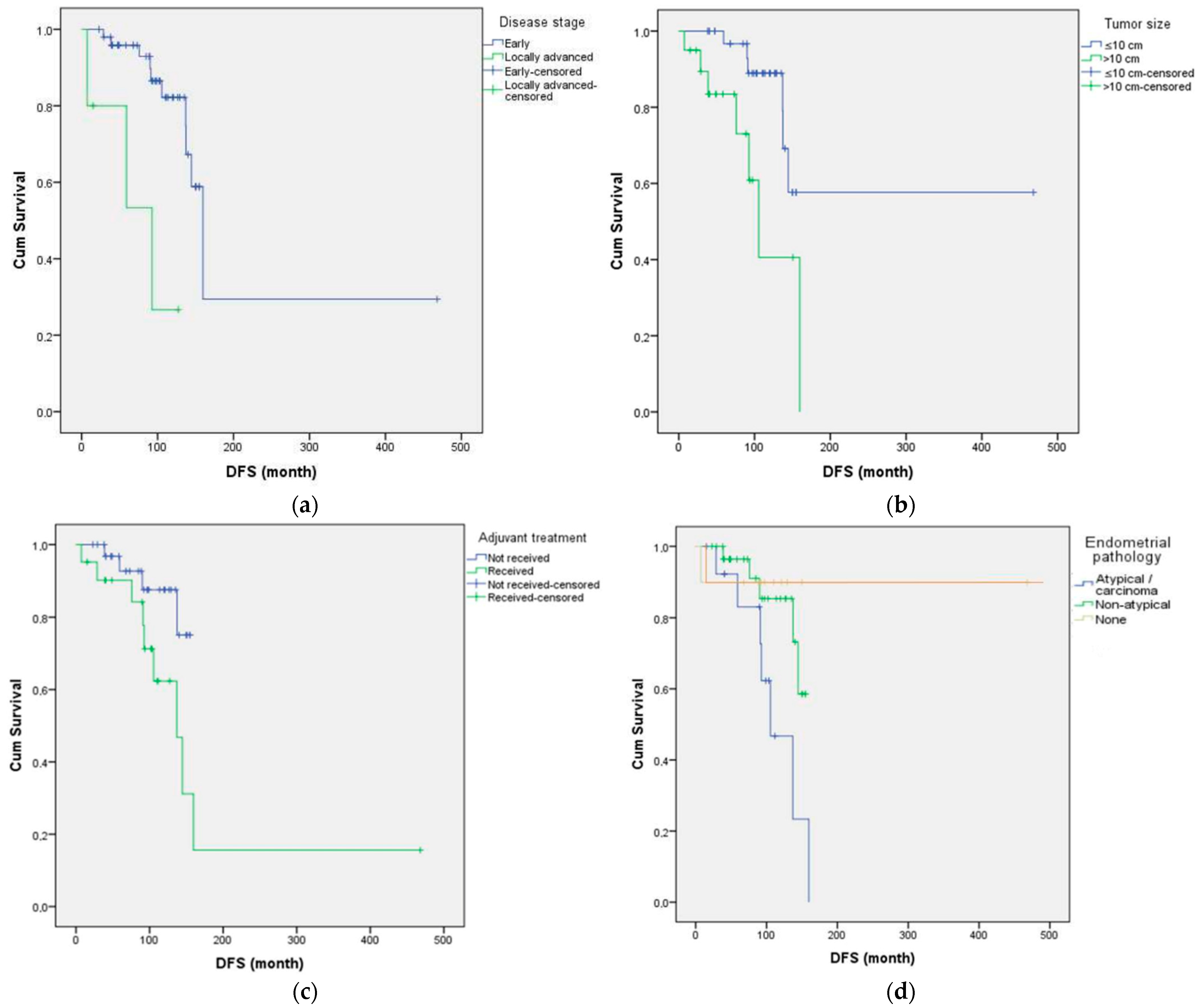
Figure 1. Kaplan–Meier curves for DFS according to key clinicopathological variables. (a) DFS by disease stage (early vs. locally advanced), (b) DFS by tumor size (≤10 cm vs. >10 cm), and (c) DFS by adjuvant chemotherapy status (received vs. not received) (d) DFS by endometrial pathology (none, non-atypical, atypical/carcinoma).
Curroncol 32 00504 g001
Figure 2. Forest plot summarizing HRs with 95% CIs from univariate and multivariate Cox regression analyses of DFS in AGCT patients. Black bullet points represent HR estimates; solid grey lines indicate 95% CIs. The vertical red dashed line marks HR = 1 (no effect).
Curroncol 32 00504 g002
Table 1. Distribution of clinical and pathological features.
VariableCategoryn (%)
Menstrual StatusPremenopausal21 (38.2%)
Perimenopausal5 (9.1%)
Postmenopausal29 (52.7%)
Comorbidity CountNone25 (45.5%)
One11 (20.0%)
≥219 (34.5%)
RecurrenceAbsent42 (76.4%)
Present13 (23.6%)
Recurrence SiteLocal recurrence4 (7.3%)
Visceral metastasis3 (5.5%)
Peritoneal6 (10.9%)
Endometrial PathologyNone28 (50.9%)
Non-atypical hyperplasia17 (30.9%)
Atypical hyperplasia7 (12.7%)
Carcinoma3 (5.4%)
Estrogen Receptor StatusPositive18 (32.7%)
Negative10 (18.2%)
Unknown27 (49.1%)
Inhibin StatusNegative9 (16.4%)
Positive46 (83.6%)
Adjuvant TreatmentNone34 (61.8%)
BEP/EP16 (29.1%)
Carboplatin–paclitaxel5 (9.1%)
ParityUnknown6 (10.9%)
≤233 (60.0%)
≥316 (29.1%)
SymptomsMenstrual irregularity8 (14.5%)
Abdominal distension21 (38.2%)
Postmenopausal bleeding18 (32.7%)
Vaginal bleeding5 (9.1%)
Acute abdomen1 (1.8%)
Incidental2 (3.6%)
Surgery TypePrimary staging51 (92.7%)
Other4 (7.2%)
Tumor Size≤10 cm35 (63.6%)
>10 cm20 (36.4%)
Tumor LocationRight25 (45.5%)
Left30 (54.5%)
Pathological StageStage 149 (89.1%)
Stage 21 (1.8%)
Stage 35 (9.1%)
Mitotic Index≤419 (34.5%)
>436 (65.5%)
Vital StatusAlive50 (90.9%)
Deceased5 (9.1%)
Table 2. Association between clinicopathological factors and recurrence status (chi-square test). p < 0.05 was considered statistically significant.
VariableCategory ComparisonChi-Square (χ2)p-Value
Endometrial pathologyNone vs. Non-atypical vs.
Atypical/Carcinoma
7.3890.025
Adjuvant chemotherapyNo vs. BEP/EP or
Carboplatin–Paclitaxel
6.9530.008
StageStage I–II vs. Stage III4.0290.045
Age group≤65 vs. >650.1010.751
Menopausal statusPre vs Peri/Post0.4580.498
Tumor size≤10 cm vs. >10 cm2.2480.134
Mitotic index≤4 vs. >40.990.32
Ki-67 index≤10% vs. >10%0.1030.749
Estrogen receptor
status
Negative vs. Positive vs Unknown0.4080.815
Inhibin expressionNegative vs Positive0.5610.454
Table 3. Univariate Cox regression analysis for DFS. p < 0.05 was considered statistically significant.
VariableUnivariate HR (95% CI)p-ValueMultivariate HR (95% CI)p-Value
Stage (Stage III vs. I–II)7.14 (1.78–28.73)0.0064.45 (1.03–19.27)0.046
Tumor size
(>10 cm vs. ≤10 cm)
3.59 (1.18–10.95)0.0252.13 (0.60–7.49)0.241
Endometrial pathology (Absent)0.343 (0.138–0.858)0.0220.51 (0.18–1.42)0.197
Adjuvant chemotherapy (Yes vs. No)3.21 (0.96–10.69)0.058--
Mitotic index
(>4 vs. ≤4)
1.40 (0.54–3.63)0.488--
Menopausal status0.866 (0.501–1.495)0.605--
Ki-67 (>10% vs. ≤10%)1.01 (0.40–2.58)0.984--
Estrogen receptor positivity (Yes vs. No)1.17 (0.45–3.06)0.746--
Tumor laterality
(Unilateral vs. Bilateral)
1.23 (0.47–3.25)0.675--
Parity (≥3 vs. ≤2)0.85 (0.33–2.21)0.740--
Inhibin positivity
(Yes vs. No)
0.77 (0.27–2.23)0.620--
Comorbidity
(Yes vs. No)
1.11 (0.43–2.86)0.820--
Disclaimer/Publisher’s Note: The statements, opinions and data contained in all publications are solely those of the individual author(s) and contributor(s) and not of MDPI and/or the editor(s). MDPI and/or the editor(s) disclaim responsibility for any injury to people or property resulting from any ideas, methods, instructions or products referred to in the content.