Influence of Vitamin D and Its Analogues in Type-B Lymphomas

 ,
,  ,
,  ,
,  , , ,
, , ,
Abstract
1. Introduction
2. Materials and Methods
3. Effects of Vitamin D and Its Analogues
3.1. Vitamin D and Its Analogues
3.2. In Vitro Effects of Vitamin D and Its Analogues
3.3. In Vivo Effects of Vitamin D
3.4. Diet and Vitamin D
4. Discussion and Conclusions
Author Contributions
Funding
Conflicts of Interest
References
- Gallamini, A.; Juweid, M. Lymphoma [Internet]; Exon Publications: Brisbane, Australia, 2021. [Google Scholar]
- Mugnaini, E.N.; Ghosh, N. Lymphoma. Prim. Care Clin. Off. Pract. 2016, 43, 661–675. [Google Scholar] [CrossRef]
- De Leval, L.; Jaffe, E.S. Lymphoma Classification. Cancer J. 2020, 26, 176–185. [Google Scholar] [CrossRef] [PubMed]
- Lewis, W.D.; Lilly, S.; Jones, K.L. Lymphoma: Diagnosis and Treatment. Am. Fam. Physician 2020, 101, 34–41. [Google Scholar] [PubMed]
- Bowzyk Al-Naeeb, A.; Ajithkumar, T.; Behan, S.; Hodson, D.J. Non-Hodgkin lymphoma. BMJ 2018, 362, k3204. [Google Scholar] [CrossRef] [PubMed]
- Bray, F.; Laversanne, M.; Sung, H.; Ferlay, J.; Siegel, R.L.; Soerjomataram, I.; Jemal, A. Global cancer statistics 2022: GLOBOCAN estimates of incidence and mortality worldwide for 36 cancers in 185 countries. CA Cancer J. Clin. 2024, 74, 229–263. [Google Scholar] [CrossRef] [PubMed]
- Thandra, K.C.; Barsouk, A.; Saginala, K.; Padala, S.A.; Barsouk, A.; Rawla, P. Epidemiology of Non-Hodgkin’s Lymphoma. Med. Sci. 2021, 9, 5. [Google Scholar] [CrossRef]
- Seifert, M.; Scholtysik, R.; Küppers, R. Origin and Pathogenesis of B Cell Lymphomas. Methods Mol. Biol. 2019, 1956, 1–33. [Google Scholar] [PubMed]
- Hang, H.; Zhou, H.; Ma, L. Prognostic factors and clinical survival outcome in patients with primary mediastinal diffuse large B-cell lymphoma in rituximab era: A population-based study. Medicine 2024, 103, e37238. [Google Scholar] [CrossRef] [PubMed]
- Hamadani, M.; Awan, F.T. Remission induction, consolidation, and novel agents in development for adults with acute myeloid leukaemia. Hematol. Oncol. 2010, 28, 3–12. [Google Scholar] [CrossRef] [PubMed]
- Kulling, P.M.; Olson, K.C.; Olson, T.L.; Feith, D.J.; Loughran, T.P., Jr. Vitamin D in hematological disorders and malignancies. Eur. J. Haematol. 2017, 98, 187–197. [Google Scholar] [CrossRef] [PubMed]
- Shah, N.N.; Fry, T.J. Mechanisms of resistance to CAR T cell therapy. Nat. Rev. Clin. Oncol. 2019, 16, 372–385. [Google Scholar] [CrossRef]
- Hohaus, S.; Tisi, M.C.; Bellesi, S.; Maiolo, E.; Alma, E.; Tartaglia, G.; Corrente, F.; Cuccaro, A.; D’Alo’, F.; Basile, U.; et al. Vitamin D deficiency and supplementation in patients with aggressive B-cell lymphomas treated with immunochemotherapy. Cancer Med. 2018, 7, 270–281. [Google Scholar] [CrossRef]
- Trump, D.L.; Deeb, K.; Johnson, C.S. Vitamin D: Considerations in the Continued Development as an Agent for Cancer Prevention and Therapy. Cancer J. 2010, 16, 1–9. [Google Scholar] [CrossRef] [PubMed]
- Li, M.; Chen, P.; Li, J.; Chu, R.; Xie, D.; Wang, H. Review: The impacts of circulating 25-hydroxyvitamin D levels on cancer patient outcomes: A systematic review and meta-analysis. J. Clin. Endocrinol. Metab. 2014, 99, 2327–2336. [Google Scholar] [CrossRef] [PubMed]
- Bandera Merchan, B.; Morcillo, S.; Martin-Nuñez, G.; Tinahones, F.J.; Macías-González, M. The role of vitamin D and VDR in carcinogenesis: Through epidemiology and basic sciences. J. Steroid Biochem. Mol. Biol. 2017, 167, 203–218. [Google Scholar] [CrossRef]
- Kim, J.H.; Park, W.H.; Suh, D.H.; Kim, K.; No, J.H.; Kim, Y.B. Calcitriol Combined With Platinum-based Chemotherapy Suppresses Growth and Expression of Vascular Endothelial Growth Factor of SKOV-3 Ovarian Cancer Cells. Anticancer Res. 2021, 41, 2945–2952. [Google Scholar] [CrossRef] [PubMed]
- Abu El Maaty, M.A.; Wölfl, S. Effects of 1,25(OH)2D3 on Cancer Cells and Potential Applications in Combination with Established and Putative Anti-Cancer Agents. Nutrients 2017, 9, 87. [Google Scholar] [CrossRef] [PubMed]
- García-Quiroz, J.; Cárdenas-Ochoa, N.; García-Becerra, R.; Morales-Guadarrama, G.; Méndez-Pérez, E.A.; Santos-Cuevas, C.; Ramírez-Nava, G.J.; Segovia-Mendoza, M.; Prado-García, H.; Avila, E.; et al. Antitumoral effects of dovitinib in triple-negative breast cancer are synergized by calcitriol in vivo and in vitro. J. Steroid Biochem. Mol. Biol. 2021, 214, 105979. [Google Scholar] [CrossRef] [PubMed]
- Wakle, K.S.; Mokale, S.N.; Sakle, N.S. Emerging perspectives: Unraveling the anticancer potential of vitamin D3. Naunyn-Schmiedeberg’s Arch. Pharmacol. 2024, 397, 2877–2933. [Google Scholar] [CrossRef] [PubMed]
- Bouillon, R.; Marcocci, C.; Carmeliet, G.; Bikle, D.; White, J.H.; Dawson-Hughes, B.; Lips, P.; Munns, C.F.; Lazaretti-Castro, M.; Giustina, A.; et al. Skeletal and Extraskeletal Actions of Vitamin D: Current Evidence and Outstanding Questions. Endocr. Rev. 2019, 40, 1109–1151. [Google Scholar] [CrossRef] [PubMed]
- Arksey, H.; O’Malley, L. Scoping studies: Towards a methodological framework. Int. J. Soc. Res. Methodol. 2005, 8, 19–32. [Google Scholar] [CrossRef]
- Munn, Z.; Peters, M.D.J.; Stern, C.; Tufanaru, C.; McArthur, A.; Aromataris, E. Systematic review or scoping review? Guidance for authors when choosing between a systematic or scoping review approach. BMC Med. Res. Methodol. 2018, 18, 143. [Google Scholar] [CrossRef] [PubMed]
- Biasucci, G.; Donini, V.; Cannalire, G. Rickets Types and Treatment with Vitamin D and Analogues. Nutrients 2024, 16, 416. [Google Scholar] [CrossRef]
- Rochel, N.; Wurtz, J.M.; Mitschler, A.; Klaholz, B.; Moras, D. The crystal structure of the nuclear receptor for vitamin D bound to its natural ligand. Mol. Cell 2000, 5, 173–179. [Google Scholar] [CrossRef] [PubMed]
- Meyer, M.B. The Vitamin D Receptor: New Paradigms for the Regulation of Gene Expression by 1,25-DihydroxyvitaminD(3). Endocrinol. Metab. Clin. N. Am. 2010, 39, 255–269. [Google Scholar]
- Charoenngam, N.; Shirvani, A.; Holick, M.F. Vitamin D for skeletal and non-skeletal health: What we should know. J. Clin. Orthop. Trauma 2019, 10, 1082–1093. [Google Scholar] [CrossRef] [PubMed]
- Paul, S.; Kaushik, R.; Chawla, P.; Upadhyay, S.; Rawat, D.; Akhtar, A. Vitamin-D as a multifunctional molecule for overall well-being: An integrative review. Clin. Nutr. ESPEN 2024, 62, 10–21. [Google Scholar] [CrossRef] [PubMed]
- World Health Organization. Vitamin D and Cancer. IARC Working Group Reports; International Agency for Research on Cancer: Geneva, Switzerland, 2008; Volume 5. [Google Scholar]
- Bird, R.P. Vitamin D and cancer. Adv. Food Nutr. Res. 2024, 109, 92–159. [Google Scholar] [PubMed]
- Qin, B.; Xu, B.; Ji, N.; Yao, S.; Pawlish, K.; Llanos, A.A.M.; Lin, Y.; Demissie, K.; Ambrosone, C.B.; Hong, C.C.; et al. Intake of vitamin D and calcium, sun exposure, and risk of breast cancer subtypes among black women. Am. J. Clin. Nutr. 2020, 111, 396–405. [Google Scholar] [CrossRef] [PubMed]
- Piatek, K.; Schepelmann, M.; Kallay, E. The Effect of Vitamin D and Its Analogs in Ovarian Cancer. Nutrients 2022, 14, 3867. [Google Scholar] [CrossRef]
- Maestro, M.A.; Molnár, F.; Carlberg, C. Vitamin D and Its Synthetic Analogs. J. Med. Chem. 2019, 62, 6854–6875. [Google Scholar] [CrossRef] [PubMed]
- Thiel, A.; Hermanns, C.; Lauer, A.A.; Reichrath, J.; Erhardt, T.; Hartmann, T.; Grimm, M.O.W.; Grimm, H.S. Vitamin D and Its Analogues: From Differences in Molecular Mechanisms to Potential Benefits of Adapted Use in the Treatment of Alzheimer’s Disease. Nutrients 2023, 15, 1684. [Google Scholar] [CrossRef]
- Kozielewicz, P.; Grafton, G.; Kutner, A.; Curnow, S.J.; Gordon, J.; Barnes, N.M. Novel vitamin D analogues; cytotoxic and anti-proliferative activity against a diffuse large B-cell lymphoma cell line and B-cells from healthy donors. J. Steroid Biochem. Mol. Biol. 2016, 164, 98–105. [Google Scholar] [CrossRef]
- Sherman, M.H.; Yu, R.T.; Engle, D.D.; Ding, N.; Atkins, A.R.; Tiriac, H.; Collisson, E.A.; Connor, F.; Van Dyke, T.; Kozlov, S.; et al. Vitamin D receptor-mediated stromal reprogramming suppresses pancreatitis and enhances pancreatic cancer therapy. Cell 2014, 159, 80–93. [Google Scholar] [CrossRef] [PubMed]
- Sehn, L.H.; Salles, G. Diffuse Large B-Cell Lymphoma. N. Engl. J. Med. 2021, 384, 842–858. [Google Scholar] [CrossRef]
- Han, J.; Tang, Y.; Zhong, M.; Wu, W. Antitumor effects and mechanisms of 1,25(OH)2D3 in the Pfeiffer diffuse large B lymphoma cell line. Mol. Med. Rep. 2019, 20, 5064–5074. [Google Scholar] [CrossRef]
- Neumann, F.; Acker, F.; Schormann, C.; Pfreundschuh, M.; Bittenbring, T.J. Determination of optimum vitamin D3 levels for NK cell-mediated rituximab- and obinutuzumab-dependent cellular cytotoxicity. Cancer Immunol. Immunother. 2018, 67, 1709–1718. [Google Scholar] [CrossRef] [PubMed]
- Bold, A.; Gross, H.; Holzmann, E.; Smetak, M.; Birkmann, J.; Bertsch, T.; Triebel, J.; Sauer, K.; Wilhelm, M.; Hoeres, T. Immune activating and inhibiting effects of calcitriol on γδ T cells and NK cells. Immunobiology 2022, 227, 152286. [Google Scholar] [CrossRef]
- Gharbaran, R.; Zhang, B.; Valerio, L.; Onwumere, O.; Wong, M.; Mighty, J.; Redenti, S. Effects of vitamin D3 and its chemical analogs on the growth of Hodgkin’s lymphoma, in vitro. BMC Res. Notes 2019, 12, 216. [Google Scholar] [CrossRef] [PubMed]
- Gleba, J.J.; Mielko, K.A.; Wietrzyk, J.; Kłopotowska, D.; Banach, J.; Turlej, E.; Gebura, K.; Bogunia-Kubik, K.; Kutner, A. Polymorphism of VDR Gene and the Sensitivity of Human Leukemia and Lymphoma Cells to Active Forms of Vitamin D. Cancers 2022, 14, 387. [Google Scholar] [CrossRef] [PubMed]
- Liu, W.; Liu, J.; Song, Y.; Wang, X.; Mi, L.; Cai, C.; Zhao, D.; Wang, L.; Ma, J.; Zhu, J. Burden of lymphoma in China, 1990–2019: An analysis of the global burden of diseases, injuries, and risk factors study 2019. Aging 2022, 14, 3175–3190. [Google Scholar] [CrossRef]
- Carpio, C.; Bouabdallah, R.; Ysebaert, L.; Sancho, J.M.; Salles, G.; Cordoba, R.; Pinto, A.; Gharibo, M.; Rasco, D.; Panizo, C.; et al. Avadomide monotherapy in relapsed/refractory DLBCL: Safety, efficacy, and a predictive gene classifier. Blood 2020, 135, 996–1007. [Google Scholar] [CrossRef] [PubMed]
- Min, D.; Lv, X.B.; Wang, X.; Zhang, B.; Meng, W.; Yu, F.; Hu, H. Downregulation of miR-302c and miR-520c by 1,25(OH)2D3 treatment enhances the susceptibility of tumour cells to natural killer cell-mediated cytotoxicity. Br. J. Cancer 2013, 109, 723–730. [Google Scholar] [CrossRef] [PubMed]
- Weeres, M.A.; Robien, K.; Ahn, Y.O.; Neulen, M.L.; Bergerson, R.; Miller, J.S.; Verneris, M.R. The effects of 1,25-dihydroxyvitamin D3 on in vitro human NK cell development from hematopoietic stem cells. J. Immunol. 2014, 193, 3456–3462. [Google Scholar] [CrossRef] [PubMed]
- Chen, J.; Bruce, D.; Cantorna, M.T. Vitamin D receptor expression controls proliferation of naive CD8+ T cells and development of CD8 mediated gastrointestinal inflammation. BMC Immunol. 2014, 15, 6. [Google Scholar] [CrossRef]
- De Martinis, M.; Allegra, A.; Sirufo, M.M.; Tonacci, A.; Pioggia, G.; Raggiunti, M.; Ginaldi, L.; Gangemi, S. Vitamin D Deficiency, Osteoporosis and Effect on Autoimmune Diseases and Hematopoiesis: A Review. Int. J. Mol. Sci. 2021, 22, 8855. [Google Scholar] [CrossRef] [PubMed]
- Bray, F.; Ferlay, J.; Soerjomataram, I.; Siegel, R.L.; Torre, L.A.; Jemal, A. Global cancer statistics 2018: GLOBOCAN estimates of incidence and mortality worldwide for 36 cancers in 185 countries. CA Cancer J. Clin. 2018, 68, 394–424. [Google Scholar] [CrossRef] [PubMed]
- Bittenbring, J.T.; Neumann, F.; Altmann, B.; Achenbach, M.; Reichrath, J.; Ziepert, M.; Geisel, J.; Regitz, E.; Held, G.; Pfreundschuh, M. Vitamin D deficiency impairs rituximab-mediated cellular cytotoxicity and outcome of patients with diffuse large B-cell lymphoma treated with but not without rituximab. J. Clin. Oncol. 2014, 32, 3242–3248. [Google Scholar] [CrossRef]
- Chen, P.; Cao, Y.; Duan, X.; Li, J.; Zhao, W.; Wang, H. Bioavailable 25(OH)D level is associated with clinical outcomes of patients with diffuse large B-cell lymphoma: An exploratory study. Clin. Nutr. 2021, 40, 157–165. [Google Scholar] [CrossRef] [PubMed]
- Wang, W.T.; Liang, J.H.; Wang, L.; Zhu, H.Y.; Xia, Y.; Fan, L.; Li, J.Y.; Xu, W. The prognostic value of 25-hydroxy vitamin D deficiency and its interaction with c-Myc expression in diffuse large B cell lymphoma. Ann. Hematol. 2020, 99, 2377–2384. [Google Scholar] [CrossRef]
- Nath, K.; Tomas, A.A.; Flynn, J.; Fein, J.A.; Alperovich, A.; Anagnostou, T.; Batlevi, C.L.; Dahi, P.B.; Fingrut, W.B.; Giralt, S.A.; et al. Vitamin D Insufficiency and Clinical Outcomes with Chimeric Antigen Receptor T-Cell Therapy in Large B-cell Lymphoma. Transplant. Cell. Ther. 2022, 28, 751.e1–751.e7. [Google Scholar] [CrossRef] [PubMed]
- Drake, M.T.; Maurer, M.J.; Link, B.K.; Habermann, T.M.; Ansell, S.M.; Micallef, I.N.; Kelly, J.L.; Macon, W.R.; Nowakowski, G.S.; Inwards, D.J.; et al. Vitamin D insufficiency and prognosis in non-Hodgkin’s lymphoma. J. Clin. Oncol. 2010, 28, 4191–4198. [Google Scholar] [CrossRef] [PubMed]
- Kelly, J.L.; Salles, G.; Goldman, B.; Fisher, R.I.; Brice, P.; Press, O.; Casasnovas, O.; Maloney, D.G.; Soubeyran, P.; Rimsza, L.; et al. Low Serum Vitamin D Levels Are Associated with Inferior Survival in Follicular Lymphoma: A Prospective Evaluation in SWOG and LYSA Studies. J. Clin. Oncol. 2015, 33, 1482–1490. [Google Scholar] [CrossRef] [PubMed]
- Tracy, S.I.; Maurer, M.J.; Witzig, T.E.; Drake, M.T.; Ansell, S.M.; Nowakowski, G.S.; Thompson, C.A.; Inwards, D.J.; Johnston, P.B.; Micallef, I.N.; et al. Vitamin D insufficiency is associated with an increased risk of early clinical failure in follicular lymphoma. Blood Cancer J. 2017, 7, e595. [Google Scholar] [CrossRef] [PubMed]
- Eicher, F.; Mansouri Taleghani, B.; Schild, C.; Bacher, U.; Pabst, T. Reduced survival after autologous stem cell transplantation in myeloma and lymphoma patients with low vitamin D serum levels. Hematol. Oncol. 2020, 38, 523–530. [Google Scholar] [CrossRef]
- Xu, D.M.; Liang, J.H.; Wang, L.; Zhu, H.Y.; Xia, Y.; Fan, L.; Li, J.Y.; Xu, W. 25-Hydroxy vitamin D deficiency predicts inferior prognosis in mantle cell lymphoma. J. Cancer Res. Clin. Oncol. 2020, 146, 1003–1009. [Google Scholar] [CrossRef] [PubMed]
- Qin, J.Q.; Yin, H.; Wu, J.Z.; Chen, R.Z.; Xia, Y.; Wang, L.; Zhu, H.Y.; Fan, L.; Li, J.Y.; Liang, J.H.; et al. 25-Hydroxy vitamin D deficiency predicts inferior prognosis in Hodgkin lymphoma. Leuk. Res. 2021, 105, 106580. [Google Scholar] [CrossRef] [PubMed]
- Borchmann, S.; Cirillo, M.; Goergen, H.; Meder, L.; Sasse, S.; Kreissl, S.; Bröckelmann, P.J.; von Tresckow, B.; Fuchs, M.; Ullrich, R.T.; et al. Pretreatment Vitamin D Deficiency Is Associated With Impaired Progression-Free and Overall Survival in Hodgkin Lymphoma. J. Clin. Oncol. 2019, 37, 3528–3537. [Google Scholar] [CrossRef]
- Zinzani, P.L.; Muñoz, J.; Trotman, J. Current and future therapies for follicular lymphoma. Exp. Hematol. Oncol. 2024, 13, 87. [Google Scholar] [CrossRef] [PubMed]
- López, C.; Silkenstedt, E.; Dreyling, M.; Beà, S. Biological and clinical determinants shaping heterogeneity in mantle cell lymphoma. Blood Adv. 2024, 8, 3652–3664. [Google Scholar] [CrossRef] [PubMed]
- Minoia, C.; Gerardi, C.; Allocati, E.; Daniele, A.; De Sanctis, V.; Bari, A.; Guarini, A. The Impact of Healthy Lifestyles on Late Sequelae in Classical Hodgkin Lymphoma and Diffuse Large B-Cell Lymphoma Survivors. A Systematic Review by the Fondazione Italiana Linfomi. Cancers 2021, 13, 3135. [Google Scholar] [CrossRef]
- Levy Yurkovski, I.; Andreazzoli, F.; Ben-Arye, E.; Attias, S.; Tadmor, T. Integrative Approaches in the Treatment of Patients Affected by Lymphoma. Curr. Oncol. Rep. 2023, 25, 1523–1534. [Google Scholar] [CrossRef] [PubMed]
- Gouni, S.; Marques-Piubelli, M.L.; Strati, P. Follicular lymphoma and macrophages: Impact of approved and novel therapies. Blood Adv. 2021, 5, 4303–4312. [Google Scholar] [CrossRef]
- Neelapu, S.S.; Locke, F.L.; Bartlett, N.L.; Lekakis, L.J.; Miklos, D.B.; Jacobson, C.A.; Braunschweig, I.; Oluwole, O.O.; Siddiqi, T.; Lin, Y.; et al. Axicabtagene Ciloleucel CAR T-Cell Therapy in Refractory Large B-Cell Lymphoma. N. Engl. J. Med. 2017, 377, 2531–2544. [Google Scholar] [CrossRef]
- Renne, C.; Benz, A.H.; Hansmann, M.L. Vitamin D3 receptor is highly expressed in Hodgkin’s lymphoma. BMC Cancer 2012, 12, 215. [Google Scholar] [CrossRef]
- Boughanem, H.; Canudas, S.; Hernandez-Alonso, P.; Becerra-Tomás, N.; Babio, N.; Salas-Salvadó, J.; Macias-Gonzalez, M. Vitamin D Intake and the Risk of Colorectal Cancer: An Updated Meta-Analysis and Systematic Review of Case-Control and Prospective Cohort Studies. Cancers 2021, 13, 2814. [Google Scholar] [CrossRef]
- Liu, J.; Dong, Y.; Lu, C.; Wang, Y.; Peng, L.; Jiang, M.; Tang, Y.; Zhao, Q. Meta-analysis of the correlation between vitamin D and lung cancer risk and outcomes. Oncotarget 2017, 8, 81040–81051. [Google Scholar] [CrossRef] [PubMed]
- Wacker, M.; Holick, M.F. Sunlight and vitamin D: A global perspective for health. Dermato-Endocrinology 2013, 5, 51–108. [Google Scholar] [CrossRef] [PubMed]
- Robsahm, T.E.; Tretli, S.; Torjesen, P.A.; Babigumira, R.; Schwartz, G.G. Serum 25-hydroxyvitamin D levels predict cancer survival: A prospective cohort with measurements prior to and at the time of cancer diagnosis. Clin. Epidemiol. 2019, 11, 695–705. [Google Scholar] [CrossRef] [PubMed]
- Adams, J.S.; Hewison, M. Extrarenal expression of the 25-hydroxyvitamin D-1-hydroxylase. Arch. Biochem. Biophys. 2012, 523, 95–102. [Google Scholar] [CrossRef] [PubMed]
- Hewison, M.; Adams, J.S. Regulation of extra-renal synthesis of 1,25(OH)2D. In Feldman and Pike’s Vitamin D, 5th ed.; Volume One: Biochemistry, Physiology and Diagnostics; Academic Press: Cambridge, MA, USA, 2024; pp. 155–187. [Google Scholar]
- Marcinkowska, E.; Wallace, G.R.; Brown, G. The Use of 1α,25-Dihydroxyvitamin D3 as an Anticancer Agent. Int. J. Mol. Sci. 2016, 17, 729. [Google Scholar] [CrossRef]
- Shallis, R.M.; Rome, R.S.; Reagan, J.L. Mechanisms of Hypercalcemia in Non-Hodgkin Lymphoma and Associated Outcomes: A Retrospective Review. Clin. Lymphoma Myeloma Leuk. 2018, 18, e123–e129. [Google Scholar] [CrossRef] [PubMed]
- Hewison, M.; Kantorovich, V.; Liker, H.R.; Van Herle, A.J.; Cohan, P.; Zehnder, D.; Adams, J.S. Vitamin D-mediated hypercalcemia in lymphoma: Evidence for hormone production by tumor-adjacent macrophages. J. Bone Miner. Res. 2003, 18, 579–582. [Google Scholar] [CrossRef] [PubMed]
- Mudde, A.H.; van den Berg, H.; Boshuis, P.G.; Breedveld, F.C.; Markusse, H.M.; Kluin, P.M.; Bijvoet, O.L.; Papapoulos, S.E. Ectopic production of 1,25-dihydroxyvitamin D by B-cell lymphoma as a cause of hypercalcemia. Cancer 1987, 59, 1543–1546. [Google Scholar] [CrossRef] [PubMed]
- Davies, M.; Hayes, M.E.; Yin, J.A.; Berry, J.L.; Mawer, E.B. Abnormal synthesis of 1,25-dihydroxyvitamin D in patients with malignant lymphoma. J. Clin. Endocrinol. Metab. 1994, 78, 1202–1207. [Google Scholar] [PubMed]
- Ogawa, M.; Morikawa, M.; Kobatake, M.; Murakami, T.; Yamamoto, Y.; Watanabe, R.; Yamada, K.; Nishiyama, K.; Yasutomo, Y.; Hara, K. Hypercalcemia Associated with the Ectopic Expression of 25-hydroxyvitamin D3-1α-hydroxylase in Diffuse Large B-cell Lymphoma. Intern. Med. 2022, 61, 2489–2495. [Google Scholar] [CrossRef] [PubMed]
- Nakayama, S.; Matsuda, M. 25-Hydroxyvitamin D3-1α-hydroxylase- and multiple cytokine-producing diffuse large B-cell lymphoma. Blood 2018, 131, 2271. [Google Scholar] [CrossRef]
- Bikle, D.D.; Patzek, S.; Wang, Y. Physiologic and pathophysiologic roles of extra renal CYP27b1: Case report and review. Bone Rep. 2018, 8, 255–267. [Google Scholar] [CrossRef]
- Giustina, A.; Bilezikian, J.P.; Adler, R.A.; Banfi, G.; Bikle, D.D.; Binkley, N.C.; Bollerslev, J.; Bouillon, R.; Brandi, M.L.; Casanueva, F.F.; et al. Consensus Statement on Vitamin D Status Assessment and Supplementation: Whys, Whens, and Hows. Endocr. Rev. 2024, 45, 625–654. [Google Scholar] [PubMed]
- Goggin, K.P.; Lu, L.; Lee, D.E.; Howell, C.R.; Srivastava, D.; Brinkman, T.M.; Armstrong, G.T.; Bhakta, N.; Robison, L.L.; Ehrhardt, M.J.; et al. Severe Sepsis During Treatment for Childhood Leukemia and Sequelae Among Adult Survivors. JAMA Netw. Open 2024, 7, e242727. [Google Scholar] [CrossRef]
- Allegra, A.; Tonacci, A.; Musolino, C.; Pioggia, G.; Gangemi, S. Secondary Immunodeficiency in Hematological Malignancies: Focus on Multiple Myeloma and Chronic Lymphocytic Leukemia. Front. Immunol. 2021, 12, 738915. [Google Scholar] [CrossRef] [PubMed]
- Aljabari, S.; Balch, A.; Larsen, G.Y.; Fluchel, M.; Workman, J.K. Severe Sepsis-Associated Morbidity and Mortality among Critically Ill Children with Cancer. J. Pediatr. Intensive Care 2019, 8, 122–129. [Google Scholar] [CrossRef] [PubMed]
- Carboo, J.A.; Dolman-Macleod, R.C.; Malan, L.; Lombard, M.J. High-dose oral vitamin D supplementation for prevention of infections in children aged 0 to 59 months: A systematic review and meta-analysis. Nutr. Rev. 2024, 82, 579–599. [Google Scholar] [CrossRef] [PubMed]
- Mora, J.R.; Iwata, M.; von Andrian, U.H. Vitamin effects on the immune system: Vitamins A and D take centre stage. Nat. Rev. Immunol. 2008, 8, 685–698. [Google Scholar] [CrossRef] [PubMed]
- Adams, J.S. Vitamin D as a defensin. J. Musculoskelet. Neuronal Interact. 2006, 6, 344–346. [Google Scholar] [PubMed]
- Ramanarayanan, P.; Heine, G.; Worm, M. Vitamin A and vitamin D induced nuclear hormone receptor activation and its impact on B cell differentiation and immunoglobulin production. Immunol. Lett. 2023, 263, 80–86. [Google Scholar] [CrossRef] [PubMed]
- Shimoni, A.; Marcus, H.; Canaan, A.; Ergas, D.; David, M.; Berrebi, A.; Reisner, Y. A Model for Human B-Chronic Lymphocytic Leukemia in Human/Mouse Radiation Chimera: Evidence for Tumor-Mediated Suppression of Antibody Production in Low-Stage Disease. Blood 1997, 89, 2210–2218. [Google Scholar] [CrossRef] [PubMed]
- Forconi, F.; Moss, P. Perturbation of the Normal Immune System in Patients with CLL. Blood 2015, 126, 573–581. [Google Scholar] [CrossRef]
- Bussel, J.B.; Cunningham-Rundles, C. Intravenous Usage of Gammaglobulin: Humoral Immunodeficiency, Immune Thrombocytopenic Purpura, and Newer Indications. Cancer Investig. 1985, 3, 361–366. [Google Scholar] [CrossRef]
- Cantwell, M.; Hua, T.; Pappas, J.; Kipps, T.J. Acquired CD40-Ligand Deficiency in Chronic Lymphocytic Leukemia. Nat. Med. 1997, 3, 984–989. [Google Scholar] [CrossRef]
- Cerutti, A.; Kim, E.C.; Shah, S.; Schattner, E.J.; Zan, H.; Schaffer, A.; Casali, P. Dysregulation of CD30+ T Cells by Leukemia Impairs Isotype Switching in Normal B Cells. Nat. Immunol. 2001, 2, 150–156. [Google Scholar] [CrossRef] [PubMed]
- Cesur, F.; Atasever, Z.; Özoran, Y. Impact of vitamin D3 supplementation on COVID-19 vaccine response and immunoglobulin G antibodies in deficient women: A randomized controlled trial. Vaccine 2023, 41, 2860–2867. [Google Scholar] [CrossRef]
- Maruki, T.; Nomoto, H.; Iwamoto, N.; Yamamoto, K.; Kurokawa, M.; Iwatsuki-Horimoto, K.; Yamayoshi, S.; Suzuki, Y.; Kawaoka, Y.; Ohmagari, N. Successful management of persistent COVID-19 using combination antiviral therapy (nirmatrelvir/ritonavir and remdesivir) and intravenous immunoglobulin transfusion in an immunocompromised host who had received CD20 depleting therapy for follicular lymphoma. J. Infect. Chemother. 2024, 30, 793–795. [Google Scholar] [CrossRef]
- Yang, J.M.; Li, Z.Q.; Zhong, Y.B.; Xie, H.Y.; Luo, Y.; Xiao, L.; Liao, J.H.; Wang, M.Y. Association Between Vitamin D and COVID-19-Related Outcomes: An Umbrella Review of Meta-Analyses. Nutr. Rev. 2025, 17, nuae225. [Google Scholar] [CrossRef] [PubMed]
- Young, J.; Welin, E.; Braeutigam, C.; Gilger, E.; Lane, A.; Salloum, R. Impact of a Vitamin D Replacement Algorithm in Children and Young Adults with Acute Lymphoblastic Leukemia. J. Pediatr. Hematol. Oncol. 2018, 40, 594–597. [Google Scholar] [CrossRef] [PubMed]
- Asif, A.; Farooq, N. Vitamin D toxicity. In StatPearls; StatPearls Publishing: Treasure Island, FL, USA, 2023. [Google Scholar]
- Galior, K.; Grebe, S.; Singh, R. Development of Vitamin D Toxicity from Overcorrection of Vitamin D Deficiency: A Review of Case Reports. Nutrients 2018, 10, 953. [Google Scholar] [CrossRef] [PubMed]
- Subramanian, A.; Burrowes, H.B.; Rumph, J.T.; Wilkerson, J.; Jackson, C.L.; Jukic, A.M.Z. Vitamin D Levels in the United States: Temporal Trends (2011–2018) and Contemporary Associations with Sociodemographic Characteristics (2017–2018). Nutrients 2024, 16, 3414. [Google Scholar] [CrossRef]
- Chen, G.C.; Lv, D.B.; Pang, Z.; Liu, Q.F. Fruits and vegetables consumption and risk of non-Hodgkin’s lymphoma: A meta-analysis of observational studies. Int. J. Cancer 2013, 133, 190–200. [Google Scholar] [CrossRef] [PubMed]
- Solans, M.; Benavente, Y.; Saez, M.; Agudo, A.; Naudin, S.; Hosnijeh, F.S.; Noh, H.; Freisling, H.; Ferrari, P.; Besson, C.; et al. Adherence to the mediterranean diet and lymphoma risk in the european prospective investigation into cancer and nutrition. Int. J. Cancer 2019, 145, 122–131. [Google Scholar] [CrossRef]
- Marini, H.R.; Facchini, B.A.; di Francia, R.; Freni, J.; Puzzolo, D.; Montella, L.; Facchini, G.; Ottaiano, A.; Berretta, M.; Minutoli, L. Glutathione: Lights and Shadows in Cancer Patients. Biomedicines 2023, 11, 2226. [Google Scholar] [CrossRef]
- Huang, J.R.; Song, J.R.; Cai, W.S.; Shao, Z.W.; Zhou, D.Y.; Song, L. Enhancing vitamin D3 bioaccessibility: Unveiling hydrophobic interactions in soybean protein isolate and vitamin D3 binding via an infant in vitro digestion model. Food Chem. 2024, 451, 139507. [Google Scholar] [CrossRef] [PubMed]
- Marwaha, R.K.; Dev, T.; Mittal, A.; Mani, K.; Narang, A.; Arora, P.; Singh, A.; Chadha, A.; Dang, N.; Goel, M.; et al. A randomised controlled trial comparing the efficacy of micellised and fat-soluble vitamin D3 supplementation in healthy adults. Br. J. Nutr. 2019, 121, 859–865. [Google Scholar] [CrossRef] [PubMed]
- Marini, H.R. Mediterranean Diet and Soy Isoflavones for Integrated Management of the Menopausal Metabolic Syndrome. Nutrients 2022, 14, 1550. [Google Scholar] [CrossRef]

 Increased;
  Decreased.
Increased; Decreased.
Decreased.
Increased; Decreased.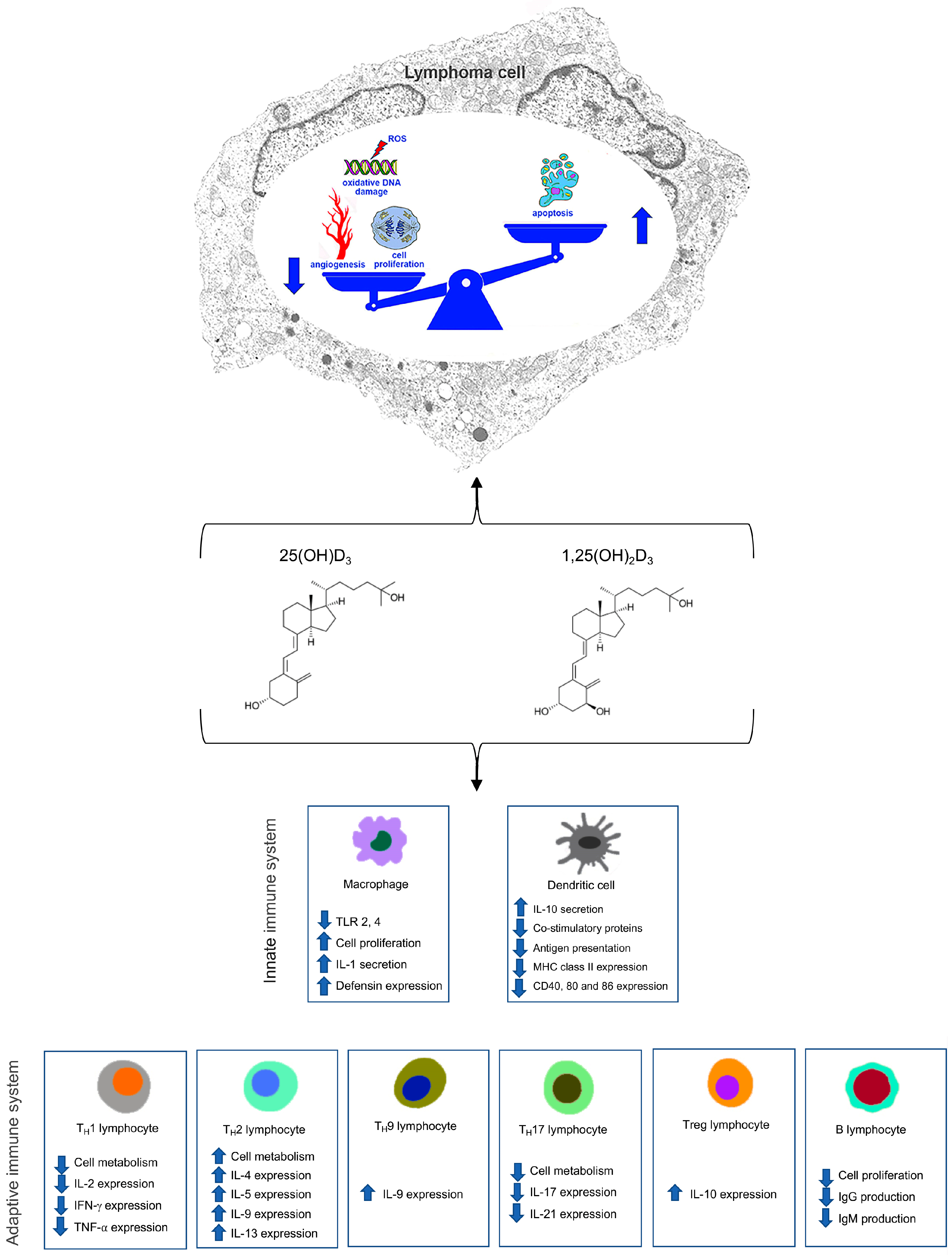

| Study | Cell Line | Results | Reference Number |
|---|---|---|---|
| Kozielewicz et al., 2016 | DOHH2 | VitD and VDAs had cytotoxic and pro-apoptotic actions, increased VDR expression, and the anti-proliferative efficacy of clomipramine. | [35] |
| Han et al., 2019 | DLBCL Pfeiffer | Calcitriol blocked proliferation and induced G1 phase, and this effect was augmented with the co-administration of RAPA; calcitriol increased autophagy by stopping the PI3K/AKT/mTOR pathway. | [38] |
| Neumann et al., 2018 | DAUDI (CD20+ Burkitt’s lymphoma cells) | DAUDI cells cultured with NK-cells obtained from healthy donors and ADCC activity were determined by lactate dehydrogenase release assay. NK-cells killed lymphoma cells in a concentration and E:T ratio-dependent manner with obinutuzumab displaying a stronger ADCC activity than rituximab. | [39] |
| Bold et al., 2022 | DAUDI, U2932 | Increased ADCC against tumor cells in cells stimulated with IL-2 and treated with highest concentration of calcitriol. | [40] |
| Gharbaran et al., 2019 | HL, HRS (Hodgkin and Reed-Sternberg cells) | Low VDR expression; reduction in HL-cell line growth after 72 h of treatment with VitD and VDAs at 10 μM concentration; this reduction was associated with an increased VDR nuclear accumulation. | [41] |
| Gleba et al., 2022 | KG-1, K562, HL-60, MV-4-11, Thp-1 Jurkat, DAUDI, Raji | MV-4-11, Thp-1, and HL-60 cell lines sensitive to calcitriol and tacalcitol inhibition on proliferation showed morphological changes. | [42] |
| Study | Number of Patients | Type of Lymphoma | Results | Reference Number |
|---|---|---|---|---|
| Bittenbring et al., 2014 | 359 | DLBCL | Worst EFS and OS in patients with VDD. | [50] |
| Chen et al., 2021 | 332 | DLBCL | Worst PFS and better response to treatment in patients with higher VitD blood levels. | [51] |
| Wang et al., 2020 | 208 | DLBCL | Worst PFS and OS in patients with VDD. | [52] |
| Nath et al., 2022 | 111 | DLBCL | Worst response to treatment and 2-year OS. In patients with VDD, no correlation between pre-treatment VDD and CAR-T-related toxicity. | [53] |
| Drake et al., 2010 | 983 | DLBCL | Worst EFS and OS in patients with VDD; no association between EFS and VDD in other NHL subtypes. | [54] |
| Kelly et al., 2015 | 1979 | FL | Worst PFS in patients with VDD. | [55] |
| Tracy et al., 2017 | 642 | FL | Worst OS and EFS at 12 months in patients with VDI. | [56] |
| Eicher et al., 2020 | 183 | Various types of lymphomas | Better PFS and OS in patients with VitD levels > 52 nmol/L. | [57] |
| Xu et al., 2020 | 70 | MCL | Worst PFS and OS in patients with VDD. | [58] |
| Qin et al., 2021 | 77 | HL | Worst PFS and OS in patients VDD. | [59] |
| Borchmann et al., 2019 | 351 | HL | VitD low levels more common in R/R patients; worst PFS in patients with VDD. | [60] |
Disclaimer/Publisher’s Note: The statements, opinions and data contained in all publications are solely those of the individual author(s) and contributor(s) and not of MDPI and/or the editor(s). MDPI and/or the editor(s) disclaim responsibility for any injury to people or property resulting from any ideas, methods, instructions or products referred to in the content. |
© 2025 by the authors. Licensee MDPI, Basel, Switzerland. This article is an open access article distributed under the terms and conditions of the Creative Commons Attribution (CC BY) license (https://creativecommons.org/licenses/by/4.0/).
Share and Cite
Basile, V.; Allegra, A.; Marini, H.R.; Berretta, M.; Granata, B.; Freni, J.; Puzzolo, D.; Stagno, F.; Midiri, P.; Urzì Brancati, V.; et al. Influence of Vitamin D and Its Analogues in Type-B Lymphomas. Curr. Oncol. 2025, 32, 135. https://doi.org/10.3390/curroncol32030135
Basile V, Allegra A, Marini HR, Berretta M, Granata B, Freni J, Puzzolo D, Stagno F, Midiri P, Urzì Brancati V, et al. Influence of Vitamin D and Its Analogues in Type-B Lymphomas. Current Oncology. 2025; 32(3):135. https://doi.org/10.3390/curroncol32030135
Chicago/Turabian StyleBasile, Valerio, Alessandro Allegra, Herbert Ryan Marini, Massimiliano Berretta, Barbara Granata, José Freni, Domenico Puzzolo, Fabio Stagno, Paola Midiri, Valentina Urzì Brancati, and et al. 2025. "Influence of Vitamin D and Its Analogues in Type-B Lymphomas" Current Oncology 32, no. 3: 135. https://doi.org/10.3390/curroncol32030135
APA StyleBasile, V., Allegra, A., Marini, H. R., Berretta, M., Granata, B., Freni, J., Puzzolo, D., Stagno, F., Midiri, P., Urzì Brancati, V., & Minutoli, L. (2025). Influence of Vitamin D and Its Analogues in Type-B Lymphomas. Current Oncology, 32(3), 135. https://doi.org/10.3390/curroncol32030135