Case Report

HRAS Q61L Mutation as a Possible Target for Non-Small Cell Lung Cancer: Case Series and Review of Literature

1
Service d’Oncologie Médicale, CHU Nantes, Nantes Université, F-44000 Nantes, France
2
UMR 1302/EMR6001, INCIT, Immunology and New Concepts in ImmunoTherapy, CNRS, INSERM, Laboratoire de Biochimie, CHU Nantes, Nantes Université, F-44000 Nantes, France
3
Pharmacie, CHU Nantes, Nantes Université, F-44000 Nantes, France
4
Department of Medical Oncology, Hospital Foch, F-92073 Suresnes, France
5
Nantes Université, Inserm UMR 1307, CNRS UMR 6075, Université d’Angers, CRCI2NA, F-44000 Nantes, France
*
Author to whom correspondence should be addressed.
Current address: Hôpital Laennec, CHU de Nantes, Boulevard Professeur Jacques Monod, 44800 Saint Herblain, France.
Curr. Oncol. 2022, 29(5), 3748-3758; https://doi.org/10.3390/curroncol29050300
Submission received: 9 March 2022 / Revised: 10 May 2022 / Accepted: 12 May 2022 / Published: 20 May 2022

Abstract

:
Introduction: Assessment of actionable gene mutations and oncogene fusions have made a paradigm shift in treatment strategies of non-small cell lung cancer (NSCLC). HRAS mutations involved around 0.2–0.8% of NSCLC patients, mostly on codon 61. For these patients, few data are available regarding clinical characteristics and response to therapies. Methods: Next-Generation Sequencing (NGS) done routinely at Nantes University Hospital was used to identify HRAS molecular alterations in NSCLC patients. We identified and described four HRAS p.GlnQ61Leu mutated patients. Literature of previously HRAS-mutant NSCLC cases was reviewed, and available data in solid tumour with the most advanced H-Ras specific inhibitor, tipifarnib, were presented. Results: Of 1614 patients diagnosed with advanced NSCLC from January 2018 to December 2020, four (0.25%) had HRAS p.Gln61Leu mutation. Three of them died during the first-line systemic therapy. Furthermore, three additional cases were identified in literature. All cases were current or former smokers, most of them had pleural or pericardial effusion at diagnosis. Conclusions: The clinical course of patients with HRAS-mutant NSCLC remains unclear. Furthers cases should be identified in order to clarify prognosis and response to therapies. Tipifarnib, a farnesyl transferase inhibitor, is a promising candidate to target HRAS-mutant tumours and should be explored in NSCLC patients.

1. Introduction

Lung cancer accounted for more than 2.2 million cases and almost 1.8 million deaths worldwide in 2020 [1]. Non-small cell lung cancer (NSCLC) is the main type of lung cancer and represents 85% of all cases. The prognosis of oncogenic-addicted NSCLC has been transformed due to the development of targeted therapies, firstly inhibiting the mutant epidermal growth factor receptor (EGFR) [2,3], then ALK [4,5,6] and ROS1 rearrangement [7]. More recently, KRAS appeared as a promising target in NSCLC patients, due to the development of efficient targeted therapies such as sotorasib targeting the KRAS p.Gly12Cys mutation [8,9,10].
The RAS genes belong to a well-described family of oncogenic drivers, encoding four small GTPase proteins, K-Ras4A and K-Ras4B (which are two splice variants of the KRAS gene), H-Ras and N-Ras [9,10,11]. These proteins have an active form, Ras-GTP, and an inactive form, Ras-GDP. Transition from one to another is regulated by Ras-Guanine nucleotide Exchange Factors (Ras-GEFs) that catalyse the exchange of GDP for GTP, and by intrinsic GTPase activity hydrolysing GTP to GDP with the assistance of Ras-GTPase Activating Proteins (Ras-GAPs). In humans, Ras proteins participate in many physiological processes related to cell growth, division, and survival [10]. Substitution mutations, mainly involving codons Gly12, Gly13, and Gln61 of the RAS genes, are found in about 20% of human cancers. They are known to cause constitutive activation of the signalling activity mainly through impairment of their intrinsic GTPase activity [12,13] and thus of the conversion of active form Ras-GTP to inactive form Ras-GDP. Ras-GTP interact with downstream effectors such as PhosphoInositide 3-Kinase (PI3K) and Mitogen Activated Protein Kinases (MAPK) pathways, that enhanced growth, proliferation, differentiation, and survival of cancer cells [10].
Based on a recent publication evaluating several leading cancer mutation databases, KRAS is by far the most commonly mutated of the three RAS genes in solid tumours (75%), followed by NRAS (17%) and HRAS (7%) [13]. HRAS is mostly altered in head and neck squamous cell carcinoma (HNSCC) (5–9%), salivary glands (15%), and bladder cancer (5–30%) [13,14]. HRAS mutations are involved around 0.2–0.8% of lung adenocarcinoma and squamous cell lung cancer patients [13]. Among HRAS mutated tumours, single-base substitutions mutations occurring most frequently on codon 12 (27–33%), 13 (25–27%), or 61 (37–40%) [13,15,16].
In this study, we reported the incidence of HRAS mutations among a large cohort of NSCLC patients, and we focused on the clinical-pathological features of four cases with HRAS p.Gln61Leu mutations. Then, we completed with literature review of previously described HRAS-mutant NSCLC cases. Last, we described available data in solid tumours (lung cancer and others) with the most advanced H-Ras specific inhibitor, tipifarnib.

2. Patients and Methods
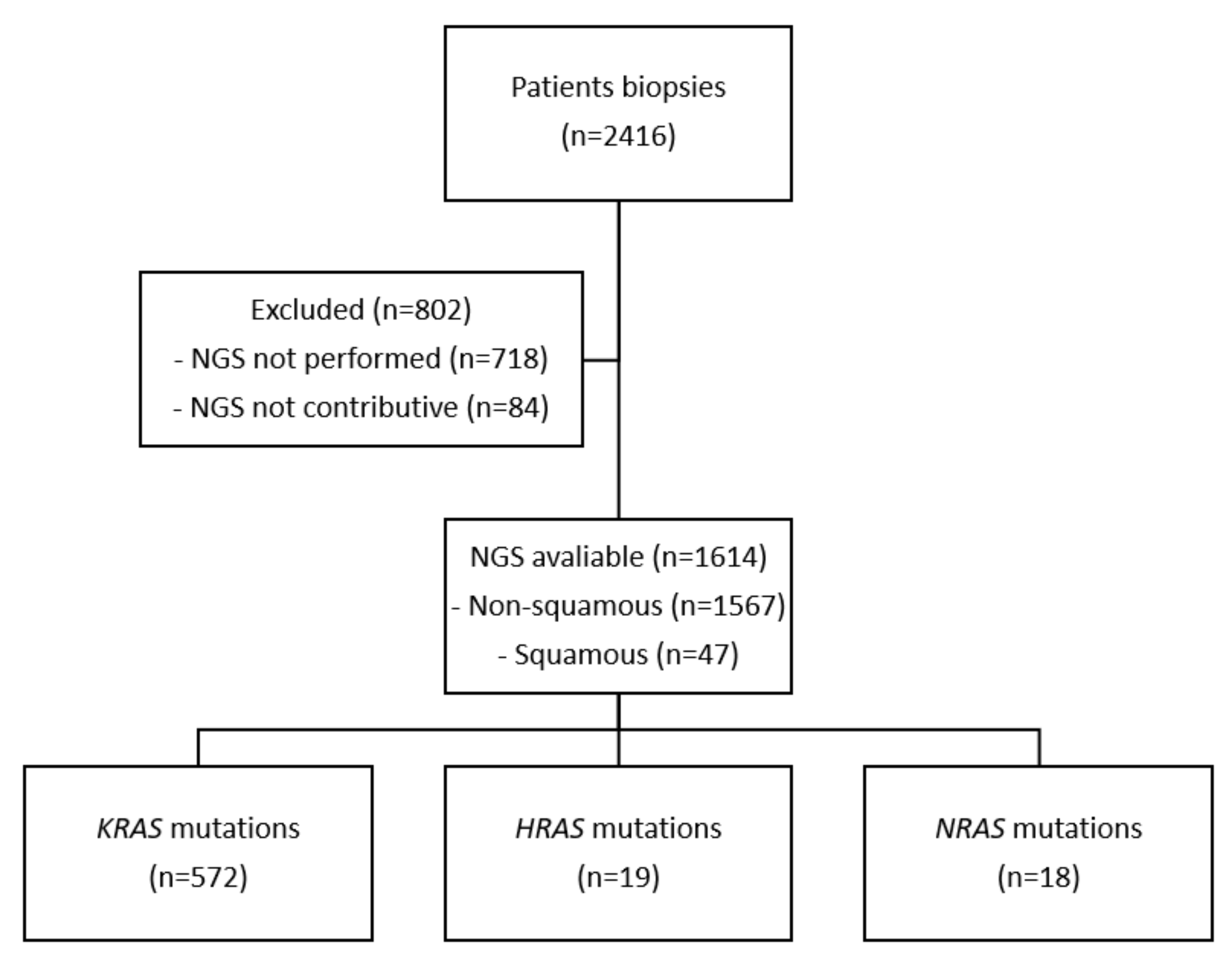
A multicentre retrospective study was performed including all newly diagnosed NSCLC patients between 1 January 2018 and 31 December 2020 with their genomic analyses performed at Nantes University Hospital. Next-generation sequencing (NGS) was systematically performed for all newly diagnosed non-squamous NSCLC patients presenting advanced disease, and never smokers squamous NSCLC patients. Briefly, DNA was extracted from formalin-fixed paraffin-embedded (FFPE) tumours using the Maxwell RSC RNA FFPE kit (Promega, Madison, WI, USA). NGS libraries were synthesized using the QIAseq Targeted DNA Custom Panel (QIAGEN, Hilden, Germany) kit, an amplicon library construction kit based on Anchored Multiplex PCR (AMP) technology. Sequencing was performed on a MiSeq sequencer (Illumina, San Diego, CA, USA), and NGS data analysis was performed using the Biomedical Genomics Workbench (QIAGEN). Panel of tested genes is shown in the Table A1. Patients with HRAS-mutant tumours were identified, and clinical data from the medical records of HRAS p.Gln61Leu mutated patients (the most frequently observed HRAS mutation) were extracted. Other cases included in the literature were reviewed from Medline database, using the keywords “non-small cell lung cancer” and “HRAS”. This study was conducted in accordance with the local regulations, and it was approved by the local independent ethics committee.

3. Results

3.1. Cases Presentation

NGS was performed on 1614 patients during recruitment (Figure 1). Nineteen (1.18%) had HRAS mutation, 572 (35.43%) had a KRAS mutation, and 18 (1.12%) had an NRAS mutation. The mutation most frequently observed was HRAS p.Gln61Leu, in four patients (Table 1). All cases are reported in Table 2.
The first one was an active smoker 50-year-old female, with a past medical history of hypertension and chronic obstructive pulmonary disease. In 2015, she underwent right lobectomy and adjuvant chemotherapy (cisplatin and pemetrexed) for a pT4N0M0 lung adenocarcinoma. In January 2019, a control computed tomography (CT) scan revealed an upper left lobe mass, mediastinal adenopathies, liver metastases, and pericardial effusion. She had no symptoms at this time. Histology of pericardial effusion after drainage showed lung adenocarcinoma cells, Tumour Proportion Score (TPS) PD-L1 < 1%. NGS showed an HRAS p.Gln61Leu mutation (variant allele fraction (VAF) 10.0%) with no other alteration. She started a first-line chemotherapy with carboplatin and pemetrexed; however, she only received three cycles of chemotherapy due to a major cachexia. A CT scan confirmed a lung, liver, and bone progression. She died four weeks later.
The second case was a 55-year-old male, with a past medical history of pulmonary embolism and diabetes mellitus. He was a former smoker of around 60 packs per year. Lung cancer was diagnosed in July 2019. The histology revealed a Not Otherwise Specified (NOS) carcinoma, TTF1 and P40 negative, TPS PD-L1 = 0%. NGS showed KRAS p.Gly12Cys (VAF 19.6%) and HRAS p.Gln61Leu mutations (VAF 27.9%). CT showed a right upper lobe mass with bronchus invasion, bilateral mediastinal and hilar lymph node invasion, with no extra-thoracic extension. Health status was preserved with a performance status (PS) score of 1. He initiated a first-line chemotherapy with four cycles of carboplatin and pemetrexed, followed by maintenance with pemetrexed. He was hospitalized one month after the beginning of maintenance therapy for altered general condition. CT revealed disease progression, and the patient died one month after.
The third case was an active smoker 63-year-old male, 35 packs per year. His only antecedent was hypertension. He was diagnosed in August 2020, in a context of headache, dizziness, and asthenia. CT showed three brain metastases, a 67mm right mediastino-hilar mass, multiple mediastinal adenopathies, and a pericardial effusion. Histology revealed a NOS carcinoma, TPS PD-L1 = 60%. NGS showed KRAS p.Gly12Ser (VAF 27.7%), TP53 c.784_809del (VAF 26.45%) and HRAS p.Gln61Leu mutations (VAF 29.1%). A whole brain radiotherapy of 30 Gy was performed leading to a neurological improvement. He was treated with a combination of chemotherapy plus immunotherapy (carboplatin, pemetrexed and pembrolizumab) on September 2020. He received four cycles followed by monotherapy with pembrolizumab. The first evaluation in December 2020 showed a partial response of 39%. The patient was still treated at the time of data closure in November 2021.
The fourth case was a 61-year-old female, without past medical history. She was a highly active smoker with 68 packs per year. The presence of a dysphonia led to a chest CT in September 2020 and revealed a large compressive mediastino-hilar mass, and moderate abundance of bilateral pleural effusion. A brain CT and a PET-CT showed no extra-thoracic metastases. Histology was an adenocarcinoma, TPS PD-L1 = 60%. NGS showed TP53 p.Ile195Thr (VAF 13.6%) and HRAS p.Gln61Leu mutations (VAF 28.1%). She was hospitalized for obstructive pneumopathy, atrial fibrillation, and cardiogenic failure. She died in November 2020.

3.2. Review of Literature

To our knowledge, only three other individual clinical cases of NSCLC patients with HRAS-mutant tumours have been described in literature. All cases were p.Gln61Leu mutations (Table 2). According to the TCGA database (pro-jects TCGA-LUAD and TCGA-LUSC), HRAS mutations were found in 3/550 (0.55%) adenocarcinomas and 8/480 (1.67%) squamous cell carcinomas (data available online: https://portal.gdc.cancer.gov/ accessed on 14 April 2022). One adenocarcinoma had HRAS p.Gln61Leu mutation. The first case was from the USA, and the two others came from China. The first patient was a former smoker 79-year-old male, with a rapid progression from stage IB disease to metastatic adenocarcinoma and death [17]. His only antecedent was meningioma. The patient underwent right upper lobectomy in September 2010, for a localized pT2aN0M0 adenocarcinoma. He received four cycles of adjuvant carboplatin and pemetrexed. He relapsed 10 months after the surgery, with brain metastases, multiple bone, liver, and bilateral adrenal lesions. He underwent craniotomy for a temporal lobe mass and stereotaxic radiosurgery of additional brain lesions. The molecular biology of the brain lesion revealed a HRAS p.Gln61Leu mutation. Other mutations, such as KRAS, TP53, and 51 other genes were negative. He was unable to receive chemotherapy and died in January 2012.
The second patient was a 58-year-old male without past medical history, who showed rapid metastatic progression and passed away due to respiratory failure after 2 weeks of systemic treatment [18]. He was an active smoker. He was diagnosed with a cavitary nodule of the right lung and malignant pleural effusion. Histology suggested a poorly differentiated carcinoma. Video-assisted thoracoscopic lobectomy and lymph node dissection was performed for palliative symptoms management. Amplification Refractory Mutation System (ARMS)- PCR assay showed a weak signal for p.Leu858Arg and p.Thr90Met of EGFR mutation, thus he was treated with cisplatin and osimertinib. He died 15 days after. A reassessment of the primary tumour and lymph nodes showed HRAS p.Gln61Leu and NRAS p.Gln61Lys mutations, but EGFR mutations were not detected.
A third case, although from China, was reported. It was a 76-year-old man with an antecedent of Lynch syndrome and 50-year smoking history [19]. Chest CT showed tumour masses in the upper lobe and hilar of right lung, with mediastinal lymphadenopathies, presenting no other distant metastases on PET-CT. Histology identified a squamous cell carcinoma, TPS PD-L1 = 50%. NGS showed HRAS p.Gln61Leu and TP53 p.Arg158Leu. Other genes such as LRP1B, DNMT3A, POLE, TCF7L2, or NTM were also mutated. A heterozygous germline mutation of MSH6 was detected, but immunohistochemistry suggested normal expression of MLH1, MSH2, MSH6, and PMS2 in tumour biopsy. He was treated with pembrolizumab and achieved stable disease as the best response. Disease progression finally occurred after eight cycles. At this moment, plasma circulating tumour DNA test revealed a STK11 mutation, which was not present at diagnosis. Subsequently, the patient received chemotherapy with albumin-bound paclitaxel.

4. Discussion

Among non-squamous NSCLC, while KRAS mutations account for around 30% of patients, HRAS and NRAS mutations account for less than 1% [13,20,21]. Concordant with previous studies, we found a low prevalence of 0.82% HRAS mutations among NSCLC patients (19/1614 patients). In our centre, according to national guidelines, NGS was not routinely performed in squamous cell carcinoma, excepted for exceptional never-smokers. Thus, we might have missed other HRAS-mutant tumours, since HRAS mutations were also rarely described in squamous cell carcinoma [13,22]. A dedicated study in this population should be performed.
The clinical course of a patient with HRAS-mutant NSCLC remains unclear. Indeed, we have found only three cases describing HRAS-mutant lung cancer patients, all of them being HRAS p.Q61L. Our four cases plus the three others described in literature found that all patients were active or former smokers. Pleural and/or pericardial effusion was described in four out of seven cases. Clinical presentation was mostly aggressive, with one newly diagnosed patient who died before any treatment and four others who died during first-line treatment. Two patients received immunotherapy: one was still under treatment after more than one year, and the other received eight cycles with a stable disease as the best response.
HRAS mutations have been mainly documented in HNSCC, salivary glands, pheochromocytoma and bladder cancers [10,13,14,20,21,23,24,25,26,27,28,29] (Figure 2). HRAS mutations were known to be oncogenic; it was demonstrated in vitro that mutant HRAS hyperactivated MAPK and mTOR pathways in various cancer cell lines including lung, bladder, and oesophageal cancer [30,31]. Thus, it might appear as a potential interesting target. Among therapeutic strategies targeting HRAS-mutant cancers, tipifarnib, a farnesyl transferase (FT) inhibitor, is one of the most promising candidates [16,32]. Considerable work on FT inhibitors have been performed since 20 years, but these treatments failed to demonstrate any benefit in unselected populations, ending the development of these treatments as pan-Ras-targeted strategy [33]. Farnesylation is a mechanism needed for RAS molecules to be integrated in plasma membranes and for its activation. Contrary to tyrosine kinase inhibitors (TKI) such as EGFR TKIs that directly inhibit enzyme, tipifarnib indirectly disrupts RAS activity through preventing appropriate intracellular localization by interfering with RAS prenylation [31]. Although all RAS isoforms are FT substrates, only H-Ras exclusively depends on farnesylation for its membrane localization. Indeed, prenylation of H-Ras mostly depends on farnesylation, whereas K-Ras (4A and 4B) and N-Ras can be integrated in plasma membranes via other mechanisms, such as geranylgeranylation [16,34]. Accordingly, blockade of FT is sufficient to inhibit the action of H-Ras. Due to the development of RAS mutations profiling, tipifarnib appeared as an interesting strategy to inhibit HRAS-mutant tumours [33]. Its indirect mechanism of action is very different from covalent KRAS inhibitors (such as sotorasib and adagrasib), which act by selectively forming a covalent bond with cysteine 12 within the switch-II pocket of KRAS-G12C protein, thereby locking KRAS in the inactive state to arrest cell proliferation [35].
Importantly, 2/4 patients with HRAS mutation in our cohort also had a co-occurring KRAS mutation on codon 12. As KRAS mutations are described as strong oncogenic mutations, it remains unclear whether HRAS is a driver mutation or a passenger one and if those patients would benefit from HRAS inhibition.
Tipifarnib activity was characterized in a wide panel of HRAS-mutant and wild-type HNSCC xenograft models, and also in HNSCC patient-derived xenografts [31]. Considering the rarity of this mutation in lung cancer, extensive pre-clinical approach seems difficult to assess. However, using CRISPR-CAS 9 technology, HRAS mutation might be introduced in an appropriate lung cancer cell line in order to mimic the clinical situation. It is important to highlight that most of the activating HRAS mutations involved codon 12, 13, 61, 117, and 146. To our knowledge, other mutations have not been characterized in NSCLC. Nonetheless, tipifarnib seemed efficient regardless of HRAS hotspot mutation, as it inhibited activity on patient derived xenograft HNSCC models harbouring Q61L, G12C, G12S, G13R, and K117N mutations [31].
We resumed published clinical trials evaluating tipifarnib in solid tumours in Table 3 [36,37,38,39,40,41,42,43,44]. Only two were specifically conducted in lung cancer, with no selection based on HRAS status. The first clinical trial was carried out in 2003 [43]. This phase II study tested the tipifarnib in first-line setting of 44 patients with a locally-advanced NSCLC, not amenable to chemo-radiotherapy. No objective responses were documented, whereas inhibition of farnesylation in vivo was consistently documented. Median time to progression was 2.7 months, and seven patients (16%) had disease stabilization for greater than 6 months. Tipifarnib was also tested in 22 patients with sensitive relapsed small-cell lung cancer in a phase II trial [42]. No significant antitumour activity was observed. Median progression-free survival was only 1.4 months.
In 2021, a phase II study enrolling 30 patients with recurrent and/or metastatic HNSCC harbouring HRAS mutation shown highly promising results. Objective response rate was 55% (11/20 patients; 95% confidence interval (CI), 31.5 to 76.9), with a median progression-free-survival of 5.6 months (95% CI, 3.6 to 16.4), and a median overall survival of 15.4 months (95% CI, 7.0 to 29.7) [36]. The most frequent adverse events were anaemia (37%) and lymphopenia (13%). The mechanistic basis of tipifarnib toxicity might be related to the inhibition of several dozen other farnesylated proteins in cells [45]. Moreover, tipifarnib was recently described as an inhibitor of the CXCL12/CXCR4 pathway, that might lead to other side effects [46]. A phase II study evaluating tipifarnib in HRAS-mutant squamous NSCLC (NCT03496766) and another phase II study of tipifarnib in HNSCC with HRAS mutations are ongoing (NCT02383927).

5. Conclusions

HRAS mutations are uncommon genomic alterations in NSCLC patients, representing less than 1% of patients. Further studies are needed to better understand clinical course and prognosis associated with these mutations. However, preliminary data seems to show an association with tabagic status, aggressive presentation, and co-occurring mutations in MAPK pathway and TP53. Interestingly, a targeted therapy against HRAS-mutant showed promising results in HNSCC patients. Whether HRAS could represent a druggable oncogene in NSCLC is still unknown and should be explored in further studies.

Author Contributions

Conceptualization, L.M. and E.P.-T.; investigation, S.R., C.F. and J.B.; resources, G.H. and M.D.; writing—original draft preparation, L.M. and E.P.-T.; writing—review and editing, E.P.-T., G.H., M.D., C.B. and L.M.; supervision, E.P.-T. All authors have read and agreed to the published version of the manuscript.

Funding

This research received no external funding.

Institutional Review Board Statement

The study was conducted in accordance with the Declaration of Helsinki, and approved by the Institutional Review Board of Nantes University Hospital (22 June 2022).

Informed Consent Statement

Informed consent was obtained from all subjects involved in the study.

Data Availability Statement

The data presented in this study are available on request from the corresponding author.

Conflicts of Interest

The authors declare they have no conflict of interest.

Appendix A

Table A1. Next-Generation Sequencing (NGS).
Sequenced Regions
AKT1 exon 3 (NM_001014431.1)
ALK exons 22 to 25 (NM_004304.1)
BRAF exons 11 and 15 (NM_004333.4)
CTNNB1 exon 3 (NM_001904.4)
DDR2 exons 5 to 10 and 14 to 19 (NM_001014796.2)
EGFR exons 18 to 21 (NM_005228.3)
ERBB2 (HER2) exons 19 to 22 (NM_004448.2)
ERBB4 codons 393 and 452 (NM_005235.2)
FGFR2 codons 252, 549 and 659 (NM_000141.4)
FGFR3 exons 6, 8 and 13 (NM_000142.4)
HRAS exons 2, 3 and 4 (NM_005343.2)
IDH1 codons 100 and 132 (NM_005896.3)
IDH2 codon 172 (NM_002168.3)
KIT exons 8, 9, 11, 13, 14, 17 and 18 (NM_000222.2)
KRAS exons 2, 3 and 4 (NM_033360.2)
MAP2K1 (MEK1) exon 2 (NM_002755.3)
MET exon 2, intron 13 and exons 14 to 20 (NM_001127500.1)
NRAS exons 2, 3 and 4 (NM_002524.3)
PDGFRA exons 12, 14 and 18 (NM_006206.4)
PIK3CA exons 10 and 21 (NM_006218.2)
RET exons 11 and 16 (NM_020975.6)
TP53 exons 2 to 11 (NM_000546.4)
Microsatellites: BAT25, BAT26, NR21, NR24, MONO27

References

  1. Sung, H.; Ferlay, J.; Siegel, R.L.; Laversanne, M.; Soerjomataram, I.; Jemal, A.; Bray, F. Global Cancer Statistics 2020: GLOBOCAN Estimates of Incidence and Mortality Worldwide for 36 Cancers in 185 Countries. CA Cancer J. Clin. 2021, 71, 209–249. [Google Scholar] [CrossRef] [PubMed]
  2. Lynch, T.J.; Bell, D.W.; Sordella, R.; Gurubhagavatula, S.; Okimoto, R.A.; Brannigan, B.W.; Harris, P.L.; Haserlat, S.M.; Supko, J.G.; Haluska, F.G.; et al. Activating Mutations in the Epidermal Growth Factor Receptor Underlying Responsiveness of Non-Small-Cell Lung Cancer to Gefitinib. N. Engl. J. Med. 2004, 350, 2129–2139. [Google Scholar] [CrossRef] [PubMed]
  3. Ramalingam, S.S.; Vansteenkiste, J.; Planchard, D.; Cho, B.C.; Gray, J.E.; Ohe, Y.; Zhou, C.; Reungwetwattana, T.; Cheng, Y.; Chewaskulyong, B.; et al. Overall Survival with Osimertinib in Untreated, EGFR-Mutated Advanced NSCLC. N. Engl. J. Med. 2020, 382, 41–50. [Google Scholar] [CrossRef] [PubMed]
  4. Peters, S.; Camidge, D.R.; Shaw, A.T.; Gadgeel, S.; Ahn, J.S.; Kim, D.W.; Ou, S.H.I.; Pérol, M.; Dziadziuszko, R.; Rosell, R.; et al. Alectinib versus Crizotinib in Untreated ALK-Positive Non-Small-Cell Lung Cancer. N. Engl. J. Med. 2017, 377, 829–838. [Google Scholar] [CrossRef]
  5. Camidge, D.R.; Kim, H.R.; Ahn, M.-J.; Yang, J.C.-H.; Han, J.-Y.; Lee, J.-S.; Hochmair, M.J.; Li, J.Y.-C.; Chang, G.-C.; Lee, K.H.; et al. Brigatinib versus Crizotinib in ALK-Positive Non-Small-Cell Lung Cancer. N. Engl. J. Med. 2018, 379, 2027–2039. [Google Scholar] [CrossRef]
  6. Shaw, A.T.; Bauer, T.M.; de Marinis, F.; Felip, E.; Goto, Y.; Liu, G.; Mazieres, J.; Kim, D.-W.; Mok, T.; Polli, A.; et al. First-Line Lorlatinib or Crizotinib in Advanced ALK-Positive Lung Cancer. N. Engl. J. Med. 2020, 383, 2018–2029. [Google Scholar] [CrossRef]
  7. Shaw, A.T.; Ou, S.-H.I.; Bang, Y.-J.; Camidge, D.R.; Solomon, B.J.; Salgia, R.; Riely, G.J.; Varella-Garcia, M.; Shapiro, G.I.; Costa, D.B.; et al. Crizotinib in ROS1-Rearranged Non-Small-Cell Lung Cancer. N. Engl. J. Med. 2014, 371, 1963–1971. [Google Scholar] [CrossRef] [Green Version]
  8. Hong, D.S.; Fakih, M.G.; Strickler, J.H.; Desai, J.; Durm, G.A.; Shapiro, G.I.; Falchook, G.S.; Price, T.J.; Sacher, A.; Denlinger, C.S.; et al. KRASG12C Inhibition with Sotorasib in Advanced Solid Tumors. N. Engl. J. Med. 2020, 383, 1207–1217. [Google Scholar] [CrossRef]
  9. Ryan, M.B.; Corcoran, R.B. Therapeutic Strategies to Target RAS-Mutant Cancers. Nat. Rev. Clin. Oncol. 2018, 15, 709–720. [Google Scholar] [CrossRef]
  10. Pylayeva-Gupta, Y.; Grabocka, E.; Bar-Sagi, D. RAS Oncogenes: Weaving a Tumorigenic Web. Nat. Rev. Cancer 2011, 11, 761–774. [Google Scholar] [CrossRef] [Green Version]
  11. Cox, A.D.; Der, C.J. Ras History: The Saga Continues. Small GTPases 2010, 1, 2–27. [Google Scholar] [CrossRef] [Green Version]
  12. Karnoub, A.E.; Weinberg, R.A. Ras Oncogenes: Split Personalities. Nat. Rev. Mol. Cell Biol. 2008, 9, 517–531. [Google Scholar] [CrossRef] [PubMed] [Green Version]
  13. Prior, I.A.; Hood, F.E.; Hartley, J.L. The Frequency of Ras Mutations in Cancer. Cancer Res. 2020, 80, 2969–2974. [Google Scholar] [CrossRef] [PubMed] [Green Version]
  14. Zehir, A.; Benayed, R.; Shah, R.H.; Syed, A.; Middha, S.; Kim, H.R.; Srinivasan, P.; Gao, J.; Chakravarty, D.; Devlin, S.M.; et al. Mutational Landscape of Metastatic Cancer Revealed from Prospective Clinical Sequencing of 10,000 Patients. Nat. Med. 2017, 23, 703–713. [Google Scholar] [CrossRef] [PubMed]
  15. Matsumoto, S.; Taniguchi-Tamura, H.; Araki, M.; Kawamura, T.; Miyamoto, R.; Tsuda, C.; Shima, F.; Kumasaka, T.; Okuno, Y.; Kataoka, T. Oncogenic Mutations Q61L and Q61H Confer Active Form-Like Structural Features to the Inactive State (State 1) Conformation of H-Ras Protein. Biochem. Biophys. Res. Commun. 2021, 565, 85–90. [Google Scholar] [CrossRef] [PubMed]
  16. Roskoski, R. Blockade of Mutant RAS Oncogenic Signaling with a Special Emphasis on KRAS. Pharmacol. Res. 2021, 172, 105806. [Google Scholar] [CrossRef] [PubMed]
  17. Cathcart-Rake, E.; Corless, C.; Sauer, D.; Lopez-Chavez, A. Elderly Former Smoker with HRAS Mutant Non-Small-Cell Lung Cancer. J. Thorac. Oncol. 2014, 9, e75–e78. [Google Scholar] [CrossRef] [Green Version]
  18. Zhao, J.; Zhang, X.; He, M.; Chen, X.; Cui, X.; Qin, T.; Niu, X.; Zhao, L. Poor Prognosis of Pulmonary Adenosquamous Carcinoma with NRAS and HRAS Double Mutation. Onco Targets Ther. 2021, 14, 1113–1116. [Google Scholar] [CrossRef]
  19. Long, Y.; Tang, Y.; Cai, C.; Yu, M.; Zhang, M.; Chen, R.; Huang, M. The Influence of STK11 Mutation on Acquired Resistance to Immunotherapy in Advanced Non-Small Cell Lung Cancer with Lynch Syndrome: A Case Report and Literature Review. Ann. Palliat. Med. 2021, 10, 7088–7094. [Google Scholar] [CrossRef]
  20. The Cancer Genome Atlas Research Network. Comprehensive Molecular Profiling of Lung Adenocarcinoma. Nature 2014, 511, 543–550. [Google Scholar] [CrossRef]
  21. Mendiratta, G.; Ke, E.; Aziz, M.; Liarakos, D.; Tong, M.; Stites, E.C. Cancer Gene Mutation Frequencies for the U.S. Population. Nat. Commun. 2021, 12, 5961. [Google Scholar] [CrossRef] [PubMed]
  22. Hammerman, P.S.; Lawrence, M.S.; Voet, D.; Jing, R.; Cibulskis, K.; Sivachenko, A.; Stojanov, P.; McKenna, A.; Lander, E.S.; Gabriel, S.; et al. Comprehensive Genomic Characterization of Squamous Cell Lung Cancers. Nature 2012, 489, 519–525. [Google Scholar] [CrossRef]
  23. Simanshu, D.K.; Nissley, D.V.; McCormick, F. RAS Proteins and Their Regulators in Human Disease. Cell 2017, 170, 17–33. [Google Scholar] [CrossRef] [PubMed] [Green Version]
  24. Lawrence, M.S.; Sougnez, C.; Lichtenstein, L.; Cibulskis, K.; Lander, E.; Gabriel, S.B.; Getz, G.; Ally, A.; Bal-asundaram, M.; Birol, I.; et al. Comprehensive Genomic Characterization of Head and Neck Squamous Cell Carcinomas. Nature 2015, 517, 576–582. [Google Scholar] [CrossRef] [Green Version]
  25. Weinstein, J.N.; Akbani, R.; Broom, B.M.; Wang, W.; Verhaak, R.G.W.; McConkey, D.; Lerner, S.; Morgan, M.; Creighton, C.J.; Smith, C.; et al. Comprehensive Molecular Characterization of Urothelial Bladder Carcinoma. Nature 2014, 507, 315–322. [Google Scholar] [CrossRef] [Green Version]
  26. Khan, A.Q.; Kuttikrishnan, S.; Siveen, K.S.; Prabhu, K.S.; Shanmugakonar, M.; Naemi, H.A.A.; Haris, M.; Dermime, S.; Uddin, S. RAS-Mediated Oncogenic Signaling Pathways in Human Malignancies. Semin. Cancer Biol. 2019, 54, 1–13. [Google Scholar] [CrossRef]
  27. Robertson, A.G.; Kim, J.; Al-Ahmadie, H.; Bellmunt, J.; Guo, G.; Cherniack, A.D.; Hinoue, T.; Laird, P.W.; Hoadley, K.A.; Akbani, R.; et al. Comprehensive Molecular Characterization of Muscle-Invasive Bladder Cancer. Cell 2017, 171, 540–556. [Google Scholar] [CrossRef] [Green Version]
  28. Stenman, A.; Welander, J.; Gustavsson, I.; Brunaud, L.; Bäckdahl, M.; Söderkvist, P.; Gimm, O.; Juhlin, C.C.; Larsson, C. HRAS Mutation Prevalence and Associated Expression Patterns in Pheochromocytoma. Genes Chromosomes Cancer 2016, 55, 452–459. [Google Scholar] [CrossRef] [Green Version]
  29. Andersson, P.; Kolaric, A.; Windahl, T.; Kirrander, P.; Söderkvist, P.; Karlsson, M.G. PIK3CA, HRAS and KRAS Gene Mutations in Human Penile Cancer. J. Urol. 2008, 179, 2030–2034. [Google Scholar] [CrossRef]
  30. Kiessling, M.K.; Curioni-Fontecedro, A.; Samaras, P.; Atrott, K.; Cosin-Roger, J.; Lang, S.; Scharl, M.; Rogler, G. Mutant HRAS as Novel Target for MEK and MTOR Inhibitors. Oncotarget 2015, 6, 42183–42196. [Google Scholar] [CrossRef] [Green Version]
  31. Gilardi, M.; Wang, Z.; Proietto, M.; Chillà, A.; Calleja-Valera, J.L.; Goto, Y.; Vanoni, M.; Janes, M.R.; Mikulski, Z.; Gualberto, A.; et al. Tipifarnib as a Precision Therapy for HRAS-Mutant Head and Neck Squamous Cell Carcinomas. Mol. Cancer Ther. 2020, 19, 1784–1796. [Google Scholar] [CrossRef] [PubMed]
  32. End, D.W.; Smets, G.; Todd, A.V.; Applegate, T.L.; Fuery, C.J.; Angibaud, P.; Venet, M.; Sanz, G.; Poignet, H.; Skrzat, S.; et al. Characterization of the Antitumor Effects of the Selective Farnesyl Protein Transferase Inhibitor R115777 In Vivo and In Vitro. Cancer Res. 2001, 61, 131–137. [Google Scholar] [PubMed]
  33. Moore, A.R.; Rosenberg, S.C.; McCormick, F.; Malek, S. RAS-Targeted Therapies: Is the Undruggable Drugged? Nat. Rev. Drug Discov. 2020, 19, 533–552. [Google Scholar] [CrossRef] [PubMed]
  34. Whyte, D.B.; Kirschmeier, P.; Hockenberry, T.N.; Nunez-Oliva, I.; James, L.; Catino, J.J.; Bishop, W.R.; Pai, J.K. K- and N-Ras Are Geranylgeranylated in Cells Treated with Farnesyl Protein Transferase Inhibitors. J. Biol. Chem. 1997, 272, 14459–14464. [Google Scholar] [CrossRef] [PubMed] [Green Version]
  35. Liu, J.; Kang, R.; Tang, D. The KRAS-G12C Inhibitor: Activity and Resistance. Cancer Gene Ther. 2021, 1–4. [Google Scholar] [CrossRef] [PubMed]
  36. Ho, A.L.; Brana, I.; Haddad, R.; Bauman, J.; Bible, K.; Oosting, S.; Wong, D.J.; Ahn, M.-J.; Boni, V.; Even, C.; et al. Tipifarnib in Head and Neck Squamous Cell Carcinoma With HRAS Mutations. J. Clin. Oncol. 2021, 39, 1856–1864. [Google Scholar] [CrossRef]
  37. Hanna, G.J.; Guenette, J.P.; Chau, N.G.; Sayehli, C.M.; Wilhelm, C.; Metcalf, R.; Wong, D.J.; Brose, M.; Razaq, M.; Pérez-Ruiz, E.; et al. Tipifarnib in Recurrent, Metastatic HRAS-Mutant Salivary Gland Cancer. Cancer 2020, 126, 3972–3981. [Google Scholar] [CrossRef]
  38. Lee, H.W.; Sa, J.K.; Gualberto, A.; Scholz, C.; Sung, H.H.; Jeong, B.C.; Choi, H.Y.; Kwon, G.Y.; Park, S.H. A Phase II Trial of Tipifarnib for Patients with Previously Treated, Metastatic Urothelial Carcinoma Harboring HRAS Mutations. Clin. Cancer Res. 2020, 26, 5113–5119. [Google Scholar] [CrossRef]
  39. Jazieh, K.; Molina, J.; Allred, J.; Yin, J.; Reid, J.; Goetz, M.; Lim, V.-S.; Kaufmann, S.H.; Adjei, A. A Phase I Study of the Farnesyltransferase Inhibitor Tipifarnib in Combination with the Epidermal Growth Factor Tyrosine Kinase Inhibitor Erlotinib in Patients with Advanced Solid Tumors. Investig. New Drugs 2019, 37, 307–314. [Google Scholar] [CrossRef]
  40. Whitehead, R.P.; McCoy, S.; Macdonald, J.S.; Rivkin, S.E.; Neubauer, M.A.; Dakhil, S.R.; Lenz, H.-J.; Tanaka, M.S.; Abbruzzese, J.L.; Southwest Oncology Group. Phase II Trial of R115777 (NSC #70818) in Patients with Advanced Colorectal Cancer: A Southwest Oncology Group Study. Investig. New Drugs 2006, 24, 335–341. [Google Scholar] [CrossRef]
  41. Lara, P.N.J.; Law, L.Y.; Wright, J.J.; Frankel, P.; Twardowski, P.; Lenz, H.J.; Lau, D.H.M.; Kawaguchi, T.; Gumerlock, P.H.; Doroshow, J.H.; et al. Intermittent Dosing of the Farnesyl Transferase Inhibitor Tipifarnib (R115777) in Advanced Malignant Solid Tumors: A Phase I California Cancer Consortium Trial. Anti-Cancer Drugs 2005, 16, 317–321. [Google Scholar] [CrossRef] [PubMed]
  42. Heymach, J.V.; Johnson, D.H.; Khuri, F.R.; Safran, H.; Schlabach, L.L.; Yunus, F.; DeVore, R.F.; De Porre, P.M.; Richards, H.M.; Jia, X.; et al. Phase II Study of the Farnesyl Transferase Inhibitor R115777 in Patients with Sensitive Relapse Small-Cell Lung Cancer. Ann. Oncol. 2004, 15, 1187–1193. [Google Scholar] [CrossRef] [PubMed]
  43. Adjei, A.A.; Mauer, A.; Bruzek, L.; Marks, R.S.; Hillman, S.; Geyer, S.; Hanson, L.J.; Wright, J.J.; Erlichman, C.; Kaufmann, S.H.; et al. Phase II Study of the Farnesyl Transferase Inhibitor R115777 in Patients with Advanced Non-Small-Cell Lung Cancer. J. Clin. Oncol. 2003, 21, 1760–1766. [Google Scholar] [CrossRef] [PubMed]
  44. Hahn, S.M.; Bernhard, E.J.; Regine, W.; Mohiuddin, M.; Haller, D.G.; Stevenson, J.P.; Smith, D.; Pramanik, B.; Tepper, J.; DeLaney, T.F.; et al. A Phase I Trial of the Farnesyltransferase Inhibitor L-778,123 and Radiotherapy for Locally Advanced Lung and Head and Neck Cancer. Clin. Cancer Res. 2002, 8, 1065–1072. [Google Scholar] [PubMed]
  45. Storck, E.M.; Morales-Sanfrutos, J.; Serwa, R.A.; Panyain, N.; Lanyon-Hogg, T.; Tolmachova, T.; Ventimiglia, L.N.; Martin-Serrano, J.; Seabra, M.C.; Wojciak-Stothard, B.; et al. Dual Chemical Probes Enable Quantitative System-Wide Analysis of Protein Prenylation and Prenylation Dynamics. Nat. Chem. 2019, 11, 552–561. [Google Scholar] [CrossRef]
  46. Gualberto, A.; Scholz, C.; Mishra, V.; Janes, M.R.; Kessler, L.; Cutsem, E.V.; Ho, A.L.; Witzig, T. Abstract CT191: Mechanism of Action of the Farnesyltransferase Inhibitor, Tipifarnib and Its Clinical Applications. Cancer Res. 2019, 79, CT191. [Google Scholar] [CrossRef]
Figure 1. Flowchart. NGS: Next-Generation Sequencing.
Curroncol 29 00300 g001
Figure 2. Frequencies of HRAS mutations among solid cancer types.
Curroncol 29 00300 g002
Table 1. HRAS mutations type among all 19 patients.
HRAS MutationsNumbers of Patients
p.Q61L (p.Gln61Leu; C.182A>T)4
p.G13V (p.Gly13Val; c.38G>T)2
p.E98K (p.Glu98Lys; c.292G>A)1
p.S89F (p.Ser89Phe; c.266C>T)1
p.A11P (p.Ala11Pro; c.31G>C)1
p.K117N (p.Lys117Asn; c.351G>T)1
p.R102L (p.Arg102Leu; c.305G>T)1
p.D107fs (p.Asp107fs; c.319del)1
p.V109L (p.Val109Leu; c.325G>T)1
p.T58I (p.Thr58Ile; c.173C>T)1
p.T148P (p.Thr148Pro; c.442A>C)1
p.R41W (p.Arg41Trp; c.121C>T)1
p.R135Q (p.Arg135Gln; c.404G>A)1
p.M72I (p.Met72Ile; c.216G>T)1
p.E76D (p.Glu76Asp; c.228G>T)1
Table 2. Clinico-pathologic features of non-small cell lung cancer patients with HRAS p.Gln61Leu mutation reported in literature. ADC: adenocarcinoma; SCC: squamous cell carcinoma; ADSQ: adenosquamous; NOS: not otherwise specified; WBRT: whole brain radiotherapy; PFS: Progression-Free-Survival; OS: Overall Survival; NA: Not Available.
ReferenceSex (Female/Male)Age at Diagnosis (Years)Smoking StatusPathologyPD-L1 (%)Other AlterationsMetastatic SiteTreatmentPFS
(Weeks)
OS
(Weeks)
CurrentF50ActiveADC<1NoneLung/Liver/Pericardial effusion
(4 years after adjuvant chemotherapy)
Carboplatin—Pemetrexed1115
CurrentM55FormerNOS<1KRAS p.Gly12CysLocally-advanced diseaseCarboplatin—Pemetrexed2230
CurrentM63ActiveNOS60KRAS p.Gly12Ser
TP53 c.784_809del
Brain/Pericardial effusionCarboplatin—Pemetrexed—Pembrolizumab
WBRT
On treatmentOn treatment
CurrentF61ActiveADC60TP53 p.Ile195ThrPleural effusionNot treatedNA3
Cathcart-Rake E., 2014 [17]M79FormerADCNANoneBrain/Bone/Liver/Adrenal
(10 months after adjuvant chemotherapy)
Adjuvant
Carboplatin—Pemetrexed
Brain surgery—Stereotactic radiosurgery
NA64
Zhao J., 2021 [18]M58ActiveADSQNAEGFR p.Leu858Arg and p.Thr790Met (only on pleural effusion), NRAS p.Gln61LysPleural effusionCisplatin—Osimertinib24
Long Y., 2021 [19]M76ActiveSCC50TP53 p.Arg158Leu
LRP1B p.Val3711Phe
LRP1B p.Leu4013Met
DNMT3A p.Wrp601 *
DDR2 p.Val336Leu
NTM p.Thr240Ile
TCF7L2 p.Arg420Pro
POLE p.Arg579Leu
Locally-advanced diseasePembrolizumab24NA
* is used for stop codon in HGSV nomenclature.
Table 3. Clinical trials evaluating Tipifarnib in solid tumours. HNSCC: Head and Neck Squamous Cell Carcinoma; NSCLC: Non-Small Cell Lung Cancer; SCLC: Small Cell Lung Cancer; NGS: Next-Generation Sequencing; PO: Per Os; ORR: Overall Response Rate; SD: Stable Disease; PFS: Progression Free Survival; OS: Overall Survival; VAF: Variant Allele Frequency; NA: Not Applicable.
ReferencePhaseTumour SiteNumber of PatientsSettingBiomarkerTipifarnib Dose & SchedulePrimary EndpointORR (%)
(SD)
Median PFS (Months)Median OS (Months)
Ho A.L., 2021 [36]2HNSCC22RelapsedMissense HRAS mutation/VAF > 20%
either in blood, primary tumour tissue, recurrent or metastatic disease
800 or 900 mg PO twice daily on days 1–7 and 15–21 of 28-day cyclesORR50
(41)
5.615.4
Haddad R., 20212
(Ongoing)
HNSCCNARelapsedR/M mHRAS VAF ≥ 20% (tumour tissue) detected by NGS600 mg PO with a meal twice a day for 7 days in alternating weeks (Days 1–7 and 15–21) of 28-day cyclesORR in High VAF population55
(NA)
NA15.4
Hanna G.J., 2020 [35]2Salivary gland carcinoma13RelapsedMissense HRAS mutation with a VAF > 20%: 54% p.Gln61Arg (tumour tissue)900 mg PO twice daily on days 1 to 7 and days 15 to 21 of a 28-dayORR8
(54)
7.018.0
Lee H.W., 2020 [36]2Urothelial carcinoma21RelapsedMissense, nonsynonymous HRAS mutations (p.Gly13Arg, n = 7; p.Gln61Arg, n = 4; p.Gly12Ser, n = 3; p.Gly12Cys, n = 2)
(tumour tissue)
900 mg PO twice daily on days 1–7 and 15–21 of 28-day6-month PFS24
(62)
4.76.1
Jazieh K., 2019 [37]1Advanced, recurrent or metastatic solid tumours27RelapsedNo selection on HRAS status Tumour tissue (diagnostic)4 dose levels, ranging from tipifarnib 200 mg PO twice daily plus erlotinib 75 mg PO once daily to tipifarnib 300 mg PO twice daily plus erlotinib 150 mg PO once dailySafety, tolerability, maximum tolerated dose7.4 (37)NANA
Whitehead R.P., 2006 [38]2Metastatic colorectal adenocarcinoma62No prior chemo: 33/55
Prior chemo: 22/55
No selection on HRAS status Tumour tissue (diagnostic)Fixed dose of 300 mg PO, twice daily, immediately after a meal, days 1–21, every 28 days, until tumour progression or toxicityConfirmed response probability2 (20)1.78.1
Lara Jr P.N., 2005 [39]1Advanced, recurrent or metastatic malignant tumours: (8) NSCLC, (6) colorectal, (3) prostate, (1) oesophagial, (1) pancreatic, (1) parotid, (1) renal21RelapsedNo selection on HRAS status
Tumour tissue (diagnostic)
Starting dose was 300 mg PO twice daily with escalation by 300 mg. Increments over six dose levels to a maximum of 1800 mg PO twice daily, on days 1–7 and 15–21 of 28-day treatment cyclesNot mentioned0
(32)
NANA
Heymach J.V., 2004 [42]2SCLC22RelapsedMissense HRAS mutation 3-week cycles at a dose of 400 mg PO twice daily for 14 consecutive days followed by 7 days off treatmentORR0
(5)
1.46.8
Adjei A., 2003 [43]2NSCLC44100% No prior chemotherapy (eligibility criteria)
9/44 had radiotherapy
No selection on HRAS status300 mg PO twice daily for 21 of every 28 daysORR0
(16)
2.77.7
Hahn S., 2002 [44]1NSCLC9No prior therapy: 7/9
Prior chemotherapy: 2/9
No selection on HRAS status
Tumour tissue (diagnostic)
Dose-escalation study of tipifarnib
Dose level, 280 mg/m2 daily PO during weeks 1, 2, 4, and 5 of radiotherapy
Dose level, 560 mg/m2 daily during weeks 1, 2, 4, 5, and 7 of radiotherapy
Maximum tolerated dose, dose limiting toxicityNANANA
Publisher’s Note: MDPI stays neutral with regard to jurisdictional claims in published maps and institutional affiliations.

