Article

Mutational Landscape of Patients Referred for Elevated Hemoglobin Level

1
Molecular Diagnostic Division, London Health Sciences Centre, London, ON N6A 5W9, Canada
2
Verspeeten Clinical Genome Centre, London Health Sciences Centre, London, ON N6A 5W9, Canada
3
Division of Hematology, Department of Medicine, Schulich School of Medicine and Dentistry, Western University, London, ON N6A 5W9, Canada
4
Division of Hematology, Department of Medicine, London Health Sciences Centre, London, ON N6A 5W9, Canada
5
Department of Pathology & Laboratory Medicine, Schulich School of Medicine and Dentistry, Western University, London, ON N6A 5W9, Canada
*
Authors to whom correspondence should be addressed.
Curr. Oncol. 2022, 29(10), 7209-7217; https://doi.org/10.3390/curroncol29100568
Submission received: 5 August 2022 / Revised: 13 September 2022 / Accepted: 26 September 2022 / Published: 30 September 2022
(This article belongs to the Section Hematology)

Abstract

:
Background: Since the identification of JAK2 V617F and exon 12 mutations as driver mutations in polycythemia vera (PV) in 2005, molecular testing of these mutations for patients with erythrocytosis has become a routine clinical practice. However, the incidence of myeloid mutations other than the common JAK2 V617F mutation in unselected patients referred for elevated hemoglobin is not well studied. This study aimed to characterize the mutational landscape in a real-world population of patients referred for erythrocytosis using a targeted next-generation sequencing (NGS)-based assay. Method: A total of 529 patients (hemoglobin levels >160 g/L in females or >165 g/L in males) were assessed between January 2018 and May 2021 for genetic variants using the Oncomine Myeloid Research Assay (ThermoFisher Scientific, Waltham, MA, USA) targeting 40 key genes with diagnostic and prognostic implications in hematological conditions (17 full genes and 23 genes with clinically relevant “hotspot” regions) and a panel of 29 fusion driver genes (>600 fusion partners). Results: JAK2 mutations were detected in 10.9% (58/529) of patients, with 57 patients positive for JAK2 V617F, while one patient had a JAK2 exon 12 mutation. Additional mutations were detected in 34.5% (20/58) of JAK2-positive patients: TET2 (11; 19%), DNMT3A (2;3.4%), ASXL1 (2; 3.4%), SRSF2 (2; 3.4%), BCOR (1; 1.7%), TP53 (1; 1.7%), and ZRSR2 (1; 1.7%). Diagnosis of PV was suspected in 2 JAK2-negative patients based on the 2016 World Health Organization (WHO) diagnostic criteria. Notably, one patient carried mutations in the SRSF2 and TET2 genes, and the other patient carried mutations in the SRSF2, IDH2, and ASXL1 genes. Three JAK2-negative patients with elevated hemoglobin who tested positive for BCR/ABL1 fusion were diagnosed with chronic myeloid leukemia (CML) and excluded from further analysis. The remaining 466 JAK2-negative patients were diagnosed with secondary erythrocytosis and mutations were found in 6% (28/466) of these cases. Conclusion: Mutations other than JAK2 mutations were frequently identified in patients referred for erythrocytosis, with mutations in the TET2, DNMT3A, and ASXL1 genes being detected in 34.5% of JAK2-positive PV patients. The presence of additional mutations, such as ASXL1 mutations, in this population has implications for prognosis. Both the incidence and mutation type identified in patients with secondary erythrocytosis likely reflects incidental, age-associated clonal hematopoiesis of indeterminate potential (CHIP).

1. Introduction

Elevated hemoglobin is a common reason for referral to hematologists, primarily to exclude polycythemia vera (PV), which, if left untreated, often has high morbidity and mortality. JAK2 V617F (in exon 14) and exon 12 mutations are major diagnostic criteria for PV, found in 99% of confirmed cases [1]. Traditional methods for testing JAK2 mutations, including qualitative and quantitative PCR-based methods and Sanger sequencing, have been in use in clinical practice since the identification of these driver mutations in 2005 [2,3]. The allele burden of JAK2 V617F has been shown to be associated with the phenotypic presentation of the disease; therefore, sensitive molecular techniques, such as quantitative polymerase chain reaction (qPCR) and droplet digital PCR (ddPCR), are used for a quantitative evaluation of JAK2 V617F mutation allele burden for diagnostic purposes and MRD monitoring [4]. Although these targeted methods can provide rapid and reliable results, they are limited in their capacities to concomitantly identify additional mutations that might influence diagnosis and prognosis in both JAK2-positive PV and the rare JAK2-negative patients [5]. In recent years, the introduction of high-throughput Next Generation Sequencing (NGS) has transformed molecular testing for hematological malignancies [6]. Simultaneous screening of multiple genes provides an opportunity to discover novel genetic mutations in genes which hold diagnostic, prognostic, and therapeutic relevance.
These NGS panels target genes that are associated with multiple hematological conditions, e.g., DNMT3A, TET2, ASXL1, and genes that are implicated in the pathogenesis of specific hematological disorders, e.g., JAK2 for myeloproliferative neoplasms (MPN). Targeted panel-based testing is now routinely performed for many hematological diseases including acute myeloid leukemia (AML), myelodysplastic syndromes (MDS), MPN, and lymphoproliferative disorders [7,8]. However, the incidence of additional somatic mutations other than JAK2 mutations in unselected patients referred for elevated hemoglobin is less well studied. Our laboratory has previously described the clinical validation and implementation of an NGS pipeline combining a DNA sequencing and RNA-based gene fusion panel for the clinical diagnosis of several hematological diseases [7,9]. In the present study, we aimed to characterize the spectrum of mutations in patients referred for elevated hemoglobin using this NGS-based assay, targeting genes with diagnostic and prognostic relevance in hematological conditions.

2. Materials and Methods

2.1. Subjects (Patient Selection Criteria)

This study was conducted at London Health Sciences Centre, which serves a population of 2.5 million in southwestern Ontario, Canada. Patients referred for elevated hemoglobin levels between January 2018 and May 2021 were included in this study. Elevated hemoglobin levels were defined as (>160 g/L in females or >165 g/L in males). Patient demographics, laboratory data, and final diagnosis were extracted from the electronic medical record. For all patients with genetic mutations, clinical diagnosis was confirmed by three independent hematologists.

2.2. Next Generation Sequencing Assay

All patients underwent molecular testing using the NGS-based Oncomine Myeloid Research Assay (ThermoFisher Scientific, Waltham, MA, USA), which has been previously validated in our laboratory and implemented at our centre [7,9]. This assay involves analyzing DNA for sequence variants in 40 key target genes (17 genes with full coverage and 23 genes with partial coverage, including clinically relevant “hotspot” regions) and RNA for 29 gene fusions including 687 fusion partners associated with hematologic disorders. This assay sequences exon 12–15 of the JAK2 gene. DNA/RNA extraction, library preparation, template preparation, sequencing, variant (DNA and RNA fusion) calling, and data analyses are performed as previously described [9]. Variants are classified and interpreted by Genome Analysts, certified clinical molecular geneticists, and/or molecular pathologists following the AMP/ASCO/CAP joint guideline [10]. All variants identified at a variant allele frequency (VAF) ≥5% were assessed and classified. Variants were classified into four tiers (Tier I to IV) based on their level of clinical significance in cancer diagnosis, prognosis, and/or therapeutics. Variants classified as Tier I and II were of strong clinical significance, Tier III variants were with unknown clinical significance due to lack of significant evidence, and Tier IV variants were benign or likely benign. Only Tier I and II variants at VAF ≥5% were reported and further analyzed in this study.
To further characterize the mutational landscape of erythrocytosis, a literature search was performed to summarize recently reported mutations in patients with non-MPN erythrocytosis. A hand search through abstracts and non-peer-reviewed reports (“grey literature”) was conducted. Articles were included if they reported genetic analysis of patients with erythrocytosis where MPN and secondary causes were ruled out. A total of 741 articles (within a period from July 2017 to July 2022) were found, and 19 [11,12,13,14,15,16,17,18,19,20,21,22,23,24,25,26,27,28,29,30] of these were included in our summary. The results are summarized in Supplementary File II (Summary of mutations associated with MPN negative erythrocytosis reported in literature between July 2017 and July 2022).

3. Results

3.1. Patient Cohort

A total of 529 patients with erythrocytosis (389 males and 140 females) were included in this study. The demographics and clinical characteristics of these patients are summarized in Table 1. Out of 529 patients, 60 patients were diagnosed with polycythemia vera, 3 with chronic myeloid leukemia, and the remaining 466 patients were diagnosed with secondary erythrocytosis. A secondary diagnosis was confirmed for patients when the cause of the abnormal blood count was not due to intrinsic blood or marrow problem but due to (secondary) other causes, including medications and underlying chronic diseases, etc. as confirmed by the independent chart review done by a hematologist for each patient. In clinical practice, the diagnosis of secondary erythrocytosis is most often made based on clinical assessment and judgment based on the exclusion of PV (mutation analysis, EPO levels, and marrow) along with risk factors for secondary causes. We chose this pragmatic clinical approach in assigning patients a diagnosis of a secondary cause for elevated hemoglobin as it best reflects actual clinical practice where every possible rare cause of secondary erythrocytosis cannot be excluded.

3.2. Tier I/II Variants Identified in JAK2-Positive Patients with Erythrocytosis

A total of 92 patients tested positive for Tier I/II variants by NGS. Mutations in the JAK2 gene were detected in 10.9% (58/529) of patients, all of whom had a confirmed diagnosis of PV. The majority of JAK2-mutated PV patients (n = 57) were positive for JAK2 V617F, while one patient had an exon 12 mutation. In patients with JAK2-positive PV the variant allele frequency was >10% for all patients (Supplementary File I (Variants identified by NGS in this study)). Mutations in other genes were detected in 34.5% (20/58) of JAK2-positive patients (Table 2 and Figure 1a). Among these, mutations in the TET2 gene are the most common, followed by mutations in the DNMT3A and ASXL1 gene.

3.3. Tier I/II Variants Identified in JAK2-Negative Patients

Interestingly, two JAK2-negative patients were clinically diagnosed as suspected PV based on the 2016 WHO diagnostic criteria by their treating physicians. Both patients were found to have non-JAK2 mutations. One patient, 73 years of age, had a variant in SRSF2 and TET2 genes. The other patient, 72 years of age, had one variant in SRSF2, IDH2, and ASXL1 genes. Three JAK2-negative patients were found to carry a BCR/ABL1 fusion and were diagnosed as CML. These JAK2-negative suspected PV patients and CML patients were excluded from further analysis. Other than BCR::ABL1, no gene fusions were detected in any other patient.
A total of 466 JAK2-negative patients were diagnosed to have secondary erythrocytosis. Tier I/II variants of multiple genes were found in 6% (28/466) of these cases (Table 2 and Figure 1b). Among these, DNMT3A is the most commonly mutated gene (in 2.6% cases). This is followed by TET2 and ASXL1 gene, which occurred in ~1% cases. All patients with JAK2-negative secondary erythrocytosis had only one myeloid gene mutation detected.

3.4. Tier III Variants (Unknown Significance)

A total of 119 patients were found to carry 136 Tier III variant (s) or variants of unknown significance. Of these, 9 patients also carried JAK2 V617F and 10 patients also had Tier I/II variants in other genes. One hundred patients had tier III variants only. Out of the 136 Tier III variants, 8 were observed in more than one patient. Tier III variants were frequently observed in SH2B3 (4%), TET2 (3%), BCOR (2.5%), ASXL1 (2.7%), DNMT3A (1.9%), and NF1 (2.1%) genes in patients with secondary erythrocytosis. The majority (>90%) of these variants were observed at a VAF (variant allele frequency) near to 50% or 100%, suggesting that they are either heterozygous or homozygous variants 4.

4. Discussion

Erythrocytosis is a common reason for referral to hematologists. Distinguishing between PV and secondary causes of erythrocytosis often relies on molecular diagnostics [31]. Although JAK2 V617F and exon 12 mutations are well described in PV, the genomic landscape is evolving with the description of additional mutations that impact prognosis and risk stratification [32] and may help guide future management. The incidence of mutations in genes associated with hematological conditions in unselected patients referred for elevated hemoglobin, however, is not as well documented. Our centre integrated NGS-based testing of a wide spectrum of genes associated with hematological malignancies into the existing diagnostic algorithms for the analysis of hematologic malignancies.
In this study, we investigated the mutational profiles in 529 patients with erythrocytosis and found clinically relevant mutations (Tier I/II) in 17.3% (92/529) of them. As expected, JAK2 was the most common mutation observed in 10.9% of patients who were diagnosed with PV. Additional mutations were identified in 34.5% of these JAK2-positive PV patients, with the highest frequencies observed in the TET2 gene (19%), followed by DNMT3A, ASXL1, and BCOR genes (3.4%). In previous studies, mutations additional to JAKV 617F have been frequently reported in PV patients (53–78%) in a variety of genes, with maximum frequency observed in the TET2 (22–30%) gene, followed by ASXL1(10 to 12%) and DNMT3A gene (13%) [32,33,34]. In a recent study by Wouters et al., 38% (51/133) of patients with erythrocytosis (defined by strict criteria described by McMullin et al.) [35] were positive for a somatic mutation, and in our study, 17.3% (92/529) patients with erythrocytosis defined by (wide WHO criteria) tested positive by NGS [15]. In this study, the number of patients with more than 1 mutation is higher (55%; 28/51 vs. 24%; 22/92 in our study), but interestingly the JAK2 mutation rate was higher in our study (10.9% vs. 5.3%). CHIP-related mutations rate was lower (6% vs. 18%) but the most commonly mutated CHIP genes were similar in the two studies (DNMT3A, TET2, and ASXL1). However, the frequency of BCOR variants (0.3%) was not significantly high in our study as reported by Wouters et al. (15%) [15]. The difference in the mutation rates in comparison with other reported studies could be attributed to differences in referral patterns and patient selection criteria, study population, coverage, and gene content of the NGS assay, and variant assessment and interpretation criteria. Analysis of somatic mutations in our populations based on other definitions of erythrocytosis, such as those by McMullin et al. [35], would be informative but were not performed. Specifically, in our study, only Tier I/II variants, classified based on the guidelines recommended by ASCO/AMP/CAP [9] were reported whereas other reports have included variants now classified as Tier III. The presence of variants in additional genes has been associated with predicting overall survival and leukemic transformation in patients with myeloproliferative neoplasm including PV [36]. ASXL1 gene mutations, as an independent prognostic marker and also in combination with age and vascular complications, are associated with inferior survival in PV patients. SRSF2 gene mutations are associated with an increased risk of leukemic transformation and reduced overall survival in JAK2-mutated PV patients. Interestingly, mutations in the SRSF2 gene were detected in three patients in our cohort; one was JAK2 V617F-positive and two were JAK2-negative. One of the JAK2-negative suspected PV patients also had IDH2 and ASXL1 gene mutation in addition to SRSF2, which are further linked with inferior overall survival and leukemic transformation in JAK2-mutated PV [33,37]. Whether the combination of SFRS2, ASLX1, and IDH2 mutations, in the absence of a JAK2 mutation, is sufficient to confer the MPN phenotype, or describes a unique MPN, is uncertain and warrants further study. Both patients, aged 72 and 73 years, with JAK2-negative suspected PV, had typical features of myeloproliferative disorders, including splenomegaly, and required repeated phlebotomy and hydroxyurea therapy. The prognostic utility of these additional mutations identified in patients with suspected JAK2-negative PV supports the role of NGS-based genetic testing for patients, and may also be helpful in elucidating other myeloid drivers for the rare patients with JAK2-negative PV, a poorly characterized subgroup requiring further research.
The nature and overall incidence of mutations in JAK2-negative patients with secondary erythrocytosis (6%) is comparable to the reported incidence of Clonal Hematopoiesis of Indeterminate Potential (CHIP) (4.3%; 746/17152 in all age groups), which indicates that these may represent incidental age-related mutations [38]. While co-occurring mutations in patients with PV have been described in literature, further studies are needed to investigate the significance of variants identified in JAK2-negative patients classified as secondary erythrocytosis, to determine if these mutations contribute to clinical phenotype or constitute background CHIP. A review of the literature on recently reported mutations in patients with non-MPN erythrocytosis, highlighted the role of some known genes associated with familial erythrocytosis, such as EPO, HBB, VHL EGLN1, EPAS1, and EPOR. Some of the less well-studied genes that have come up in the literature review are SH2B3, HFE, BPGM, and other CHIP-associated genes, including DNMT3A, TET2, BCOR, and ASXL1.
We also investigated Tier III variants, or variants of undetermined significance (VUSs), identified in JAK2-negative patients with erythrocytosis. These variants had insufficient evidence as being clinically relevant either due to lack of functional evidence and/or being observed in the general population at <1%. Notably, 4% (19/466) of JAK2-negative patients had variants of unknown significance in the SH2B3 gene, which may hold relevance in patients with erythrocytosis as a negative regulator of normal hematopoiesis [39]. We are further investigating the potential clinical significance of specific Tier III SH2B3 variants in another ongoing study.
In this study we explored the mutation spectrum of patients with erythrocytosis using targeted myeloid NGS assay, and found that genetic variants in genes other than JAK2 are frequently observed in patients with erythrocytosis. However, the precise role of these variants is not clear and further studies are warranted to understand the clinical impact in these patients.

Supplementary Materials

The following supporting information can be downloaded at: https://www.mdpi.com/article/10.3390/curroncol29100568/s1, Supplementary File I: Variants identified by NGS in this study; Supplementary File II: Summary of mutations associated with MPN negative erythrocytosis reported between July 2017 and July 2022.

Author Contributions

P.B., B.C.-Y., I.C. and M.M. analyzed and interpreted the data. V.P. performed the search and review of the literature. P.B., B.C.-Y., I.C.-Y., and V.P. wrote the manuscript. C.C.H., I.C.-Y., and B.C.-Y. contributed to providing patients’ clinical information. M.A.L., A.F., A.S., and H.L., analyzed and compiled laboratory data. B.S., M.A.L. and A.S. contributed to the validation and implementation of the NGS-based assay. B.S., I.C.-Y., P.B., and B.C.-Y. planned and executed this study and contributed significantly to writing this manuscript. B.S., C.C.H., and I.C.-Y. reviewed the manuscript. All authors have read and agreed to the published version of the manuscript.

Funding

The London Health Sciences Centre Molecular Diagnostics Innovation and Development Fund.

Institutional Review Board Statement

Ethical review and approval were waived for this study as this study was deemed as a quality improvement project.

Informed Consent Statement

Informed consent was obtained from all subjects involved in the quality improvement study as part of standard clinical testing.

Data Availability Statement

All data generated or analyzed during this study are included in this published article and its supplementary information files.

Acknowledgments

Authors would like to thank all the laboratory staff at Pathology and Laboratory Medicine (PALM), and staff at hematology department, London Health Science Center for their significant contributions towards this study.

Conflicts of Interest

The authors declare no conflict of interest.

References

  1. Scott, L.M.; Tong, W.; Levine, R.L.; Scott, M.A.; Beer, P.A.; Stratton, M.R.; Futreal, P.A.; Erber, W.N.; McMullin, M.F.; Harrison, C.N.; et al. JAK2 exon 12 mutations in polycythemia vera and idiopathic erythrocytosis. N. Engl. J. Med. 2007, 356, 459–468. [Google Scholar] [CrossRef] [PubMed]
  2. Baxter, E.J.; Scott, L.M.; Campbell, P.J.; East, C.; Fourouclas, N.; Swanton, S.; Vassiliou, G.S.; Bench, A.J.; Boyd, E.M.; Curtin, N.; et al. Acquired mutation of the tyrosine kinase JAK2 in human myeloproliferative disorders. Lancet 2005, 365, 1054–1061, Erratum in Lancet 2005, 366, 122. [Google Scholar] [CrossRef]
  3. Gong, J.Z.; Cook, J.R.; Greiner, T.C.; Hedvat, C.; Hill, C.E.; Lim, M.S.; Longtine, J.A.; Sabath, D.; Wang, Y.L.; Association for Molecular Pathology. Laboratory practice guidelines for detecting and reporting JAK2 and MPL mutations in myeloproliferative neoplasms: A report of the Association for Molecular Pathology. J. Mol. Diagn. 2013, 15, 733–744. [Google Scholar] [CrossRef] [PubMed]
  4. Link-Lenczowska, D.; Pallisgaard, N.; Cordua, S.; Zawada, M.; Czekalska, S.; Krochmalczyk, D.; Kanduła, Z.; Sacha, T. A comparison of qPCR and ddPCR used for quantification of the JAK2 V617F allele burden in Ph negative MPNs. Ann. Hematol. 2018, 97, 2299–2308. [Google Scholar] [CrossRef]
  5. Williams, D.M.; Kim, A.H.; Rogers, O.; Spivak, J.L.; Moliterno, A.R. Phenotypic variations and new mutations in JAK2 V617F-negative polycythemia vera, erythrocytosis, and idiopathic myelofibrosis. Exp. Hematol. 2007, 35, 1641–1646. [Google Scholar] [CrossRef] [PubMed]
  6. Taylor, J.; Xiao, W.; Abdel-Wahab, O. Diagnosis and classification of hematologic malignancies on the basis of genetics. Blood 2017, 130, 410–423. [Google Scholar] [CrossRef]
  7. Bhai, P.; Hsia, C.C.; Schenkel, L.C.; Hedley, B.D.; Levy, M.A.; Kerkhof, J.; Santos, S.; Stuart, A.; Lin, H.; Broadbent, R.; et al. Clinical Utility of Implementing a Frontline NGS-Based DNA and RNA Fusion Panel Test for Patients with Suspected Myeloid Malignancies. Mol. Diagn. Ther. 2022, 26, 333–343. [Google Scholar] [CrossRef]
  8. Ikbal Atli, E.; Gurkan, H.; Atli, E.; Yalcintepe, S.; Demir, S.; Eker, D.; Kalkan, R.; Muzaffer Demir, A. Targeted massively parallel sequencing in the management of cytogenetically normal lymphoid malignancies. J. BU ON 2021, 26, 1540–1548. [Google Scholar]
  9. Levy, M.A.; Santos, S.; Kerkhof, J.; Stuart, A.; Aref-Eshghi, E.; Guo, F.; Hedley, B.; Wong, H.; Rauh, M.; Feilotter, H.; et al. Implementation of an NGS-based sequencing and gene fusion panel for clinical screening of patients with suspected hematologic malignancies. Eur. J. Haematol. 2019, 103, 178–189. [Google Scholar] [CrossRef]
  10. Li, M.M.; Datto, M.; Duncavage, E.J.; Kulkarni, S.; Lindeman, N.I.; Roy, S.; Tsimberidou, A.M.; Vnencak-Jones, C.L.; Wolff, D.J.; Younes, A.; et al. Standards and Guidelines for the Interpretation and Reporting of Sequence Variants in Cancer: A Joint Consensus Recommendation of the Association for Molecular Pathology, American Society of Clinical Oncology, and College of American Pathologists. J. Mol. Diagn. 2017, 19, 4–23. [Google Scholar] [CrossRef]
  11. Zmajkovic, J.; Lundberg, P.; Nienhold, R.; Torgersen, M.L.; Sundan, A.; Waage, A.; Skoda, R.C. A Gain-of-Function Mutation in EPO in Familial Erythrocytosis. N. Engl. J. Med. 2018, 378, 924–930. [Google Scholar] [CrossRef]
  12. Tavakoli, J.; Ho, G.; Kavecansky, J.; Pai, A.P. A New High Affinity Hemoglobin Variant: Hb San Francisco-KP (HBB: c.104T>C). Hemoglobin 2021, 45, 154–156. [Google Scholar] [CrossRef] [PubMed]
  13. Negro, A.; Graiani, G.; Nicoli, D.; Farnetti, E.; Casali, B.; Verzicco, I.; Tedeschi, S.; Ghirarduzzi, A.; Cannone, V.; Marco, L.D.; et al. Concurrent heterozygous Von-Hippel–Lindau and transmembrane-protein-127 gene mutation causing an erythropoietin-secreting pheochromocytoma in a normotensive patient with severe erythrocytosis. J. Hypertens. 2020, 38, 340–346. [Google Scholar] [CrossRef] [PubMed]
  14. Doma, S.A.; Kristan, A.; Debeljak, N.; Zupan, I.P. Congenital erythrocytosis-A condition behind recurrent thromboses: A case report and literature review. Clin. Hemorheol. Micro. 2021, 79, 417–421. [Google Scholar]
  15. Wouters, H.J.; Mulder, R.; van Zeventer, I.A.; Schuringa, J.J.; van der Klauw, M.M.; van der Harst, P.; Diepstra, A.; Mulder, A.B.; Huls, G. Erythrocytosis in the general population: Clinical characteristics and association with clonal hematopoiesis. Blood Adv. 2020, 4, 6353–6363. [Google Scholar] [CrossRef]
  16. Núñez-Martínez, P.M.; Taja-Chayeb, L.; Ramírez-Otero, M.A.; Fragoso-Ontiveros, V.; Wegman-Ostrosky, T.; Cruz-Robles, D.; Vidal Millán, S. Familial erythrocytosis 2 and von Hippel-Lindau disease in the same pediatric patient. Boletín Médico Hosp. Infant. México 2021, 78, 341–345. [Google Scholar] [CrossRef]
  17. Kristan, A.; Gašperšič, J.; Režen, T.; Kunej, T.; Količ, R.; Vuga, A.; Fink, M.; Žula, Š.; Anžej Doma, S.; Preložnik Zupan, I.; et al. Genetic analysis of 39 erythrocytosis and hereditary hemochromatosis-associated genes in the Slovenian family with idiopathic erythrocytosis. J. Clin. Lab. Anal. 2021, 35, e23715. [Google Scholar] [CrossRef]
  18. Lenglet, M.; Robriquet, F.; Schwarz, K.; Camps, C.; Couturier, A.; Hoogewijs, D.; Buffet, A.; Knight, S.J.; Gad, S.; Couvé, S.; et al. Identification of a new VHL exon and complex splicing alterations in familial erythrocytosis or von Hippel-Lindau disease. Blood 2018, 132, 469–483. [Google Scholar] [CrossRef]
  19. Kristan, A.; Pajič, T.; Maver, A.; Režen, T.; Kunej, T.; Količ, R.; Vuga, A.; Fink, M.; Žula, Š.; Podgornik, H.; et al. Identification of Variants Associated with Rare Hematological Disorder Erythrocytosis Using Targeted Next-Generation Sequencing Analysis. Front. Genet. 2021, 12, 689868. [Google Scholar] [CrossRef]
  20. Gangat, N.; Szuber, N.; Pardanani, A.; Tefferi, A. JAK2 unmutated erythrocytosis: Current diagnostic approach and therapeutic views. Leukemia 2021, 35, 2166–2181. [Google Scholar] [CrossRef]
  21. Sinnema, M.; Song, D.; Guan, W.; Janssen, J.W.; van Wijk, R.; Navalsky, B.E.; Peng, K.; Donker, A.E.; Stegmann, A.P.; Lee, F.S. Loss-of-function zinc finger mutation in the EGLN1 gene associated with erythrocytosis. Blood 2018, 132, 1455–1458. [Google Scholar] [CrossRef] [PubMed]
  22. Pasquier, F.; Marty, C.; Balligand, T.; Verdier, F.; Grosjean, S.; Gryshkova, V.; Raslova, H.; Constantinescu, S.N.; Casadevall, N.; Vainchenker, W.; et al. New pathogenic mechanisms induced by germline erythropoietin receptor mutations in primary erythrocytosis. Haematologica 2018, 103, 575–586. [Google Scholar] [CrossRef] [PubMed] [Green Version]
  23. Jalowiec, K.A.; Vrotniakaite-Bajerciene, K.; Capraru, A.; Wojtovicova, T.; Joncourt, R.; Rovó, A.; Porret, N.A. NGS Evaluation of a Bernese Cohort of Unexplained Erythrocytosis Patients. Genes 2021, 12, 1951. [Google Scholar] [CrossRef] [PubMed]
  24. Nabhani, I.A.; Aneke, J.C.; Verhovsek, M.; Eng, B.; Kuo, K.H.; Rudinskas, L.C.; Waye, J.S. Novel High Oxygen Affinity Hemoglobin Variant in a Patient with Polycythemia: Hb Kennisis [β85(F1)Phe→Leu (TTT>TTG); HBB: c.258T>G]. Hemoglobin 2020, 44, 10–12. [Google Scholar] [CrossRef]
  25. Loganathan, S.E.; Kattaru, S.; Chandrasekhar, C.; Vengamma, B.; Sarma, P.V.G.K. Novel mutations in EPO-R and oxygen-dependent degradation (ODD) domain of EPAS1 genes-a causative reason for Congenital Erythrocytosis. Eur. J. Med. Genet. 2022, 65, 104493. [Google Scholar] [CrossRef]
  26. Chandrasekhar, C.; Pasupuleti, S.K.; Sarma, P.V.G.K. Novel mutations in the EPO-R, VHL and EPAS1 genes in the Congenital Erythrocytosis patients. Blood Cells Mol. Dis. 2020, 85, 102479. [Google Scholar] [CrossRef]
  27. Remenyi, G.; Bereczky, Z.; Gindele, R.; Ujfalusi, A.; Illes, A.; Udvardy, M. rs779805 Von Hippel-Lindau Gene Polymorphism Induced/Related Polycythemia Entity, Clinical Features, Cancer Association, and Familiar Characteristics. Pathol. Oncol. Res. 2021, 27, 1609987. [Google Scholar] [CrossRef]
  28. Ediriwickrema, K.; Wilson, A.J.; O’Nions, J.; Sekhar, M.; Ahmed, S.; Roble, A.; Shambrook, E.L.; Lambert, J.; Alimam, S. Single Centre Analysis of JAK2 V617F and Exon 12 Negative Idiopathic Erythrocytosis. Blood 2021, 138 (Suppl. 1), 4620. [Google Scholar] [CrossRef]
  29. Mallik, N.; Jamwal, M.; Sharma, R.; Singh, N.; Sharma, P.; Bansal, D.; Trehan, A.; Chhabra, S.; Das, R. Ultra-rare Hb Regina (HBB:c.289C>G) with coinherited β-thalassaemia trait: Solving the puzzle for extreme erythrocytosis. J. Clin. Pathol. 2022, 1–2. [Google Scholar] [CrossRef]
  30. Lazana, I.; Mohamedali, A.; Smith, F.; Lavallade, H.; McLornan, D.; Raj, K. Uniparental disomy (UPD) of a novel bisphosphoglycerate mutase (BPGM) mutation leading to erythrocytosis. Br. J. Haematol. 2021, 192, 220–223. [Google Scholar] [CrossRef]
  31. Chin-Yee, B.; Cheong, I.; Matyashin, M.; Lazo-Langner, A.; Chin-Yee, I.; Bhayana, V.; Bhai, P.; Lin, H.; Sadikovic, B.; Hsia, C.C. Serum erythropoietin levels in 696 patients investigated for erythrocytosis with JAK2 mutation analysis. Am. J. Hematol. 2022, 97, E150–E153. [Google Scholar] [CrossRef] [PubMed]
  32. Tefferi, A.; Lasho, T.L.; Guglielmelli, P.; Finke, C.M.; Rotunno, G.; Elala, Y.; Pacilli, A.; Hanson, C.A.; Pancrazzi, A.; Ketterling, R.P.; et al. Targeted deep sequencing in polycythemia vera and essential thrombocythemia. Blood Adv. 2016, 1, 21–30. [Google Scholar] [CrossRef] [PubMed] [Green Version]
  33. Andréasson, B.; Pettersson, H.; Wasslavik, C.; Johansson, P.; Palmqvist, L.; Asp, J. ASXL1 mutations, previous vascular complications and age at diagnosis predict survival in 85 WHO-defined polycythaemia vera patients. Br. J. Haematol. 2020, 189, 913–919. [Google Scholar] [CrossRef]
  34. Tefferi, A.; Guglielmelli, P.; Lasho, T.L.; Coltro, G.; Finke, C.M.; Loscocco, G.G.; Sordi, B.; Szuber, N.; Rotunno, G.; Pacilli, A.; et al. Mutation-enhanced international prognostic systems for essential thrombocythaemia and polycythaemia vera. Br. J. Haematol. 2020, 189, 291–302. [Google Scholar] [CrossRef]
  35. McMullin, M.F.; Reilly, J.T.; Campbell, P.; Bareford, D.; Green, A.R.; Harrison, C.N.; Conneally, E.; National Cancer Research Institute, Myeloproliferative Disorder Subgroup; Ryan, K.; British Committee for Standards in Haematology. Amendment to the guideline for diagnosis and investigation of polycythaemia/erythrocytosis. Br. J. Haematol. 2007, 138, 821–822. [Google Scholar] [CrossRef] [PubMed]
  36. Palumbo, G.A.; Stella, S.; Pennisi, M.S.; Pirosa, C.; Fermo, E.; Fabris, S.; Cattaneo, D.; Iurlo, A. The Role of New Technologies in Myeloproliferative Neoplasms. Front. Oncol. 2019, 9, 321. [Google Scholar] [CrossRef]
  37. Loscocco, G.G.; Guglielmelli, P.; Vannucchi, A.M. Impact of Mutational Profile on the Management of Myeloproliferative Neoplasms: A Short Review of the Emerging Data. OncoTargets Ther. 2020, 13, 12367–12382. [Google Scholar] [CrossRef]
  38. Jaiswal, S.; Fontanillas, P.; Flannick, J.; Manning, A.; Grauman, P.V.; Mar, B.G.; Lindsley, R.C.; Mermel, C.H.; Burtt, N.; Chavez, A.; et al. Age-related clonal hematopoiesis associated with adverse outcomes. N. Engl. J. Med. 2014, 371, 2488–2498. [Google Scholar] [CrossRef]
  39. Maslah, N.; Cassinat, B.; Verger, E.; Kiladjian, J.J.; Velazquez, L. The role of LNK/SH2B3 genetic alterations in myeloproliferative neoplasms and other hematological disorders. Leukemia 2017, 31, 1661–1670. [Google Scholar] [CrossRef]
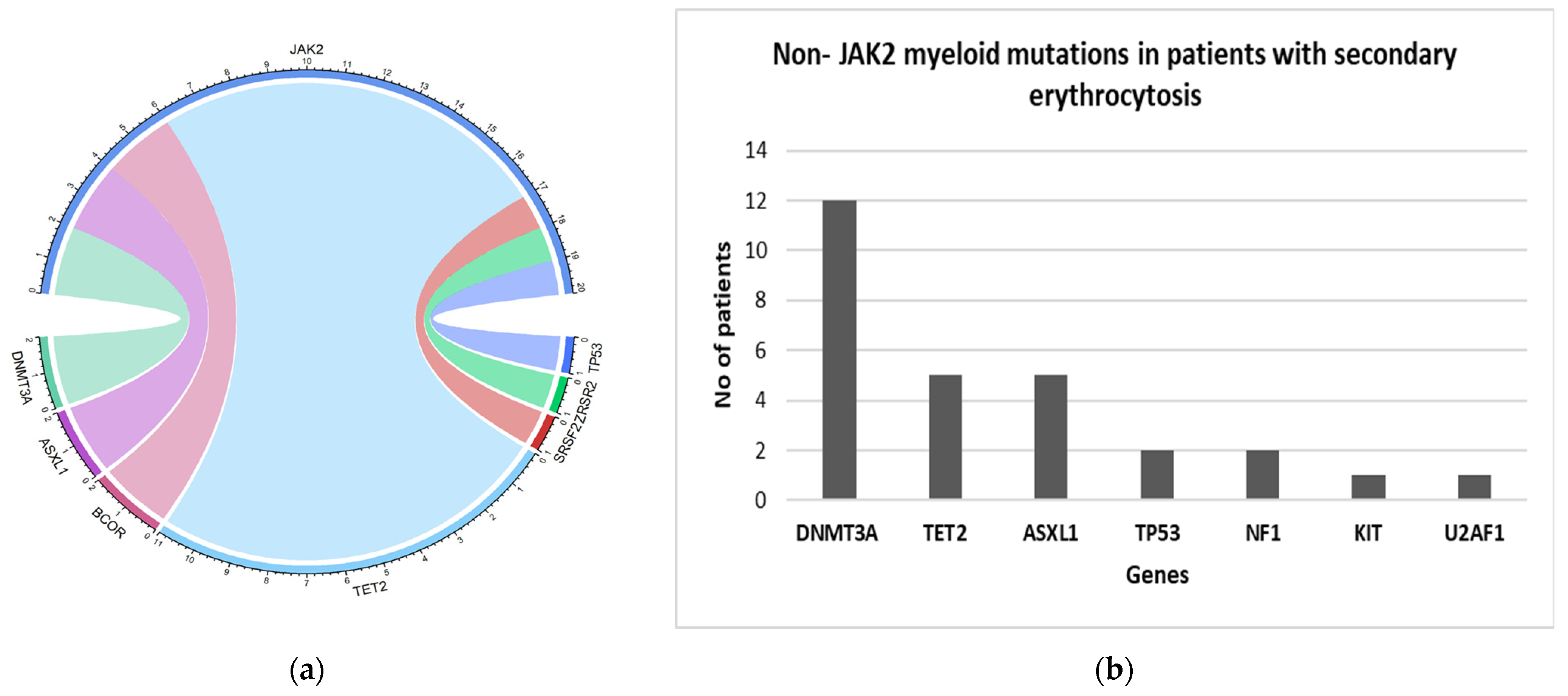
Figure 1. (a) Circos plots showing co-occurrence of additional mutation in JAK2 positive PV patients; (b) Bar Graph showing myeloid mutations in JAK2 negative patients with secondary erythrocytosis.
Curroncol 29 00568 g001
Table 1. Patients with erythrocytosis.
Total PatientsMaleFemales
Total Patients, number (%)529 (100)390 (73.7)139 (26.3)
Age; median (range) years60.6 (18–95)60.4 (18–95)61.6 (24–85)
* Height; median (range) cm 173 (125–198)177 (125–198)160.5 (141–180)
° Weight; median (range) kg91.4 (42–227.7)96.1(52.7–227.7)79.1 (42–150.3)
** BMI; median (range) kg/m230.5 (14.9–68.7) 30.9 (17.9–68.7)30.4 (14.9–53.8)
Hemoglobin; median (range) g/L175 (160–240)178 (165–240)167 (160–217)
Leukocytes; median (range) × 109/L8.3 (2.2–43.6)8 (2.2–43.6)8.8 (4.4–31.7)
Platelet count; median (range) × 109/L232.5 (31–1498)220 (31–1161)278 (50–1498)
Hematocrit; median (range) L/L0.52 (0.45–0.73)0.53 (0.46–0.73)0.51 (0.45–0.67)
# EPO levels; median (range) mIU/mL8 (1.1–456.1)8.1 (1.1–456.1)8 (1.2–111.7)
* Height was recorded for 436/529 patients; ° Weight recorded for 453/529 patients; ** BMI recorded for 436/529 patients; # Erythropoietin (EPO) levels recorded for 364 patients. mIU/mL—milli-international units per milliliter.
Table 2. Mutations identified in JAK2 positive and negative cohorts.
.Total Patients
n = 529 (%)
JAK2 Positive Cohort
n = 58 (%)
JAK2 Negative Cohort
n = 471(%)
Hemoglobin; median (range) g/L175 (160–240)174.5(161–217)175(160–240)
Leukocytes; median (range) × 109/L8.3(2.2–43.6)10.9(4.3–31.7)8(2.2–43.6)
Platelet count; median (range) × 109/L232.5(31–1498)602.5(50–1498)224(31–566)
Hematocrit; median (range) L/L0.52(0.45–0.73)0.54(0.48–0.67)0.52(0.45–0.73)
EPO levels; median (range) mIU/mL8(1.1–456.1)1.7(1.1–6.5)8.6(1.3–456.1)
Positive for Tier I/II variants by NGS91 (17.2)5833 (7)
JAK2 V617F57 (10.7)57 (98.3)0
JAK2 exon 12 mutation1 (0.2)1 (1.7)0
Clinical Diagnosis
Diagnosis of PV *6058 (100)2 $ (0.4)
Diagnosis of CML303 (0.6)
Secondary erythrocytosis4660466 (99.0)
Non-JAK2 mutationsTotal Patients
n = 529 (%)
JAK2 positive cohort
n = 58 (%)
JAK2 negative with secondary erythrocytosis
n = 466 (%)
TET216 (3)11 (19.0)5 (1.1)
DNMT3A14 (2.6)2 (3.4)12 (2.5)
ASXL17 (1.3)2 (3.4)5 (1.1)
BCOR2 (0.3)2 (1.7)0
SRSF21 (0.2)1 (1.7)0
TP531(0.2)1 (1.7)2 (0.4)
ZRSR21(0.2)10
NF12 (0.3)02 (0.4)
KIT1 (0.2)01 (0.2)
U2AF11(0.2)01 (0.2)
Total non-JAK2 mutations48 (9)20 (34.5)28 (6)
* Diagnosis of polycythemia vera defined by the World Health Organization 2017 classification; $ Out of 2 JAK2 negative patients with suspected PV, one patient had variants in SRSF2, TET2 gene and the other had variants in SRSF2, IDH2, ASXL1 gene CML—chronic myeloid leukemia; NGS—next generation sequencing.
Publisher’s Note: MDPI stays neutral with regard to jurisdictional claims in published maps and institutional affiliations.