Article

Lung Metastasis Probability in Ewing Sarcoma: A Nomogram Based on the SEER Database

Department of Orthopaedic Surgery, The First Affiliated Hospital of Zhengzhou University, Zhengzhou 450052, China
*
Author to whom correspondence should be addressed.
Curr. Oncol. 2021, 28(1), 69-77; https://doi.org/10.3390/curroncol28010009
Submission received: 8 July 2020 / Revised: 17 November 2020 / Accepted: 1 December 2020 / Published: 5 December 2020

Abstract

:
Background. Up to now, an accurate nomogram to predict the lung metastasis probability in Ewing sarcoma (ES) at initial diagnosis is lacking. Our objective was to construct and validate a nomogram for the prediction of lung metastasis in ES patients. Methods. A total of 1157 patients with ES from the Surveillance, Epidemiology, and End Results (SEER) database were retrospectively collected. The predictors of lung metastasis were identified via the least absolute shrinkage and selection operator (LASSO) and multivariate logistic analysis. The discrimination and calibration of the nomogram were validated by receiver operating characteristic (ROC) curve and calibration curve. Decision curve analysis (DCA) was used to evaluate the clinical usefulness and net benefits of the prediction model. Results. Factors including age, tumor size, primary site, tumor extension, and other site metastasis were identified as the ultimate predictors for the nomogram. The calibration curves for the training and validation cohorts both revealed good agreement, and the Hosmer–Lemeshow test identified that the model was well fitted (p > 0.05). In addition, the area under the ROC curve (AUC) values in the training and validation cohorts were 0.732 (95% confidence interval, CI: 0.607–0.808) and 0.741 (95% CI: 0.602–0.856), respectively, indicating good predictive discrimination. The DCA showed that when the predictive metastasis probability was between 1% and 90%, the nomogram could provide clinical usefulness and net benefit. Conclusion. The nomogram constructed and validated by us could provide a convenient and effective tool for clinicians that can improve prediction of the probability of lung metastasis in patients with ES at initial diagnosis.

1. Introduction

Ewing sarcoma (ES) is the second most common malignant primary osseous neoplasm, accounting for 8% of all cases in children and adolescents [1,2]. With the development of multidisciplinary therapy, the 5-year overall survival (OS) of ES has gradually improved from 10% to 75% [3]. Despite the proven effectiveness of the treatment of localized disease, the 5-year OS of ES patients with metastasis is below 30%, suggesting that these patients still fare poorly [4]. It is worth noting that most patients already have micrometastases at initial diagnosis [5]; however, only 20–28% of patients present with metastasis at initial diagnosis, and the most common site is the lung (50%) [4,6]. Although patients with lung metastasis alone have better survival than those with metastases at other sites, their mortality at 5 years is still approximately 60–70% [7,8,9,10]. The survival outcomes of patients with multiple metastases within the lung are even worse [11]. The early and accurate diagnosis of metastasis is of great significance for the targeted treatment of ES [12]. Nevertheless, because of the characteristics of micrometastases and the insufficient ability of current radiological techniques (multidetector row CT) to detect small lung nodules [13,14], improving the accuracy in detecting lung metastasis at initial diagnosis is necessary.
Some studies have investigated potential risk factors for metastasis to facilitate early diagnosis [11,15]. However, these studies analyzed only as single factor to evaluate metastasis in patients with ES. A predictive tool such as a nomogram, which can integrate multiple significant risk features to comprehensively predict lung metastasis probability, is urgently needed. Nomograms have been confirmed to provide superior individual disease risk estimation and enable accurate treatment decisions [16].
We analyzed the Surveillance, Epidemiology, and End Results (SEER) database, which collects data from seventeen geographically variable cancer registries and represents approximately 26% of the U.S. population [17], to identify independent risk factors for lung metastasis in ES at initial diagnosis; in addition, we constructed and validated a nomogram to predict lung metastasis probability.

2. Materials and Methods

2.1. Patient Cohort

The inclusion criteria were as follows: (1) diagnosed as ES of the bones with ICD-O-3/WHO 2008 morphology codes 9260 after 2010 from the SEER database; (2) microscopically confirmed, positive histology confirmed or positive exfoliative cytology confirmed.
The exclusion criteria were as follows: (1) unknown metastasis status; (2) unknown race; (3) unknown tumor size.
The clinicopathological features of the patients were categorized as follows: (1) age (<20 years old, 20 to 50 years old and >50 years old), sex (male or female), race (white, black, or other (Native American/Alaskan Native or Asian/Pacific Islander)); (2) tumor size (<5 cm, 5 to 10 cm, or >10 cm), tumor extension (inside the periosteum or beyond the periosteum), primary site (extremity (long or short bones of the upper or lower extremities), axial (skull, pelvis, spine, or ribs) or other locations), and metastasis (lung metastasis or other site metastasis).
No personal identifying information was used in the study. Hence, we did not require Institutional Review Board approval or patient informed consent. Informed consent was not required because of the retrospective nature of the study.

2.2. Statistical Analysis

We randomly divided all patients (n = 1157) into a training cohort (n = 812) and a validation cohort (n = 345). The baseline clinicopathological features were compared via the chi-square test between the two groups. To select the initial factors and prevent overfitting of the multifactor models, least absolute shrinkage, and selection operator (LASSO) regression was performed [18]. Furthermore, we used multivariate logistic regression to identify the ultimate predictive factors for the nomogram.
Using the training and validation cohorts, we validated the nomogram internally and externally. The predictive discrimination of the nomogram was assessed via a receiver operating characteristic (ROC) curve and the area under the curve (AUC), and the concordance of the nomogram was validated with a calibration plot and the Hosmer–Lemeshow test. Moreover, we utilized decision curve analysis (DCA) to assess the clinical usefulness and net benefits of the nomogram [19,20].
The chi-square test was performed via SPSS statistics software version 22.0 (IBM Corporation, Armonk, NY, USA), and the remaining statistical analyses were performed and the graphics generated by R software (3.6.3) and R studio software (1.2.5033). A two-sided p value < 0.05 was considered to have statistical significance.

3. Results

According to the inclusion and exclusion criteria, a total of 1157 ES patients, which were assigned to the training cohort (n = 812, for the construction and internal validation of the nomogram) or the validation cohort (n = 345, for the external validation of the nomogram), were identified. Most of the patients were below 20 years old, and the total proportion of patients with lung metastasis at initial diagnosis was 10.2% (Table 1). The chi-square test showed no significant differences between the two cohorts in lung metastasis, age, sex, race, tumor size, tumor extension, primary site, or other site metastasis (Table 1, p > 0.05).
To avoid overfitting, the LASSO regression selected six features with nonzero coefficients when lung metastasis was the endpoint, including age, race, tumor size, tumor extension, other site metastasis and primary site in the training cohort (Figure 1). The multivariate logistic regression analysis demonstrated that age (>50 years old, OR = 2.059, 95% CI = 1.459–4.886, p = 0.003), tumor size (5–10 cm, OR = 2.620, 95% CI = 1.494–4.823, p = 0.003; >10 cm, OR = 1.478, 95% CI = 0.814–2.800, p = 0.000), primary site (Axial, OR = 1.535, 95% CI = 1.064–2.218, p = 0.022), tumor extension (beyond periosteum, OR = 0.398, 95% CI = 0.269–0.581, p = 0.000) and other site metastasis (yes, OR = 2.610, 95% CI = 1.677–4.072, p = 0.000) were independent risk factors for lung metastasis in patients with ES (Table 2).
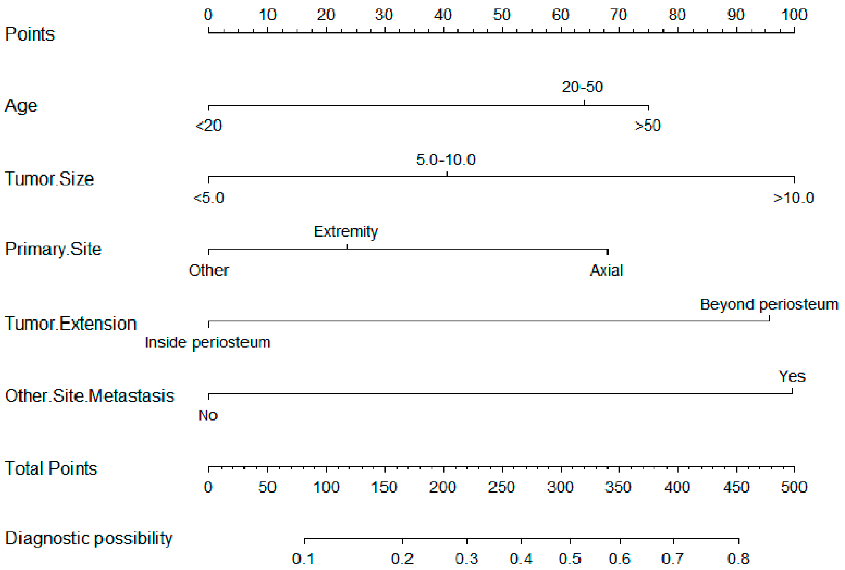
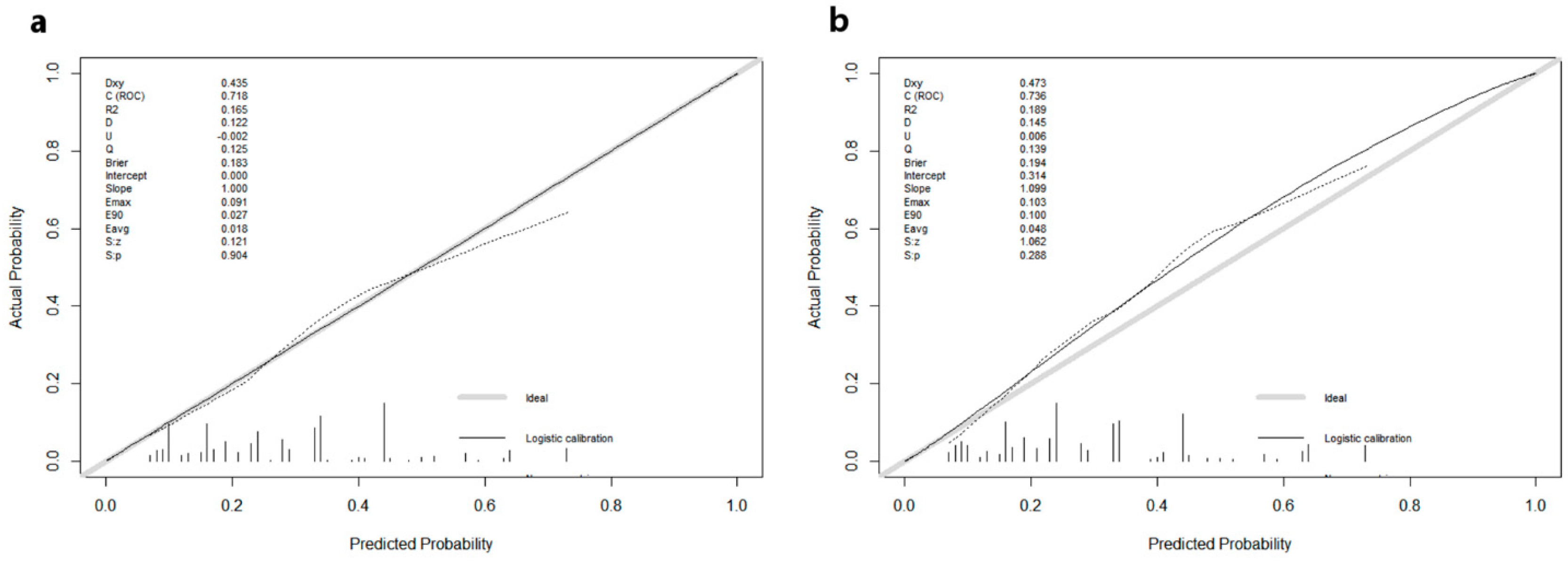
The nomogram was constructed and is presented in Figure 2. The calibration curves for the training (Figure 3a) and (Figure 3b) validation cohorts both revealed good agreement, and the Hosmer–Lemeshow test identified that the model was well fitted (p > 0.05). In addition, the area under the ROC curve (AUC) values in the training and validation cohorts were 0.732 (95% CI: 0.607–0.808) and 0.741 (95% CI: 0.602–0.856), respectively (Figure 4a), indicating good predictive discrimination. The DCA showed that when the predictive metastasis probability was between 1% and 90%, the nomogram could provide clinical usefulness and net benefit (Figure 4b).

4. Discussion

Lung metastasis in patients with ES can be affected by multiple risk factors [11,15,21,22,23]. Pathways related to platelet-derived growth factor (PDGF) signaling, Wnt signaling, apoptosis signaling, TP53, Notch signaling, and angiogenesis have been found to be of importance for the occurrence and development of metastasis in ES. Some genes have also been identified to contribute to the lung metastasis of ES. Na et al. found that CXC-chemokine receptor 6 (CXCR6) and CXC-chemokine ligand 16 (CXCL16) expression in tumor cells significantly correlated with a central location and the occurrence of lung metastasis [23]. Von et al. reported that chondromodulin 1 (CHM1) expression was increased in patients with ES lung metastases [22]. However, the clinical risk factors that affect lung metastasis in patients with ES have not been fully described. Previous clinical studies have mainly investigated all metastasis rather than lung metastasis at initial diagnosis [11,15]. In addition, previous studies did not integrate these factors, instead focusing on a single predictive index, which may have a limited effect on predicting an individual instance of lung metastasis. In recent years, nomograms have been recognized as efficient tools that can integrate all independent risk factors for diagnosis or survival outcome [24,25]. However, previous nomograms associated with ES only estimated individual patient survival outcomes, and a nomogram to predict lung metastasis in patients with ES has not yet been reported. Thus, we generated a novel nomogram to fulfill this aim. To our knowledge, this is the first study to describe a nomogram to predict lung metastasis in patients with ES.
In this study, LASSO regression and multivariate logistic regression analyses were performed to screen for risk factors and to identify independent risk factors. Variables, including age at diagnosis, tumor size, tumor extension, primary site, and other site metastasis, were independent risk factors for lung metastasis in patients with ES. As an independent risk factor, the influence of age on metastasis has been investigated in previous research findings. Ye et al. reported that ES patients between 18 and 59 years old had a high likelihood of metastatic disease at initial diagnosis [11]. Karski et al. and Ramkumar et al. found that advanced age may increase the metastasis probability of ES [26,27]. Our analyses also demonstrated that age beyond 50 years old was an independent risk factor for lung metastasis (OR = 2.059, 95% CI = 1.459–4.886, p = 0.003).
In addition, we also found that large tumor size was an independent predictor for the presentation of lung metastasis in ES patients at initial diagnosis. Large tumor size has been consistently reported as a contributor to the poor prognosis of ES patients [7,15,28,29], and it also has a major influence on metastasis in ES. Hense et al. identified that increased tumor size was positively associated with metastasis in patients with ES [30]. Ramkumar et al. showed that a tumor size greater than 118 mm caused the metastasis risk in ES patients to triple [27]. Analogously, tumors larger than 80 mm were confirmed to be more likely to have metastasis by Ye et al. [11]. Considering that increased tumor size can increase the difficulty in entirely removing the tumor and acquiring proper margins, this relationship between large tumors and metastasis seems logical. In addition, we found that tumors with a primary site in axial bones were more likely to have metastatic diseases at initial diagnosis than tumors with primary sites in other locations, which was also supported by previous results [11,15,27]. Given their nature, axial tumors are more likely to extend into the visceral cavities, thus resulting in noticeable symptoms later than tumors at other locations [31,32]. In such cases, when patients notice relevant symptoms and go to the hospital, the tumors usually are already large, and distant metastasis may have already occurred.
In the present study, the other identified predictor of lung metastasis was tumor extension. Tumor extension beyond the periosteum generally means higher malignancy and higher odds of distant metastasis. In addition, in the lung metastasis subgroup of this study, approximately 37.3% (44/118) of patients had other site metastasis at initial diagnosis. Once multiple metastases occur, metastases in the lung become very likely [6,11]. Thus, regarding metastasis at other sites as a predictive factor for lung metastasis is rational and necessary.
Undoubtedly, compared with general treatment, personalized treatment is more rational and specific [33]. As a concise but visualizable predictive model, a nomogram can be tailored according to the individual profile of the patient [34]. Such predictive tools can help clinicians optimize early diagnosis and develop personalized treatment strategies. For example, consider a 60-year-old ES patient with a tumor greater than 10 cm and tumor extension beyond the periosteum with a primary tumor site in the spine. For this patient, we could use the nomogram to connect each risk factor and obtain the patient’s total points (Figure 2). By adding up the points of each risk factor, we would obtain his ultimate score of 345 and thus conclude his lung metastatic probability is approximately 60%. According to the DCA, our nomogram would provide clinical usefulness and net benefit for our patient, as his metastasis probability is within the range of 1% to 90% (Figure 4b). Based on his result from the nomogram, we may advise that the patient be monitored for lung metastasis and consider performing detailed examinations, such as high-resolution CT or PET/CT, if necessary [35].
It is also important to consider the potential limitations of the present study. First, the retrospective nature of this study may have resulted in potential bias. Second, we validated the nomogram internally and externally with data from the same center, and, if possible, the nomogram should be validated with data from different centers to be more reliable. Finally, the SEER database did not include variables such as tumor markers and the expression of genes. Future studies could try to add these factors and develop a more comprehensive predictive model for lung metastasis of ES.

5. Conclusions

A nomogram to predict lung metastasis in patients with ES was constructed and validated based on independent factors, including age, tumor size, tumor extension, primary site, and other site metastasis. We believe this nomogram is a convenient and effective tool for clinicians that can improve prediction of the probability of lung metastasis in patients with ES at initial diagnosis.

Author Contributions

Conceptualization, J.W.; Methodology, Y.F.; software, J.W.; validation, J.W.; formal analysis, J.W.; investigation, J.W.; resources, J.W.; data curation, J.W.; writing—original draft preparation, J.W.; writing—review and editing, J.W.; visualization, Y.F.; supervision, L.X.; project administration, L.X.; funding acquisition, L.X. All authors have read and agreed to the published version of the manuscript.

Funding

This research did not receive any sources of support in the form of grants, equipment, drugs, or all of these. The authors certify that no financial and/or material support was received for this research or the creation of this work.

Acknowledgments

The authors would like to express their gratitude to the Surveillance, Epidemiology, and End Results (SEER) database, for the data from it in this study.

Conflicts of Interest

We have read and understood Current Oncology’s policy on disclosing conflicts of interest and declare that we have none.

Abbreviations

ESEwing sarcoma
OSoverall survival
SEERthe Surveillance, Epidemiology, and End Results
LASSOleast absolute shrinkage and selection operator
ROCreceiver operating characteristic
AUCarea under the curve
DCAdecision curve analysis
CXCR6CXC-chemokine receptor 6
CXCL16CXC-chemokine ligand 16
CHM1chondromodulin 1

References

  1. Ranft, A.; Seidel, C.; Hoffmann, C.; Paulussen, M.; Warby, A.C.; van den Berg, H.; Ladenstein, R.; Rossig, C.; Dirksen, U.; Rosenbaum, D.; et al. Quality of survivorship in a rare disease: Clinicofunctional outcome and physical activity in an observational cohort study of 618 long-term survivors of ewing sarcoma. J. Clin. Oncol. Off. J. Am. Soc. Clin. Oncol. 2017, 35, 1704–1712. [Google Scholar] [CrossRef]
  2. Stiller, C.A.; Bielack, S.S.; Jundt, G.; Steliarova-Foucher, E. Bone tumours in European children and adolescents, 1978–1997. Report from the Automated Childhood Cancer Information System project. Eur. J. Cancer 2006, 42, 2124–2135. [Google Scholar] [CrossRef]
  3. Balamuth, N.J.; Womer, R.B. Ewing’s sarcoma. Lancet Oncol. 2010, 11, 184–192. [Google Scholar] [CrossRef]
  4. Gaspar, N.; Hawkins, D.S.; Dirksen, U.; Lewis, I.J.; Ferrari, S.; Le Deley, M.C.; Kovar, H.; Grimer, R.; Whelan, J.; Claude, L.; et al. Ewing sarcoma: Current management and future approaches through collaboration. J. Clin. Oncol. Off. J. Am. Soc. Clin. Oncol. 2015, 33, 3036–3046. [Google Scholar] [CrossRef]
  5. Whelan, J.S.; Burcombe, R.J.; Janinis, J.; Baldelli, A.M.; Cassoni, A.M. A systematic review of the role of pulmonary irradiation in the management of primary bone tumours. Ann. Oncol. Off. J. Eur. Soc. Med. Oncol. 2002, 13, 23–30. [Google Scholar] [CrossRef]
  6. Esiashvili, N.; Goodman, M.; Marcus, R.B., Jr. Changes in incidence and survival of Ewing sarcoma patients over the past 3 decades: Surveillance epidemiology and end results data. J. Pediatric Hematol. Oncol. 2008, 30, 425–430. [Google Scholar] [CrossRef]
  7. Cotterill, S.J.; Ahrens, S.; Paulussen, M.; Jürgens, H.F.; Voûte, P.A.; Gadner, H.; Craft, A.W. Prognostic factors in Ewing’s tumor of bone: Analysis of 975 patients from the European Intergroup Cooperative Ewing’s Sarcoma Study Group. J. Clin. Oncol. Off. J. Am. Soc. Clin. Oncol. 2000, 18, 3108–3114. [Google Scholar] [CrossRef]
  8. Cangir, A.; Vietti, T.J.; Gehan, E.A.; Burgert, E.O., Jr.; Thomas, P.; Tefft, M.; Nesbit, M.E.; Kissane, J.; Pritchard, D. Ewing’s sarcoma metastatic at diagnosis. Results and comparisons of two intergroup Ewing’s sarcoma studies. Cancer 1990, 66, 887–893. [Google Scholar] [CrossRef]
  9. Sandoval, C.; Meyer, W.H.; Parham, D.M.; Kun, L.E.; Hustu, H.O.; Luo, X.; Pratt, C.B. Outcome in 43 children presenting with metastatic Ewing sarcoma: The St. Jude Children’s Research Hospital experience, 1962 to 1992. Med. Pediatric Oncol. 1996, 26, 180–185. [Google Scholar] [CrossRef]
  10. Paulussen, M.; Ahrens, S.; Burdach, S.; Craft, A.; Dockhorn-Dworniczak, B.; Dunst, J.; Fröhlich, B.; Winkelmann, W.; Zoubek, A.; Jürgens, H. Primary metastatic (stage IV) Ewing tumor: Survival analysis of 171 patients from the EICESS studies. European Intergroup Cooperative Ewing Sarcoma Studies. Ann. Oncol. Off. J. Eur. Soc. Med. Oncol. 1998, 9, 275–281. [Google Scholar] [CrossRef] [PubMed]
  11. Ye, C.; Dai, M.; Zhang, B. Risk factors for metastasis at initial diagnosis with Ewing sarcoma. Front. Oncol. 2019, 9, 1043. [Google Scholar] [CrossRef]
  12. Raciborska, A.; Bilska, K.; Rychłowska-Pruszyńska, M.; Duczkowski, M.; Duczkowska, A.; Drabko, K.; Chaber, R.; Sobol, G.; Wyrobek, E.; Michalak, E. Management and follow-up of Ewing sarcoma patients with isolated lung metastases. J. Pediatric Surg. 2016, 51, 1067–1071. [Google Scholar] [CrossRef]
  13. Meybaum, C.; Graff, M.; Fallenberg, E.M.; Leschber, G.; Wormanns, D. Contribution of CAD to the sensitivity for detecting lung metastases on thin-section CT—A prospective study with surgical and histopathological correlation. RoFo Fortschr. Geb. Rontgenstrahlen Nukl. 2020, 192, 65–73. [Google Scholar] [CrossRef] [Green Version]
  14. Ciccarese, F.; Bazzocchi, A.; Ciminari, R.; Righi, A.; Rocca, M.; Rimondi, E.; Picci, P.; Reggiani, M.L.B.; Albisinni, U.; Zompator, M. The many faces of pulmonary metastases of osteosarcoma: Retrospective study on 283 lesions submitted to surgery. Eur. J. Radiol. 2015, 84, 2679–2685. [Google Scholar] [CrossRef]
  15. Shi, J.; Yang, J.; Ma, X.; Wang, X. Risk factors for metastasis and poor prognosis of Ewing sarcoma: A population based study. J. Orthop. Surg. Res. 2020, 15, 88. [Google Scholar] [CrossRef] [Green Version]
  16. Shariat, S.F.; Karakiewicz, P.I.; Suardi, N.; Kattan, M.W. Comparison of nomograms with other methods for predicting outcomes in prostate cancer: A critical analysis of the literature. Clin. Cancer Res. Off. J. Am. Assoc. Cancer Res. 2008, 14, 4400–4407. [Google Scholar] [CrossRef] [Green Version]
  17. Cronin, K.A.; Ries, L.A.; Edwards, B.K. The surveillance, epidemiology, and end results (SEER) program of the National Cancer Institute. Cancer 2014, 120, 3755–3757. [Google Scholar] [CrossRef]
  18. Pavlou, M.; Ambler, G.; Seaman, S.; De Iorio, M.; Omar, R.Z. Review and evaluation of penalised regression methods for risk prediction in low-dimensional data with few events. Stat. Med. 2016, 35, 1159–1177. [Google Scholar] [CrossRef]
  19. Rousson, V.; Zumbrunn, T. Decision curve analysis revisited: Overall net benefit, relationships to ROC curve analysis, and application to case-control studies. BMC Med. Inform. Decis. Mak. 2011, 11, 45. [Google Scholar] [CrossRef] [Green Version]
  20. Vickers, A.J.; Elkin, E.B. Decision curve analysis: A novel method for evaluating prediction models. Med. Decis. Mak. Int. J. Soc. Med. Decis. Mak. 2006, 26, 565–574. [Google Scholar] [CrossRef] [Green Version]
  21. Mikulić, D.; Ilić, I.; Cepulić, M.; Giljević, J.S.; Orlić, D.; Zupancić, B.; Fattorini, I.; Seiwerth, S. Angiogenesis and Ewing sarcoma—Relationship to pulmonary metastasis and survival. J. Pediatric Surg. 2006, 41, 524–529. [Google Scholar] [CrossRef]
  22. Von Heyking, K.; Calzada-Wack, J.; Göllner, S.; Neff, F.; Schmidt, O.; Hensel, T.; Schirmer, D.; Fasan, A.; Esposito, I.; Muller-Tidow, C.; et al. The endochondral bone protein CHM1 sustains an undifferentiated, invasive phenotype, promoting lung metastasis in Ewing sarcoma. Mol. Oncol. 2017, 11, 1288–1301. [Google Scholar] [CrossRef] [Green Version]
  23. Na, K.Y.; Kim, H.S.; Jung, W.W.; Sung, J.Y.; Kalil, R.K.; Kim, Y.W.; Park, Y.K. CXCL16 and CXCR6 in Ewing sarcoma family tumor. Hum. Pathol. 2014, 45, 753–760. [Google Scholar] [CrossRef]
  24. Zhang, J.; Pan, Z.; Yang, J.; Yan, X.; Li, Y.; Lyu, J. A nomogram for determining the disease-specific survival in Ewing sarcoma: A population study. BMC Cancer 2019, 19, 667. [Google Scholar] [CrossRef]
  25. Kim, S.H.; Shin, K.H.; Kim, H.Y.; Cho, Y.J.; Noh, J.K.; Suh, J.S.; Yang, W.I. Postoperative nomogram to predict the probability of metastasis in Enneking stage IIB extremity osteosarcoma. BMC Cancer 2014, 14, 666. [Google Scholar] [CrossRef] [Green Version]
  26. Karski, E.E.; Matthay, K.K.; Neuhaus, J.M.; Goldsby, R.E.; Dubois, S.G. Characteristics and outcomes of patients with Ewing sarcoma over 40 years of age at diagnosis. Cancer Epidemiol. 2013, 37, 29–33. [Google Scholar] [CrossRef] [Green Version]
  27. Ramkumar, D.B.; Ramkumar, N.; Miller, B.J.; Henderson, E.R. Risk factors for detectable metastatic disease at presentation in Ewing sarcoma—An analysis of the SEER registry. Cancer Epidemiol. 2018, 57, 134–139. [Google Scholar] [CrossRef]
  28. Zhou, Q.; Wu, Z.Y.; Lin, Z.Q. A nomogram to predict prognosis in Ewing sarcoma of bone. J. Bone Oncol. 2019, 15, 100223. [Google Scholar] [CrossRef]
  29. Chen, L.; Long, C.; Liu, J.; Xing, F.; Duan, X. Characteristics and prognosis of pelvic Ewing sarcoma: A SEER population-based study. PeerJ 2019, 7, e7710. [Google Scholar] [CrossRef] [Green Version]
  30. Hence, H.W.; Ahrens, S.; Paulussen, M.; Lehnert, M.; Jurgens, H. Factors associated with tumor volume and primary metastases in Ewing tumors: Results from the (EI)CESS studies. Ann. Oncol. Off. J. Eur. Soc. Med. Oncol. 1999, 10, 1073–1077. [Google Scholar] [CrossRef]
  31. Miller, B.J.; Cram, P.; Lynch, C.F.; Buckwalter, J.A. Risk factors for metastatic disease at presentation with osteosarcoma: An analysis of the SEER database. J. Bone Jt. Surg. Am. Vol. 2013, 95, e89. [Google Scholar] [CrossRef] [Green Version]
  32. Duchman, K.R.; Gao, Y.; Miller, B.J. Prognostic factors for survival in patients with Ewing’s sarcoma using the surveillance, epidemiology, and end results (SEER) program database. Cancer Epidemiol. 2015, 39, 189–195. [Google Scholar] [CrossRef]
  33. Thewes, B.; Husson, O.; Poort, H.; Custers, J.A.E.; Butow, P.N.; McLachlan, S.A.; Prins, J.B. Fear of Cancer Recurrence in an Era of Personalized Medicine. J. Clin. Oncol. Off. J. Am. Soc. Clin. Oncol. 2017, 35, 3275–3278. [Google Scholar] [CrossRef]
  34. Iasonos, A.; Schrag, D.; Raj, G.V.; Panageas, K.S. How to build and interpret a nomogram for cancer prognosis. J. Clin. Oncol. Off. J. Am. Soc. Clin. Oncol. 2008, 26, 1364–1370. [Google Scholar] [CrossRef]
  35. Guimarães, J.B.; Rigo, L.; Lewin, F.; Emerick, A. The importance of PET/CT in the evaluation of patients with Ewing tumors. Radiol. Bras. 2015, 48, 175–180. [Google Scholar] [CrossRef]
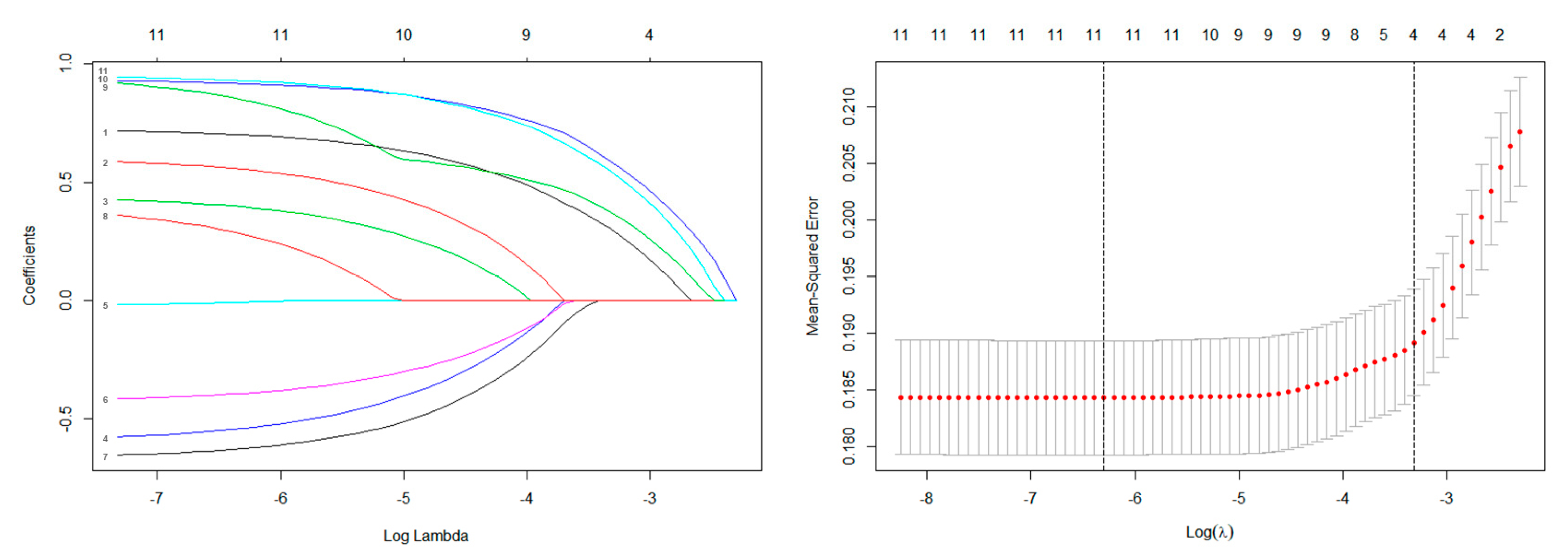
Figure 1. The results of the least absolute shrinkage and selection operator (LASSO) regression.
Curroncol 28 00009 g001
Figure 2. The nomogram for predicting the probability of lung metastasis.
Curroncol 28 00009 g002
Figure 3. Calibration curves for the training (a) and validation (b) cohorts.
Curroncol 28 00009 g003
Figure 4. The area under the ROC curve (AUC) values in the training and validation cohorts were 0.732 (95% CI: 0.607–0.808) and 0.741 (95% CI: 0.602–0.856), respectively (a), indicating good predictive discrimination. The decision curve analysis (DCA) showed that when the predictive metastasis probability was between 1% and 90%, the nomogram could provide clinical usefulness and net benefit (b).
Curroncol 28 00009 g004
Table 1. Distribution of demographic and clinical information.
VariablesTotal Population
(N = 1157; 100.0%)
Training Cohort
(N = 812; 70.1%)
Validation Cohort
(N = 345; 29.9%)
p-Value
N%N%N%
Lung Metastasis 0.616
No103989.871688.230889.3
Yes11810.29611.83710.7
Age (years) 0.376
2075164.953766.121462.0
20–5033629.022928.210731.0
50706.1465.7247.0
Race 0.619
White102988.972689.430387.8
Black443.8313.8133.8
Other847.3556.8298.4
Sex 0.719
Male72262.450462.121863.2
Female43537.630837.912736.8
Primary Site 0.893
Axial40635.128735.311934.5
Extremity50643.735643.815043.5
Other24521.216920.87622.0
Tumor Size(cm) 0.088
<517615.211313.96318.3
5–1041035.428435.012636.5
>1057149.441551.115645.2
Tumor Extension 0.160
Inside periosteum39734.328935.610831.3
Beyond periosteum76065.752364.423768.7
Other Sites Metastases 0.233
No99185.770286.528983.8
Yes16614.311013.55616.2
Chi-square test: these values are statistically significant at a p value of < 0.05.
Table 2. Multivariate logistic regression for analyzing the metastasis associated factors in the training cohort.
VariablesTraining Cohort
(N = 812)
OR (95% CI)p-Value
Age
201 (reference)
20–501.852 (0.944–5.320)0.068
502.059 (1.459–4.886)0.003 *
Race
White1 (reference)
Black0.352 (0.120–1.013)0.075
Other0.640 (0.288–1.463)0.053
Tumor Size(cm)
51 (reference)
5–102.620 (1.494–4.823)0.001 *
101.478 (0.814–2.800)0.000 *
Primary Site
Other1 (reference)
Extremity0.798 (0.496–1.267)0.344
Axial1.535 (1.064–2.218)0.022 *
Tumor Extension
Inside periosteum1 (reference)
Beyond periosteum0.398 (0.269–0.581)0.000 *
Other Sites Metastases
No1 (reference)
Yes2.610 (1.677–4.072)0.000 *
Multivariate logistic regression: these values are statistically significant (*) at a p value of < 0.05.
Publisher’s Note: MDPI stays neutral with regard to jurisdictional claims in published maps and institutional affiliations.