Next Article in Journal
Anticoagulation in Atrial Fibrillation
Previous Article in Journal
Adipozyten: Endokrine Hochleistungsfabriken
 
 
Cardiovascular Medicine is published by MDPI from Volume 28 Issue 1 (2025). Previous articles were published by another publisher in Open Access under a CC-BY (or CC-BY-NC-ND) licence, and they are hosted by MDPI on mdpi.com as a courtesy and upon agreement with Editores Medicorum Helveticorum (EMH).
Interesting Images

Asymptomatic Bland-White-Garland Syndrome in a 13-Year-Old Girl

by
Jean-Pierre Pfammatter
1,*,
Mladen Pavlovic
1 and
Stephan Windecker
2
1
Pediatric Cardiology, Inselspital, Berne, Switzerland
2
Cardiology, Inselspital, Berne, Switzerland
*
Author to whom correspondence should be addressed.
Cardiovasc. Med. 2007, 10(5), 179; https://doi.org/10.4414/cvm.2007.01247
Submission received: 25 February 2007 / Revised: 25 March 2007 / Accepted: 25 April 2007 / Published: 25 May 2007

Case report

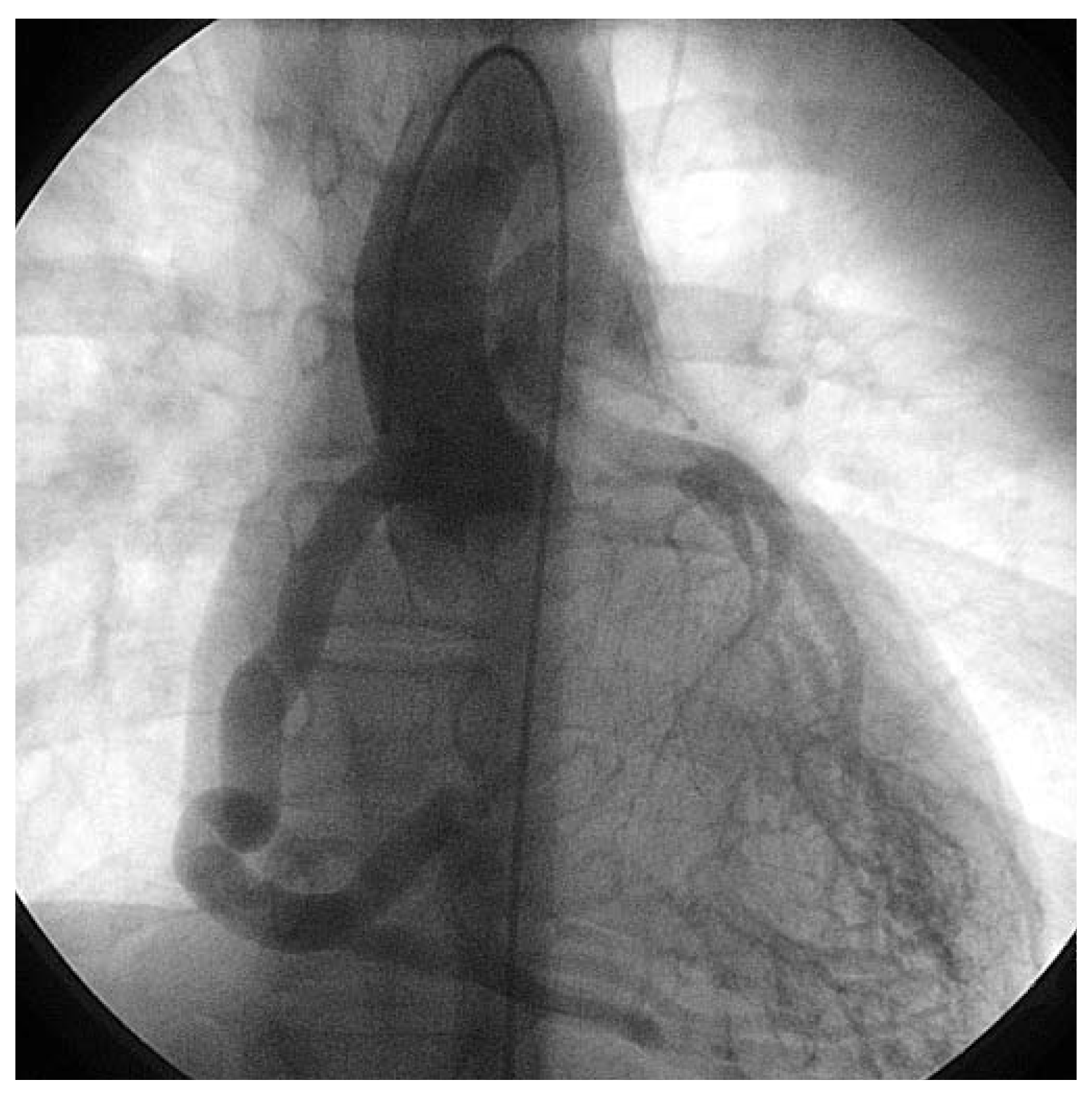
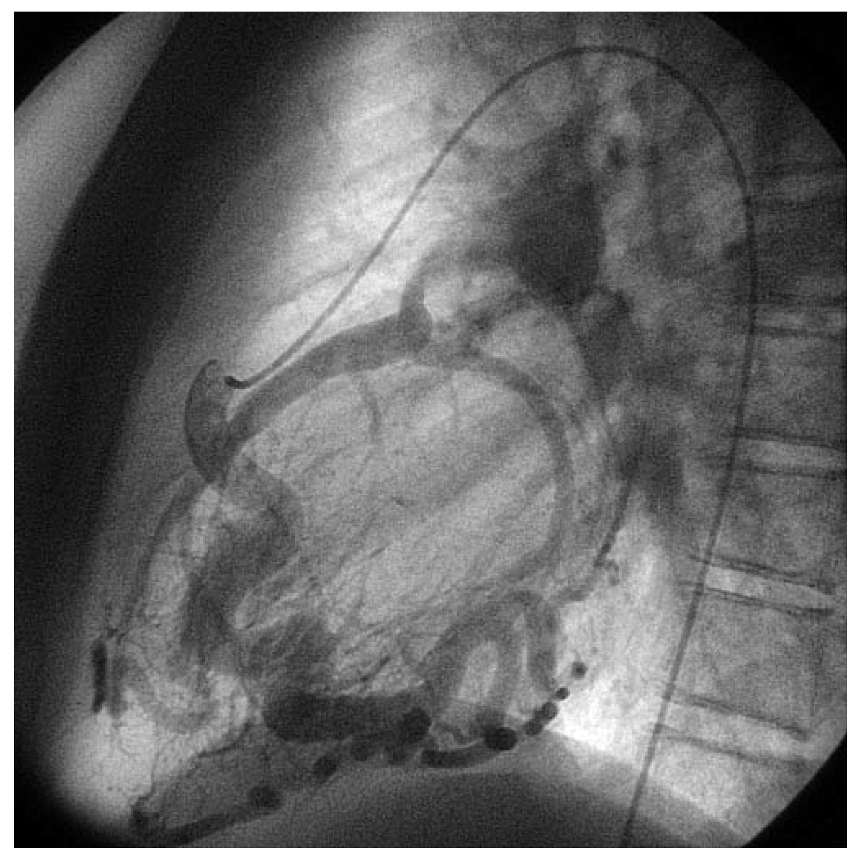
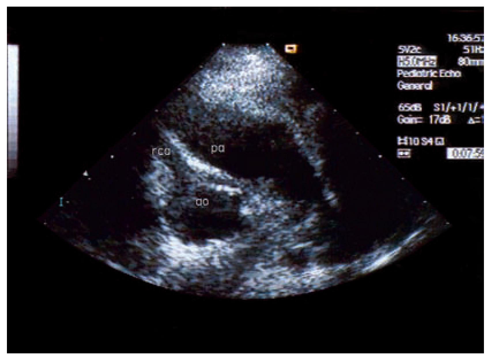
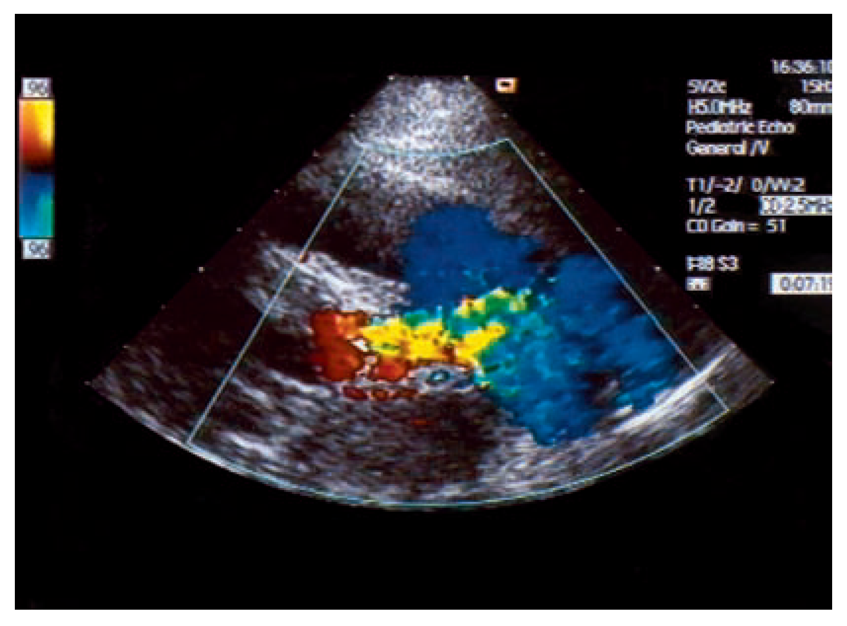
An otherwise healthy 13-year-old girl was referred to the pediatric cardiology outpatient clinic for evaluation of a newly observed systolic murmur. She was normally physically active without symptoms on exertion except for one episode of syncope two months earlier. She then was riding a bike and on climbing off the bike she was observed to hyperventilate, had parestesias of both hands followed by a short loss of consciousness. Family history was negative for any cardiac event. Clinically the systolic murmur was judged a functional ejection murmur but an Echo was done and revealed an enlarged (6 mm) right coronary artery (Figure 3) with huge collaterals especially in the septal region (Figure 4), the origin of the left coronary artery was seen in the pulmonary trunk with turbulent retrograde flow in the color Doppler (Figure 5). LV ejection fraction was 68% but the LV was slightly dilated with a mild mitral regurgitation. Cardiac catheterisation confirmed the suspected anomalous origin of the left coronary artery from the pulmonary trunk (Figure 1 and Figure 2), the patient was advised to undergo surgical correction with reimplantation of the left coronary artery in the aorta.
Figure 1. Anteroposterior view of aortography: late phase still shows dilated RCA with tortuous course and retrograde flow through the left coronary artery with drainage into the pulmonary artery.
Cardiovascmed 10 00179 g001
Figure 2. Lateral view of selective right coronary artery injection: the very tortuous course of the RCA is seen as well as the opacification of the pulmonary artery trunk with the left coronary artery ostium at its inferior aspect.
Cardiovascmed 10 00179 g002
Figure 3. Superior short axis echo view showing origin of the massively dilated right coronary artery from the aorta in usual position.
Cardiovascmed 10 00179 g003
Figure 4. Modified parasternal long axis echo view showing several turbulent flows in projection on the interventricular septum.
Cardiovascmed 10 00179 g004
Figure 5. Superior short axis echo view showing turbulent flow draining into the pulmonary artery trunk above the valve and originating from the apparent region of the left coronary artery.
Cardiovascmed 10 00179 g005

Discussion

Anomalous left coronary artery origin from the pulmonary artery (ALCAPA or Bland-White-Garland syndrome) is a very rare congenital anomaly which usually becomes symptomatic in the first months of life. The infants then show signs of congestive heart failure and the picture of dilative left ventricular cardiomyopathy. In the presymptomatic period (first weeks of life) the babies typically exhibit a chronic mild troponinaemia and a slowly progressive dilatation and dysfunction of the left ventricle [1]. A few case reports described the very unusual observation of asymptomatic survival of this disease into adulthood [2]. Once diagnosis is made, surgical correction is recommended for the risk of sudden cardiac death (documented in asymptomatic patients) and for the left to right shunting with left heart volume overload as also observed in our patient.

References

  1. Bolt, I.; Spalinger, J.; Pfammatter, J.P. Cardiac features in the presymptomatic period in a neonate with anomalous left coronary artery arising from the pulmonary artery. Cardiol Young. 2003, 13, 579–581. [Google Scholar] [CrossRef] [PubMed]
  2. Barbetakis, N.; Efstathiou, A.; Efstathiou, N.; Papagiannopolou, P.; Soulountsi, V.; et al. A long term survivor of Bland-WhiteGarland syndrome with systemic collateral supply: Case report and review of the literature. BMC Surg. 2005, 5, 23. [Google Scholar] [CrossRef] [PubMed]