Next Article in Journal
...und Immer Wieder Dieses Herzrasen!
Previous Article in Journal
Should We Hastily Adopt Public Access Defibrillation?
 
 
Cardiovascular Medicine is published by MDPI from Volume 28 Issue 1 (2025). Previous articles were published by another publisher in Open Access under a CC-BY (or CC-BY-NC-ND) licence, and they are hosted by MDPI on mdpi.com as a courtesy and upon agreement with Editores Medicorum Helveticorum (EMH).
Interesting Images

Straight into the Heart: Sinus of Valsalva Injury

by
Frank Enseleit
*,
Thomas Largiadèr
and
Rolf Jenni
Cardiovascular Center Cardiology, University Hospital, Zurich, Switzerland
*
Author to whom correspondence should be addressed.
Cardiovasc. Med. 2006, 9(6), 238; https://doi.org/10.4414/cvm.2006.01180
Submission received: 30 March 2006 / Revised: 30 April 2006 / Accepted: 30 May 2006 / Published: 30 June 2006

Case description

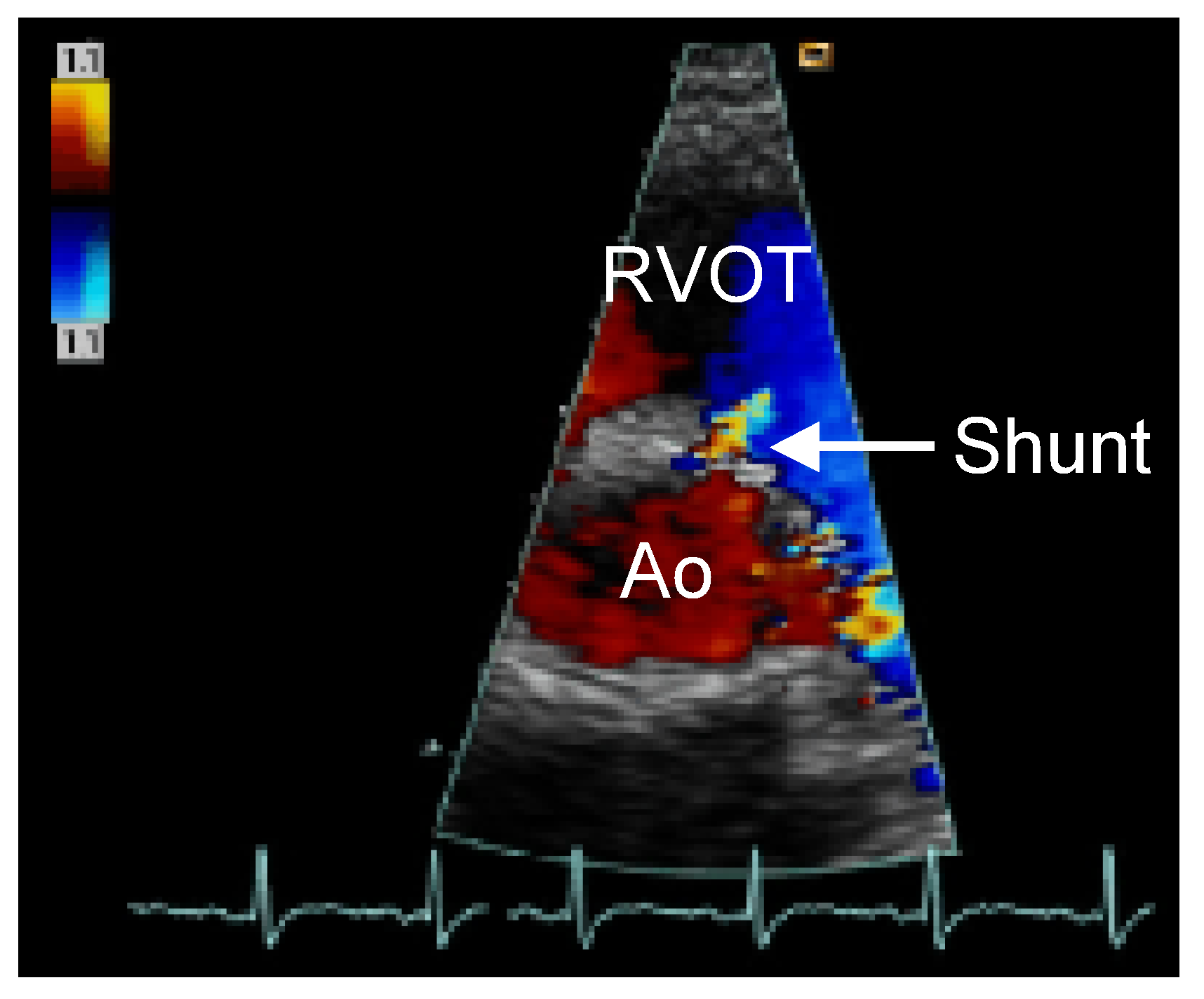
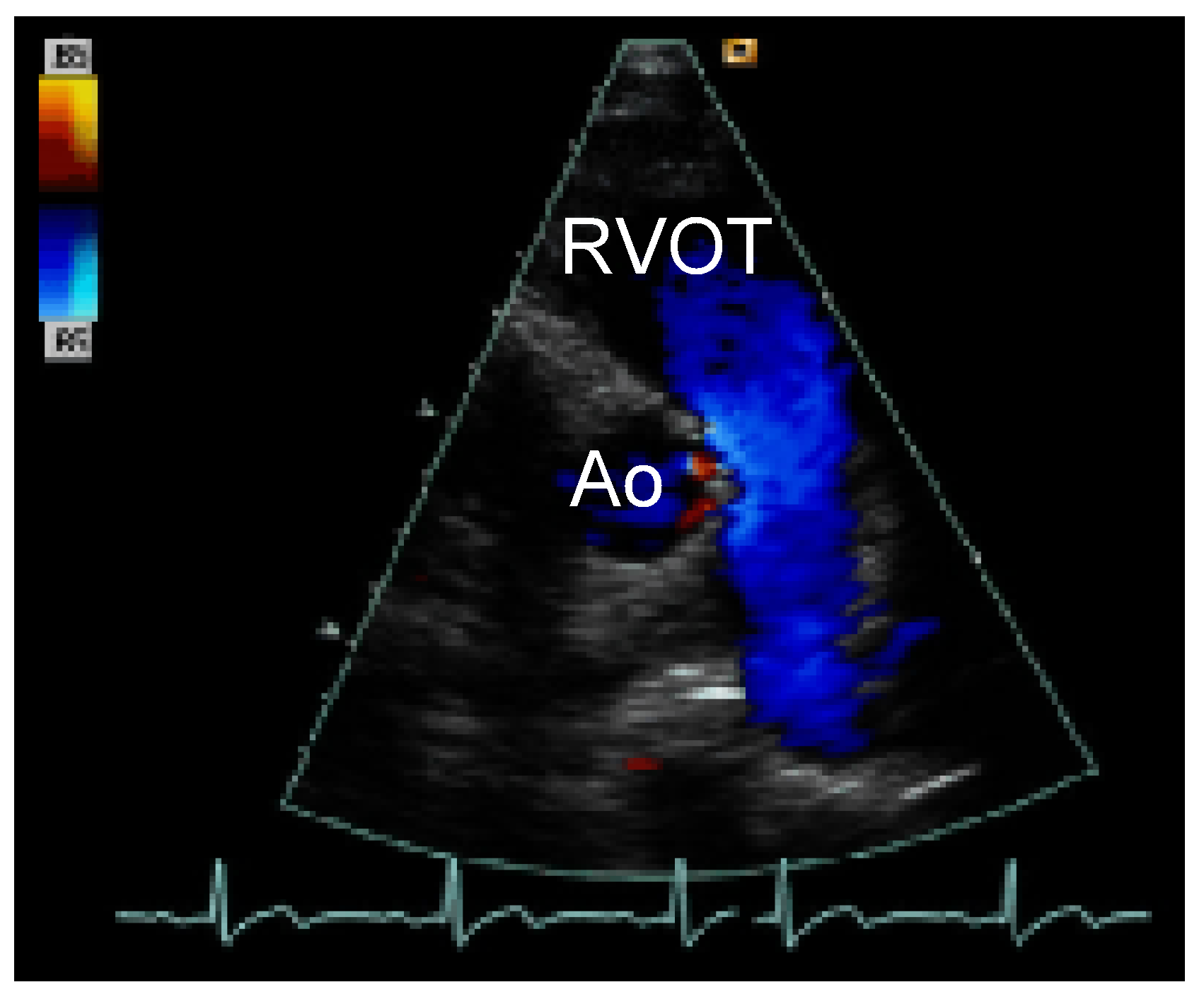
A healthy 19-year-old patient was admitted to the emergency department after a knife attack. In the physical examination of the patient a new systolic-diastolic murmur and three deep thoracic knife wounds were found. The CT-scan showed possible myocardial damage. Left-sided anterior-lateral thoracotomy was performed and a 2 cm long wound directed from the apex of the right ventricle to the pulmonary trunc was sewed. Postoperative Doppler-echocardiography revealed a shunt from the right sinus of valsalva of the aortic root (Ao) into the right ventricular outflow tract (RVOT) at the level of the commissure between the right and the left coronary cusp with a high-pressure gradient (Figure 1). Decision was made to follow-up the patient three month after discharge. In the physical examination during follow-up the systolic-diastolic murmur was no longer evident and the shunt was no longer detectable with Doppler-echocardiography, suggesting spontaneous closure of the shunt (Figure 2). This is the first description of a spontaneous closure of a left-right shunt originating from the sinus of valsalva into the right ventricular outflow tract.
Figure 1. Transthoracic short-axis view of the heart showing the shunt (Arrow) between the aortic root (Ao) and the right ventricular outflow tract (RVOT) at the level of the commissure.
Cardiovascmed 09 00238 g001
Figure 2. Transthoracic short-axis view of the heart showing that the shunt between the aortic root (Ao) and the right ventricular outflow tract (RVOT) at the level of the commissure is not longer detectable.
Cardiovascmed 09 00238 g002