Next Article in Journal
Kardiale Resynchronisationstherapie (CRT): Mit Oder Ohne ICD-Back-Up?
Previous Article in Journal
Directive 2005 Concernant les Contrôles de Patients Porteurs de Défibrillateurs Automatiques Implantable
 
 
Cardiovascular Medicine is published by MDPI from Volume 28 Issue 1 (2025). Previous articles were published by another publisher in Open Access under a CC-BY (or CC-BY-NC-ND) licence, and they are hosted by MDPI on mdpi.com as a courtesy and upon agreement with Editores Medicorum Helveticorum (EMH).
Case Report

A Difficult Pacemaker ECG Resulting in an Unnecessary Intervention

Department of Cardiology, University Hospital, Basel, Switzerland
*
Author to whom correspondence should be addressed.
Cardiovasc. Med. 2006, 9(6), 227; https://doi.org/10.4414/cvm.2006.01177
Submission received: 30 March 2006 / Revised: 30 April 2006 / Accepted: 30 May 2006 / Published: 30 June 2006

Summary

We report on a 67-year-old patient with coronary artery disease, presenting with dyspnea and chest pain. The pacemaker ECG of the patient was difficult to read in the emergency setting. Sudden changes in the heart rate were misinterpreted as a non-sustained ventricular tachycardia, and the paced ventricular rhythm was misread as ST-segment elevation. The combination of an inadequately programmed pacemaker, a patient with panic attacks and a misleading ECG interpretation lead to an unnecessary coronary angiography. We discuss how erroneous episodes of incorrect mode switching due to atrial far field sensing with rate drops can be avoided by correct pacemaker testing.

Introduction

Modern pacemaker systems offer a wide variety of programming possibilities and memory evaluation. However, these features can lead to misinterpretation by physicians not familiar with this technology. Even cardiologists who perform pacemaker tests on a regular basis might sometimes be challenged. In emergency situations the correct interpretation of a pacemaker ECG is crucial for the management of patients [1].

Case presentation

A 67-year-old male patient with a history of coronary artery disease underwent urgent coronary angiography due to acute dyspnea and chest pain at rest, presumed inferior STsegment elevation and intermittent episodes of non-sustained ventricular tachycardia on a three-lead ECG (Figure 1) taken in the ambulance.
Six months ago the patient had suffered from unstable angina and had received PCI and stenting of the distal circumflex artery while both the left anterior descending and the right coronary artery showed only insignificant stenosis. Due to haemodynamically relevant bradycardia with type II second-degree AV block, an external transvenous pacemaker was placed. Over the next two days the patient had several episodes of atypical chest pain without dynamic ECG-changes or troponin elevation. He then told his treating physicians that he had suffered from similar episodes for years, mostly occurring at night and promptly resolving after alprazolam (Xanax) intake.
Even though these episodes were suggestive for panic attacks, a psychosomatic evaluation was not undertaken. Due to the persistence of the second-degree AV block, the patient received a permanent VDD pacemaker (St. Jude 5456 Verity). Over the next six months, the clinical course was uneventful.
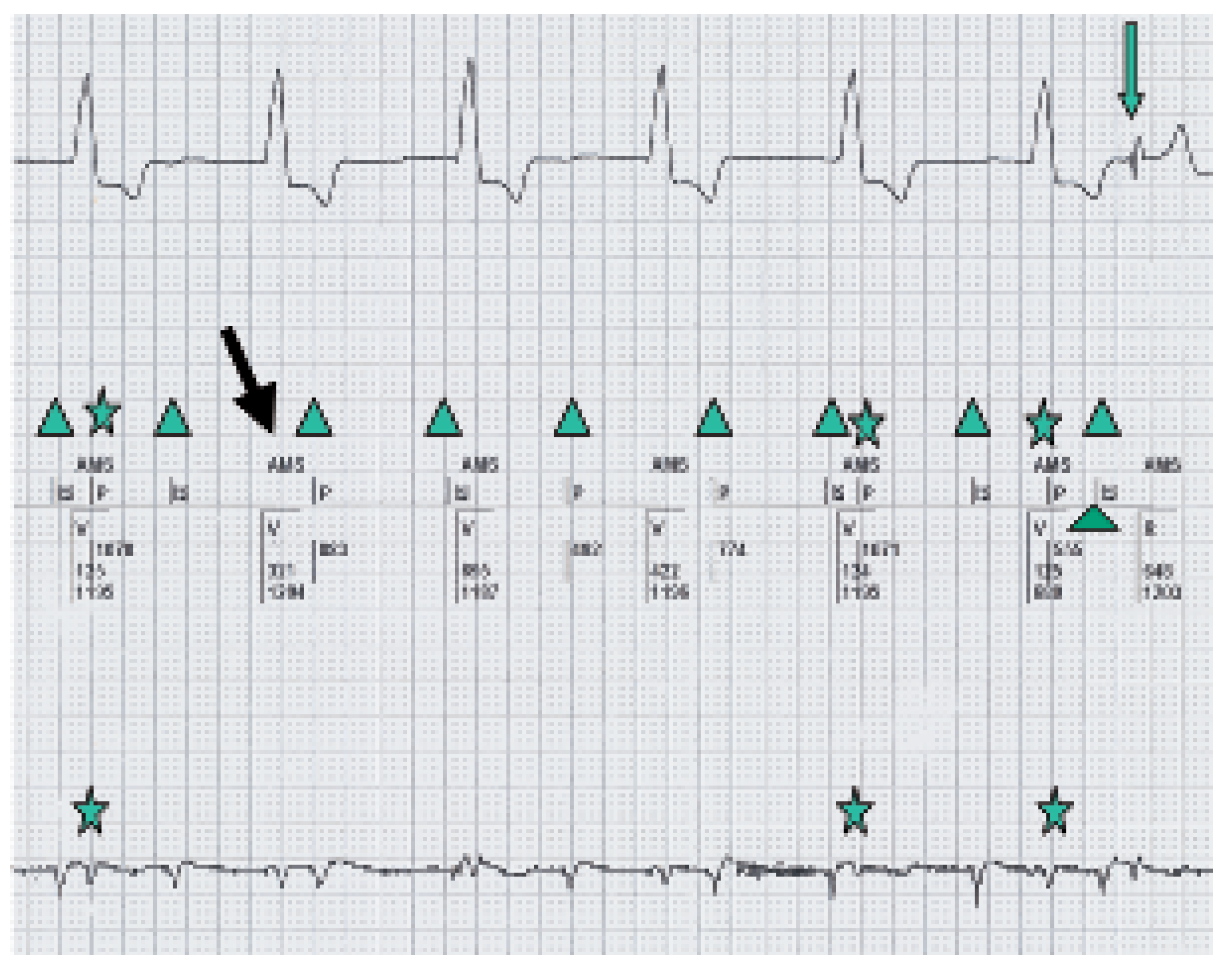
Emergency angiography was performed, but revealed neither a progression of CAD nor in-stent-restenosis. Also during angiography sudden changes in heart rate were observed, and the pacemaker was interrogated after the procedure. Evaluation of the marker channel (Figure 2) revealed that the episodes with a broad QRS complex and a heart rate around 75/min (mistakenly counted by the ECG software as a frequency of 150/min due to T-wave oversensing and misinterpreted as a ventricular tachycardia by the anaesthetist) corresponded to a correct atrial sensing/ventricular pacing mode (ASVP). The observed rate drops were caused by intermittent atrial oversensing of the QRS complex, ie atrial far field sensing (AFFS). This lead to a subsequent beat-to-beat modeswitch and a fallback into the VVI mode at a rate of 50/min. In the pacemaker memory more than 75 000 mode-switch episodes (7% of all time) were stored over a period of three months, indicating that the oversensing was a relevant problem. To prevent further episodes of inadequate mode switching, the post-ventricular atrial blanking period (PVAB) was modified from 100 to 165 ms, since the measured V-A-interval (time period between marker signals of ventricular pacing and the atrial oversensing) was around 130 ms. After that specific time adjustment no further modeswitching was observed.
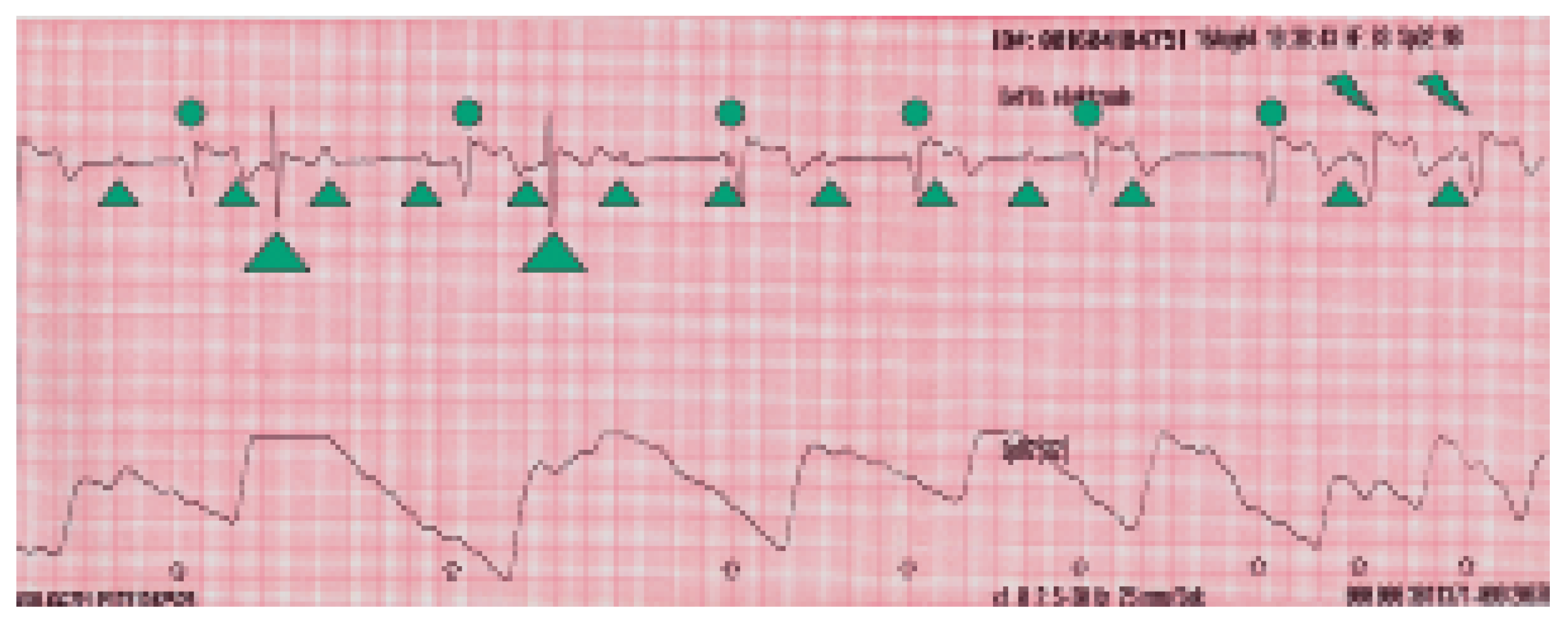
In retrospective it appears that the rapid changes in heart rate had irritated and frightened the patient, thus triggering a panic attack with dyspnea and chest pain. This cascade of events combined with the misinterpretation of the underlying pacemaker ECG (heart rate changes due to intermittent modeswitching were misread as intermittent nonsustained ventricular tachycardia) had ultimately lead to an unnecessary coronary angiography. The retrospective interpretation of the ECG strip is shown in Figure 3.

Discussion

This case shows possible diagnostic problems with modern multiprogrammable pacemaker systems in emergency situations. Atrial farfield sensing (AFFS) can be seen in any modern dual chamber pacemaker if atrial sensing is programmed to a minimal value and the PVAB is set to a minimum value of 50 ms. For correct management, at first retrograde V-A conduction, which could mimic AFFS, has to be excluded. The V-A-interval in AFFS is usually less than 150 ms, whereas in cases of retrograde conduction (exceptions are accessory pathways or a dual AV node with fast conduction properties) it is usually longer [2]. Obviously in such cases, the post-ventricular atrial refractory period (PVARP) has to be extended to avoid a pacemaker-mediated tachycardia.
To prevent inappropriate mode-switch episodes due to AFFS, the following tests (at least during the first two pacemaker controls) should be performed:
-
minimise the AV/PV interval in order to ensure ventricular pacing
-
set atrial sensing to the lowest programmable value
-
shorten the PVAB to less than 100 ms
-
check for AFFS and, if positive, measure the V-A time interval
-
set the PVAB to a value at least 50 ms above the measured value
Because amplitude and time intervals of AFFS are only minimally dependent on body position or adrenergic stimulation [3], this test can be performed in the supine position. It has been shown that the amplitude of AFFS signals is around 0.5 mV and that it is higher in paced than in intrinsic ventricular complexes [4]. That means that just increasing the atrial sensitivity level is not a good option, at least not in VDD pacemakers, where such a programming would eventually lead to atrial undersensing, consecutive AV-dyssynchrony and VVI backup pacing.
Furthermore it is advisable to program mode-switch pacing to VVIR 70/min, instead of VVI 50/min as demonstrated in our case. Thus, heart rate drops from 85 to 50/min would have been prevented, though not resolving the technical problem of AFFS.

References

  1. Brady, W.J.; Lentz, B.; Barlotta, K.; Harrigan, R.; Chan, T. ECG Patterns confounding the ECG diagnosis of acute coronary syndrome. Emerg Med Clin N 2005, 23, 999–1025. [Google Scholar] [CrossRef] [PubMed]
  2. Nowak, B.; Kramm, B.; Schwaier, H.; Miamaitiming, A.; Geil, S.; Zellerhoff, C.; et al. Is atrial sensing of ventricular far-field signals important in single-lead VDD pacing? Pacing Clin Electrophysiol 1998, 21, 2236–2239. [Google Scholar] [CrossRef] [PubMed]
  3. Cools, F.J.; van Twembeke, R.R.; Backers, J.; Verpooten, G.A. Stability of far field R wave signals in different conditions. Europace 2003, 5, 175–183. [Google Scholar] [CrossRef] [PubMed]
  4. Frohlig, G.; Helwani, Z.; Kusch, O.; Berg, M.; Schieffer, H. Bipolar ventricular far-field signals in the atrium. Pacing Clin Electrophysiol 1999, 22, 1604–1613. [Google Scholar] [CrossRef] [PubMed]
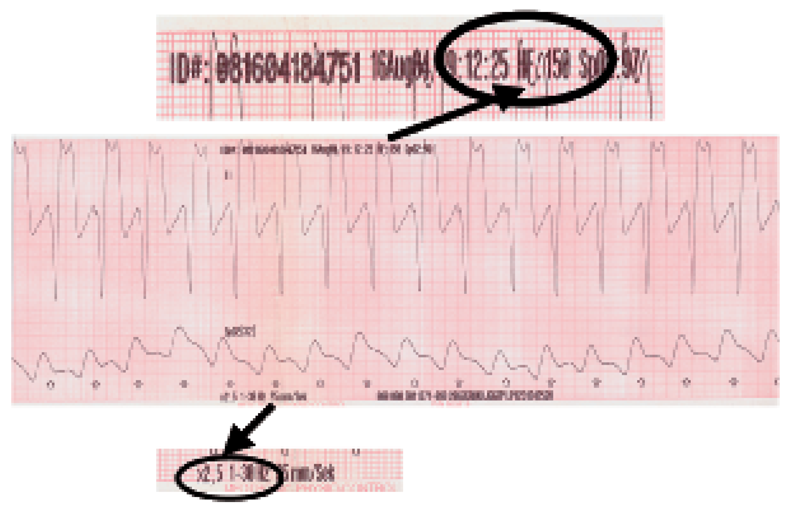
Figure 1. VDD mode with 1:1 conduction and a heart rate of 75. Due to T-wave oversensing the ECG software is measuring a heart rate of 150/min (upper bar). Compared to Figure 2 (amplification of the signal by factor 1), this rhythm strip is written in a 2.5 amplification (lower bar) and the paced ventricular complexes mimic STEMI.
Cardiovascmed 09 00227 g001
Figure 2. Interpretation of the marker channel during pacemaker control: Cardiovascmed 09 00227 i001 AMS with VVI back up mode is shown. Cardiovascmed 09 00227 i002 P-waves noted in the marker channel (middle line). Cardiovascmed 09 00227 i003 Intermittent Far Field sensing seen in the bottom line (signal measured between Atip and Aring) and in the marker channel (middle line). Cardiovascmed 09 00227 i004 Intrinsic ventricular beat.
Cardiovascmed 09 00227 g002
Figure 3. Retrospective interpretation of the ECG taken in the ambulance: Cardiovascmed 09 00227 i005 Intrinsic ventricular complexes. Cardiovascmed 09 00227 i006 P-waves. Cardiovascmed 09 00227 i007 Paced ventricular complexes in AMS with VVI back up mode of 50/min. Cardiovascmed 09 00227 i008 Switch from VVI to VDD mode with 1:1 conduction.
Cardiovascmed 09 00227 g003