Next Article in Journal
Das Herz Beim Metabolischen Syndrom
Previous Article in Journal
Dissolution of a Saphenous Vein Graft Thrombus with Systemic Glycoprotein IIb/IIIa Receptor Inhibitor Prior to Percutaneous Coronary Intervention
 
 
Cardiovascular Medicine is published by MDPI from Volume 28 Issue 1 (2025). Previous articles were published by another publisher in Open Access under a CC-BY (or CC-BY-NC-ND) licence, and they are hosted by MDPI on mdpi.com as a courtesy and upon agreement with Editores Medicorum Helveticorum (EMH).
Proceeding Paper

The Challenge of Vulnerable Plaque Detection in the Cardiac Catheterization Laboratory

by
Pierfrancesco Agostoni
,
Johannes A. Schaar
and
Patrick W. Serruys
*
ThoraxCenter, Erasmus MC Rotterdam, Room BD 404 Postbus 2040, NL-3000 CA Rotterdam, The Netherlands
*
Author to whom correspondence should be addressed.
Cardiovasc. Med. 2004, 7(10), 349; https://doi.org/10.4414/cvm.2004.01049
Submission received: 27 July 2004 / Revised: 27 August 2004 / Accepted: 27 September 2004 / Published: 27 October 2004

Summary

Rupture of vulnerable plaques is the main cause of acute coronary syndromes and myocardial infarctions. Identification of these vulnerable plaques is therefore essential to enable the development of treatment modalities to stabilise them. Several intra-vascular technologies, investigating coronary areas that will be responsible for future events, are underlined in this review. The ideal technique would provide morphological, mechanical and biochemical information, however, despite several imaging techniques are currently under development, none of them provides alone such all-embracing assessment. OCT has the advantage of high resolution, thermography has the potential to measure metabolism and spectroscopy obtains information on chemical components. IVUS and IVUS-palpography are easy to perform and assess morphology and mechanical instability. Shear stress is an important mechanical parameter deeply influencing vascular biology. Nevertheless all techniques are still under investigation and at present, none of them can completely identify a vulnerable plaque and, most importantly, predict its further development. From a clinical point of view, most techniques currently assess only one feature of the vulnerable plaque. Thus the combination of several modalities will be of importance in the future to ensure a high sensitivity and specificity in detecting vulnerable plaques.

Introduction

Rupture of vulnerable plaques is the main cause of acute coronary syndromes and myocardial infarctions. Identification of these vulnerable plaques is therefore essential to enable the development of treatment modalities to modify them. Because myocardial infarction (MI) and its consequences are so important, we must investigate options to identify those coronary areas that will be responsible for future events. A wide variety exists in the structure and function of coronary atherosclerotic plaques. Most plaques may cause no symptoms for decades, however, a few plaques disrupt and cause thrombosis. These rare but dangerous thrombosis-prone plaques are called vulnerable [1]. Thus, a vulnerable plaque is a plaque assumed to be at high short-term risk of thrombosis, causing an acute coronary syndrome (ACS).
There are three forms of vulnerable plaques, all documented by pathologic studies:
A.
Thin-cap fibro-atheroma (TCFA): in about 65% of all symptomatic coronary thrombotic events, rupture of an inflamed TCFA was evident. The major components of such TCFA are: an atheromatous core (usually >40% of the entire plaque), a thin fibrous cap with macrophage and lymphocyte infiltration and decreased smooth muscle cell content, and expansive remodeling [2].
B.
Erosion: in about 30% of all events, the endothelium overlying the plaque was found injured at the place where a thrombus has formed. Usually, these plaques are rich in proteoglycans [3].
C.
A calcified nodule: in 5% of all events, thrombosis covering a calcified nodule suggests that the plaque appeared to be heavily calcified with a calcified nodule projecting into the lumen [4].
The terms vulnerable plaque, high-risk plaque and thrombosis-prone plaque can be used identically [1].
Currently, there is no widely accepted diagnostic method to prospectively identify such vulnerable plaques. The challenge for the future is to identify vulnerable plaques before the thrombus forms. Many of the imaging techniques used to assess coronary artery disease are able to detect different features of the rupture-prone type of vulnerable plaques.

Angiography

Coronary angiography has been so far the gold standard to assess the severity of obstructive luminal narrowing. Furthermore it serves as a decision tool to direct therapy such as percutaneous coronary interventions or coronary artery bypass surgery. Using coronary angiography we can assess the lumen boundaries, but no information is given on plaque burden, plaque delineation and plaque components. Actually, angiography is able to detect complex lesions, which are considered vulnerable plaques at an advanced stage. Complex lesions have some peculiar angiographic features: intra-luminal filling defects (consisting with thrombus), presence of contrast and hazy contour beyond the vessel lumen (consisting with plaque ulceration), irregular margins and overhanging hedges (consisting with plaque irregularity and, possibly, fracture) and impaired flow with evident lumen reduction [5]. The presence of multiple complex lesions in patients after a MI has been associated with increased incidence of ACS [5]. However, angiography is a crude technique to assess the presence and burden of vulnerable lesions, as the majority of ulcerated plaques are not big enough to be detected by angiography, but can be well assessed pathologically [6]. Indeed, about 70% of acute coronary occlusions are in areas that were previously angiographically normal, and only a minority occurs where there was severe stenosis [7,8]. Furthermore, we have to take into account that the predictive power of angiography is strongly dependent on the time interval between the angiogram and the MI, because both time and interim therapy can influence atherosclerosis. In one study, the angiograms were performed between 1 and 77 months before the event [7] and showed that atherosclerosis can be a rapidly progressive process. Another study evaluated angiograms made one week before acute MI showing that signs of thrombosis and rupture were present in the majority of patients [9]. Thus, patients with silent non-obstructive coronary atherosclerosis, harbor vulnerable plaques which cannot be detected by angiograms, but are associated with adverse clinical outcomes. If a disrupted ulcerated plaque is seen on angiography (fig. 1), the existence of additional rupture prone plaques is to be expected. Angiography therefore, has a low discriminatory power to identify the vulnerable plaque.

Angioscopy

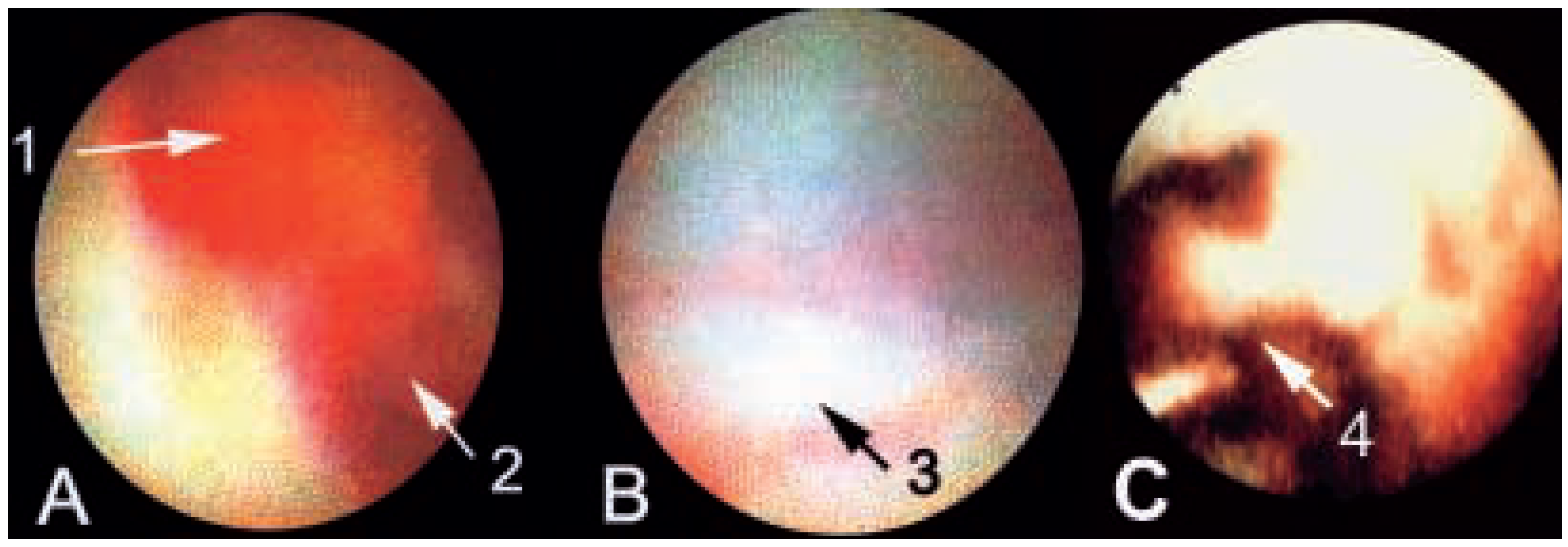
Angioscopy is using fiberoptics to visualise thrombi and plaque surface (fig. 2). Vulnerable plaque features, such as ruptured caps and red discoloration (intra-plaque hemorrhage), can be detected. In patients with acute MI, angioscopy showed diffuse disease in all the three coronary arteries, with multiple yellow plaques [10]. Furthermore, in a 12-month follow-up study of patients with stable angina, ACS occurred more frequently in patients with yellow plaques than in those with white plaques [11]. These results suggest that yellow plaques, which may be visualised by angioscopy but not by angiography, may be more prone to rupture than white plaques. However, this technique has several drawbacks. Indeed, only a limited part of the vessel tree can be investigated, due to the size of the device. Furthermore, information about the degree of plaque extension into the vessel wall is not provided. Finally, to enable clear visualisation of the vessel wall, the vessel has to be occluded and the remaining blood flushed away with saline, thereby potentially inducing ischaemia.

Intra-vascular ultrasound

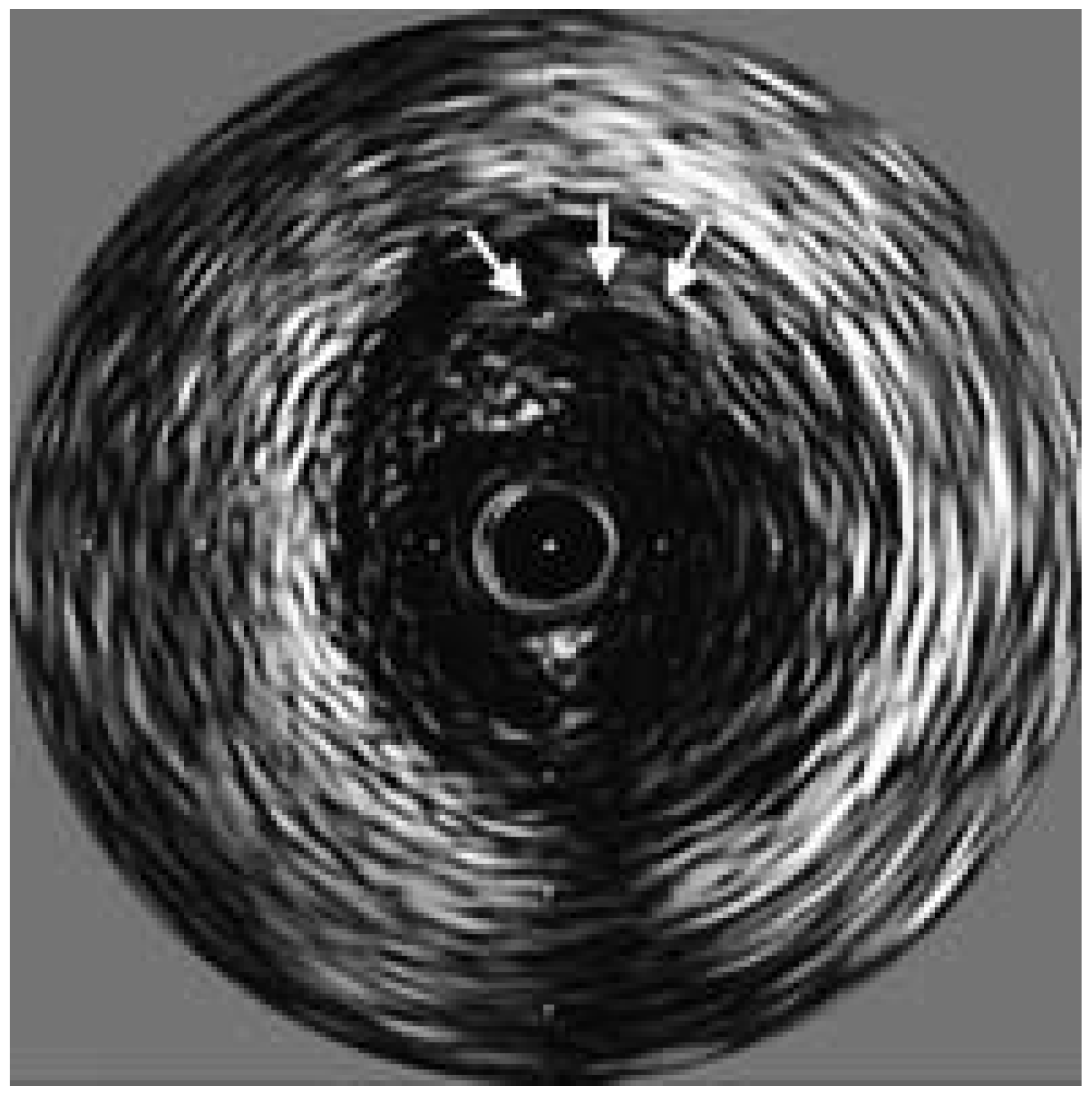
Intra-vascular coronary ultrasound (IVUS) provides real-time high-resolution images of the vessel wall and lumen. Depending on the distance of the vessel wall from the catheter, the axial resolution is about 150 microns, the lateral 300 microns. The images appear real time. Features of the vessel can be detected based on the echogenicity of different tissue types. Small structures can be visualised, however only those sized over 160 microns can be estimated accurately. The normal thickness of the media is about 125—350 mm. IVUS provides some insight into the composition of coronary plaques. In IVUS images, calcification is characterised by a bright echo signal with distal shadowing which hides plaque components and deeper vessel structures. In comparative studies between histology and IVUS, plaque calcification can be detected with a sensitivity of between 86 and 97% [12]. The sensitivity to detect micro-calcification ranges around 60% [13]. In IVUS images lipid depositions are described as echolucent zones and can be detected with a sensitivity of between 78% and 95% and specificity of 30% [14]. This sensitivity is dependent on the amount of lipid and can drop down further if the echo-lucent area is smaller than a quarter of the plaque. Echolucent zones can also be caused by loose tissue and shadowing from calcium, which makes the interpretation of these areas difficult. The sensitivity to differentiate between fibrous and fatty tissue is between 39 and 52% [15]. The detection of vulnerable plaques by IVUS is mainly based on a series of case reports. The main focus of these reports is the detection of already ruptured plaques. To evaluate the role of IVUS in detecting plaque rupture, a study was performed in patients with angina. Ruptured plaques were characterised by a cavity (echolucent area within the plaque) and a tear of the thin fibrous cap (fig. 3). Plaque rupture was confirmed by an injection of contrast medium with subsequent filling of the plaque cavity, seen on IVUS. Ruptured plaques were identified in 74% of patients presenting with unstable angina. Of the patients without plaque rupture, only 18% had unstable angina. The echolucent area (cavity) to total plaque area ratio was larger in the unstable group than in the stable group. The thickness of the fibrous cap in the unstable group was also found to be smaller than in the stable group [16]. Other studies showed that multiple plaque ruptures may be diffusely present in all the coronary arteries of patients with ACS [17], but not all of them produce symptoms. Indeed, plaque ruptures causing acute symptoms were associated with a smaller minimum lumen area and a greater thrombotic burden [18]. Major limitations of these studies are their retrospective nature and the lack of follow-up. Only Yamagishi et al. have performed a prospective study with a follow-up period of about two years. Large eccentric plaques containing an echolucent zone by IVUS were found to be at increased risk of instability even though the lumen area was preserved at the time of initial study [19]. IVUS assessment of vascular remodeling may help to classify plaques with the highest probability of spontaneous rupture. It has been demonstrated that ruptured plaques are associated with positive remodeling [20]. A number of groups have investigated the potential of ultrasound radiofrequency signal analysis for tissue characterisation [21,22]. In particular, virtual histology is the first attempt for detailed tissue characterisation. This technique is based on backscatter analysis of the radio-frequency signals produced by the IVUS unit. Spectral parameters derived from the backscatter analysis are used to develop classification schemes, which allow differentiation between four general tissue types (lipid, lipid-fibrous, calcified, calcified-necrotic), validated by exvivo histology [23]. The value of this technique is currently tested in several clinical trials.

Intra-vascular elastography/palpography

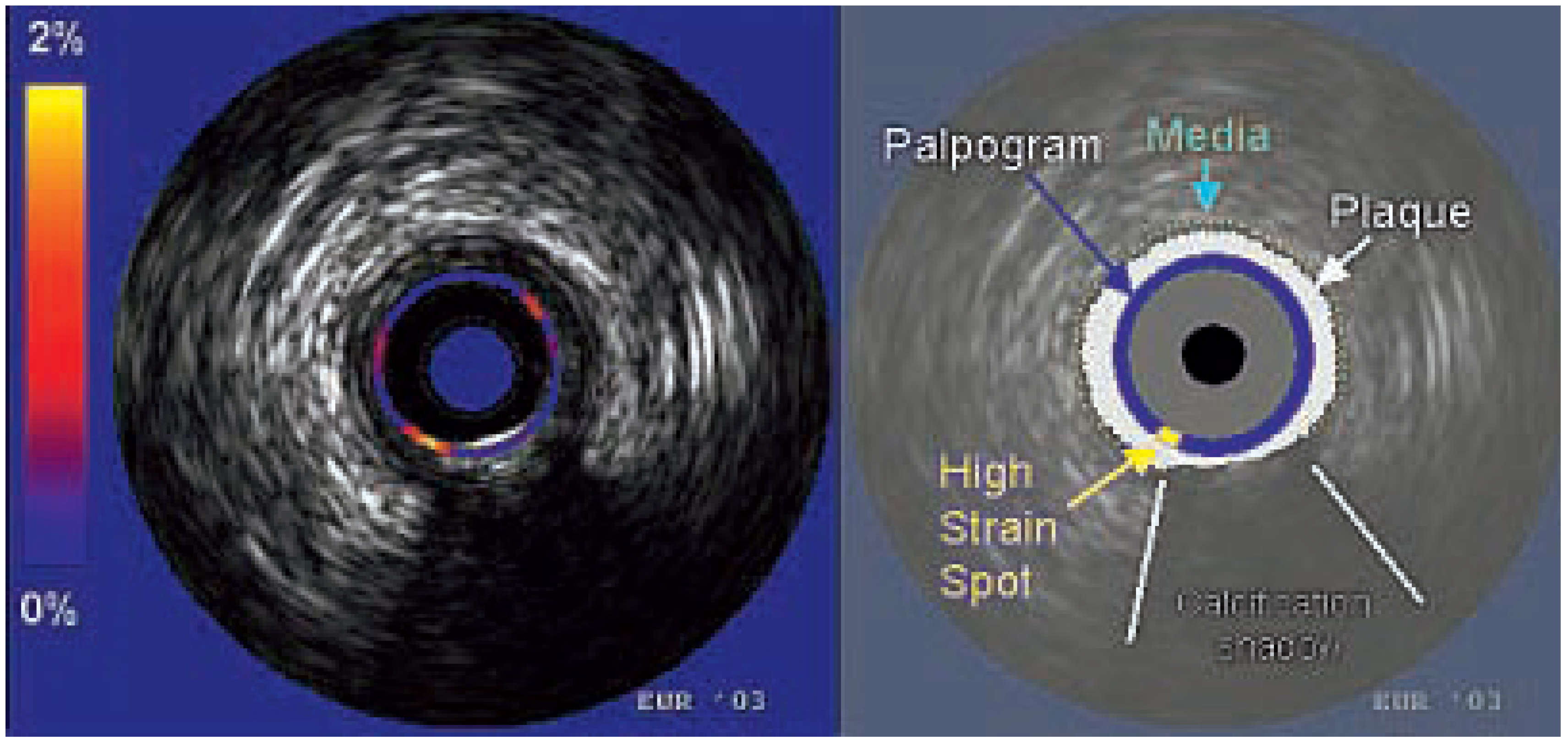
In 1991, a new technique was introduced to measure the mechanical properties of tissue using ultrasound: elastography [24]. The underlying concept is that upon uniform loading, the local relative amount of deformation (strain) of a tissue is related to the local mechanical properties of that tissue. If we apply this concept to determine the local properties of arterial tissue, blood pressure acts as a stressor. At a given pressure difference, soft plaque components will deform more than hard components. Measurement of local plaque deformation in the radial direction can be obtained with ultrasound. In-vitro studies with histological confirmation have shown that there are differences of strain normalised to pressure between fibrous, fibro-fatty and fatty components of the plaque of coronary as well as femoral arteries [25]. This difference was mainly evident between fibrous and fatty tissue. Interestingly, these plaque types could not be differentiated by echo-intensity differences on the IVUS echogram. In another invitro study, post-mortem coronary arteries were investigated with elastography and then processed for histology. The sensitivity and specificity of elastography to detect TCFA were, respectively, 88% and 89%. Furthermore, there was a high correlation between the strain in the cap and the amount of macrophages [26]. For intra-vascular purposes, a derivate of elastography called palpography may be a suitable tool [27]. In this approach, one strain value per angle is determined and plotted as a color-coded contour at the lumen vessel boundary. Since radial strain is obtained, the technique may have the potential to detect regions with elevated stress: increased circumferential stress results in an increased radial deformation of the plaque components. It is feasible to apply intra-vascular palpography during catheterisation procedures. The systemic pressure is used to strain the tissue, and the strain is determined using cross-correlation analysis of sequential frames acquired at different pressures. A likelihood function is determined to obtain the frames with minimal motion of the catheter in the lumen since motion of the catheter impairs accuracy of strain estimation. Minimal motion is mainly observed near the end of the passive filling phase. Reproducible strain estimates are obtained within one pressure cycle and over several pressure cycles. Palpography has been shown to detect, in human coronary arteries, strain patterns typical of deformable plaques (fig. 4). Furthermore, the number of deformable plaques per patient correlated positively with the clinical presentation and with the serum level of C-reactive protein [28]. Palpography provides additional information to IVUS. The differentiation between hard and soft tissue may be important for the detection of a vulnerable plaque. Since palpography is based on clinically available IVUS catheters, the technique can be easily introduced into the catheterisation laboratory. The clinical value of this technique is currently under investigation in the Integrated Biomarkers and Imaging Study (IBIS) trial.

Thermography

Since atherosclerosis is an inflammatory disease [29] and inflammation determines an elevation in temperature, hypothetically a temperature rise should be measured at the surface of a plaque. Furthermore, as vulnerable plaque is a very active metabolic area, it has been postulated that even higher temperature could be found, due to heat released by activated macrophages either on the plaque surface or under a thin cap. The pioneering paper by Casscells et al. reported that carotid plaques taken at endoatherectomy have temperature heterogeneity. The temperature difference (measured outside the body, at room temperature) between different areas was up to 2.2 5C, and correlated with cell density (mainly macrophages) [30]. Stefanadis et al. performed studies in humans. Patients with stable angina, unstable angina, and acute MI were studied. Temperature was constant within the arteries of the control subjects, whereas most atherosclerotic plaques showed higher temperatures compared with healthy vessel wall. Temperature differences between atherosclerotic plaque and healthy vessel wall increased progressively from stable angina to acute MI with a maximum difference of 1.5 ± 0.7 5C [31]. Furthermore, a high temperature gradient (>0.5 5C) between the atherosclerotic plaque in the culprit vessel and the healthy vessel wall was shown to be an independent predictor of adverse events after a percutaneous coronary intervention [32]. These data has yet to be confirmed prospectively in other centers, and the influence of parameters such as coronary blood flow or catheter design has to be studied in the future.

Optical coherence tomography

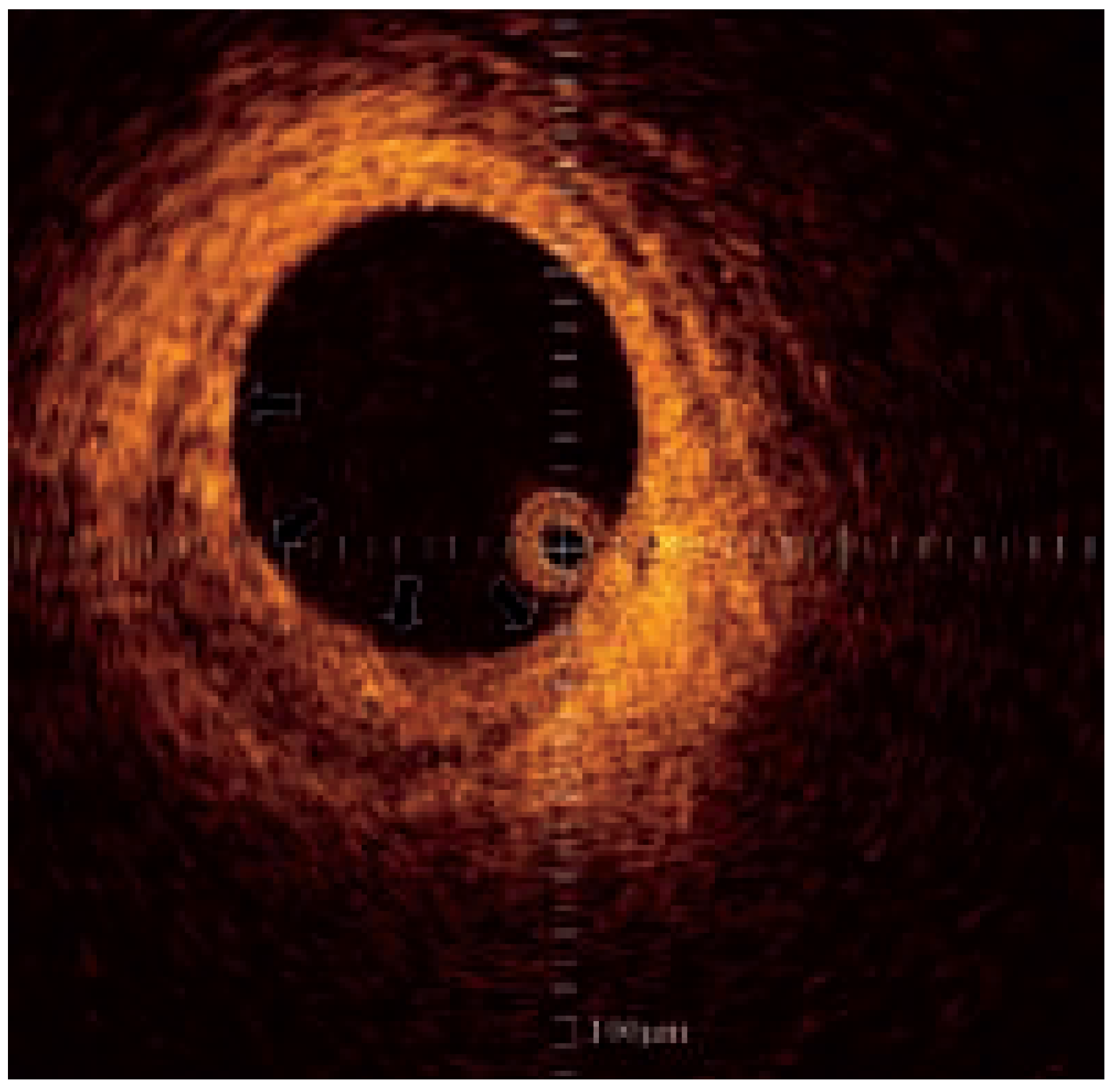
Optical coherence tomography (OCT) can provide images with ultra-high resolution. The technique measures the intensity of back-reflected light in a similar way as IVUS measures acoustic waves. Light is split into two signals: one is sent into the tissue and the other to a reference arm with a mirror. Both signals are reflected and cross correlated by interfering the light beams. To achieve cross correlation at incremental penetration depths in the tissue, the mirror is dynamically translated. The intensity of the interfering signals at a certain mirror position represents backscattering at a corresponding depth. Images with a extremely high resolution, ranging from 4–20 mm, can be achieved with a penetration depth up to 2 mm. Images can be acquired real time (fig. 5). Early attempts were made to validate OCT using histology. A lipid pool generates decreased signal areas with poorly delineated borders, a fibro-calcific plaque shows a sharply delineated region with a signal-poor interior and a fibrous plaque produces a homogenous signal rich lesion [33]. The first in-vivo comparison of OCT with IVUS demonstrated superior delineation by OCT of structural details like thin caps or tissue proliferation [34]. However, OCT has several limitations: the low penetration depth, which hinders studying large vessels, and the light absorbance by blood which currently needs to be overcome by saline infusion or balloon occlusion with associated potential for ischaemia.

Spectroscopy

Using fiberoptic technology, coronary plaques can be illuminated in-situ and the reflected light can be collected and launched into a spectrometer. Spectroscopy is based on the property that different chemical compounds absorb and scatter different amounts of energy at different wavelengths, so each tissue, due to its chemical composition (lipid, collagen, calcium, etc.), has a unique pattern of light absorbance, leaving a unique chemical (molecular) fingerprint. Different approaches are under development. Raman spectroscopy uses high-energy laser light, it has a high molecular sensitivity but its tissue penetration is as low as 0.3 mm. Near-infrared (NIR) spectroscopy (with wavelengths from 750 to 2500 nm) has greater penetration (2 mm) but lower molecular sensitivity and therefore relies on pattern recognition for plaque typing. Intracoronary spectroscopy has not yet been tested clinically [35].

ANGUS and shear stress

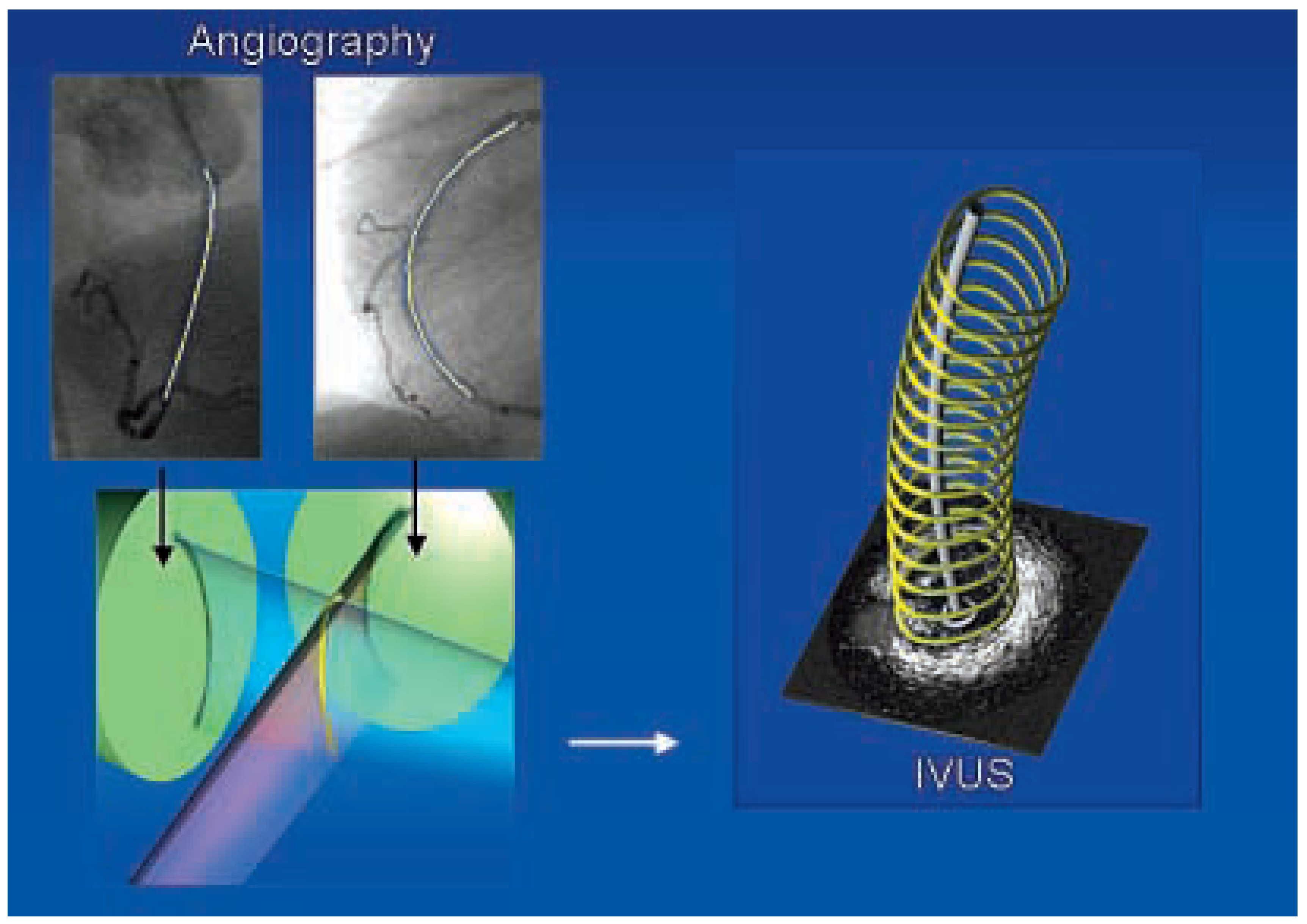
High-resolution reconstruction of three-dimensional (3D) coronary lumen and wall morphology is obtained by combining angiography and IVUS [36]. Briefly, a biplane angiogram of a sheath-based IVUS catheter taken at end-diastole allows reconstruction of the 3D pullback trajectory of the catheter. Combining this path with lumen and wall information derived from IVUS images that are successively acquired during catheter pullback at end-diastole gives accurate 3D lumen and wall reconstruction with resolution determined by IVUS (fig. 6). The use of computation flow dynamics allows calculation of detailed blood velocity profile in the lumen and shear stress on the vessel walls [37]. For this purpose absolute flow and blood viscosity need to be provided as boundary conditions. From the blood velocity profile local wall shear stress on the endothelium can be accurately derived. Wall shear stress is the frictional force, normalised to surface area that is induced by the blood passing the wall. Although from a mechanical point of view shear stress is of a very small magnitude compared to blood pressure-induced tensile stress, it has a profound influence on vascular biology and explains the localisation of atherosclerotic plaque in the presence of systemic risk factors. Many of these biological processes also influence the stability of the vulnerable plaque including inflammation, thrombogenicity, vessel remodeling, intimal thickening or regression and smooth muscle cell proliferation. Therefore, the study of this parameter as derived by image based modeling is of utmost importance.

Conclusions

Assessment of atherosclerosis by imaging techniques is essential for in-vivo identification of vulnerable plaques. The ideal technique would provide morphological, mechanical and biochemical information, however, despite several imaging techniques are currently under development, none of them provides alone such all-embracing assessment.
OCT has the advantage of high resolution, thermography has the potential to measure metabolism and spectroscopy obtains information on chemical components. IVUS and IVUS-palpography are easy to perform and assess morphology and mechanical instability. Shear stress is an important mechanical parameter deeply influencing vascular biology. Nevertheless all techniques are still under investigation and at present, none of them can completely identify a vulnerable plaque and, most importantly, predict its further development. This is related to fundamental methodological insufficiencies that may be resolved in the future. From a clinical point of view, most techniques currently assess only one feature of the vulnerable plaque. Thus the combination of several modalities will be of importance in the future to ensure a high sensitivity and specificity in detecting vulnerable plaques.

Funding

This research received no external funding.

Conflicts of Interest

The authors declare no conflict of interest.
Cardiovascmed 07 00349 i001

References

  1. Schaar, J.A.; Muller, J.E.; Falk, E.; Virmani, R.; Fuster, V.; Serruys, P.W.; et al. Terminology for high-risk and vulnerable coronary artery plaques. Eur Heart J 2004, 25, 1077–1082. [Google Scholar] [CrossRef]
  2. Kolodgie, F.D.; Burke, A.P.; Farb, A.; Gold, H.K.; Yuan, J.; Narula, J.; et al. The thin-cap fibroatheroma: a type of vulnerable plaque: the major precursor lesion to acute coronary syndromes. Curr Opin Cardiol 2001, 16, 285–292. [Google Scholar] [CrossRef]
  3. van der Wal, A.C.; Becker, A.E.; van der Loos, C.M. Das PK. Site of intimal rupture or erosion of thrombosed coronary atherosclerotic plaques is characterized by an inflammatory process irrespective of the dominant plaque morphology. Circulation 1994, 89, 36–44. [Google Scholar] [CrossRef]
  4. Virmani, R.; Kolodgie, F.D.; Burke, A.P.; Farb, A.; Schwartz, S.M. Lessons from sudden coronary death: a comprehensive morphological classification scheme for atherosclerotic lesions. Arterioscler Thromb Vasc Biol 2000, 20, 1262–1275. [Google Scholar] [CrossRef] [PubMed]
  5. Goldstein, J.A.; Demetriou, D.; Grines, C.L.; Pica, M.; Shoukfeh, M.; O’Neill, W.W. Multiple complex coronary plaques in patients with acute myocardial infarction. N Engl J Med 2000, 343, 915–922. [Google Scholar] [CrossRef] [PubMed]
  6. Frink, R.J. Chronic ulcerated plaques: new insights into the pathogenesis of acute coronary disease. J Invasive Cardio 1004, 6, 173–185. [Google Scholar]
  7. Little, W.C.; Constantinescu, M.; Applegate, R.J.; Kutcher, M.A.; Burrows, M.T.; Kahl, F.R.; et al. Can coronary angiography predict the site of a subsequent myocardial infarction in patients with mild-to-moderate coronary artery disease? Circulation 1988, 78, 1157–1166. [Google Scholar] [CrossRef]
  8. Ambrose, J.A.; Tannenbaum, M.A.; Alexopoulos, D.; HjemdahlMonsen, C.E.; Leavy, J.; Weiss, M.; et al. Angiographic progression of coronary artery disease and the development of myocardial infarction. J Am Coll Cardiol 1988, 12, 56–62. [Google Scholar]
  9. Ojio, S.; Takatsu, H.; Tanaka, T.; Ueno, K.; Yokoya, K.; Matsubara, T.; et al. Considerable time from the onset of plaque rupture and/or thrombi until the onset of acute myocardial infarction in humans: coronary angiographic findings within 1 week before the onset of infarction. Circulation 2000, 102, 2063–2069. [Google Scholar] [CrossRef]
  10. Asakura, M.; Ueda, Y.; Yamaguchi, O.; Adachi, T.; Hirayama, A.; Hori, M.; et al. Extensive development of vulnerable plaques as a pan-coronary process in patients with myocardial infarction: an angioscopic study. J Am Coll Cardiol 2001, 37, 1284–1288. [Google Scholar] [CrossRef]
  11. Uchida, Y.; Nakamura, F.; Tomaru, T.; Morita, T.; Oshima, T.; Sasaki, T.; et al. Prediction of acute coronary syndromes by percutaneous coronary angioscopy in patients with stable angina. Am Heart J 1995, 130, 195–203. [Google Scholar] [CrossRef]
  12. Di Mario, C.; The, S.H.; Madretsma, S.; van Sluyten, R.J.; Wilson, R.A.; Bom, N.; et al. Detection and characterization of vascular lesions by intravascular ultrasound: an in vitro study correlated with histology. J Am Soc Echocardiogr 1992, 5, 135–146. [Google Scholar] [CrossRef]
  13. Friedrich, G.J.; Moes, N.Y.; Muhlberger, V.A.; Gabl, C.; Mikuz, G.; Hausmann, D.; et al. Detection of intralesional calcium by intracoronary ultrasound depends on the histologic pattern. Am Heart J 1994, 128, 435–441. [Google Scholar] [CrossRef] [PubMed]
  14. Potkin, B.N.; Bartorelli, A.L.; Gessert, J.M.; Neville, R.F.; Almagor, Y.; Roberts, W.C.; et al. Coronary artery imaging with intravascular high-frequency ultrasound. Circulation 1990, 81, 1575–1585. [Google Scholar] [CrossRef]
  15. Hiro, T.; Leung, C.Y.; Russo, R.J.; Karimi, H.; Farvid, A.R.; Tobis, J.M. Variability of a three-layered appearance in intravascular ultrasound coronary images: a comparison of morphometric measurements with four intravascular ultrasound systems. Am J Card Imaging 1996, 10, 219–227. [Google Scholar]
  16. Ge, J.; Chirillo, F.; Schwedtmann, J.; Gorge, G.; Haude, M.; Baumgart, D.; et al. Screening of ruptured plaques in patients with coronary artery disease by intravascular ultrasound. Heart 1999, 81, 621–627. [Google Scholar] [CrossRef]
  17. Riouful, G.; Finet, G.; Ginon, I.; Andre-Fouet, X.; Rossi, R.; Vialle, E.; et al. Multiple atherosclerotic plaque rupture in acute coronary syndrome: a three-vessel intravascular ultrasound study. Circulation 2002, 106, 804–808. [Google Scholar] [CrossRef]
  18. Fujii, K.; Kobayashi, Y.; Mintz, G.S.; Takebayashi, H.; Dangas, G.; Moussa, I.; et al. Intravascular ultrasound assessment of ulcerated ruptured plaques: a comparison of culprit and nonculprit lesions of patients with acute coronary syndromes and lesions in patients without acute coronary syndromes. Circulation 2003, 108, 2473–2478. [Google Scholar] [CrossRef]
  19. Yamagishi, M.; Terashima, M.; Awano, K.; Kijima, M.; Nakatani, S.; Daikoku, S.; et al. Morphology of vulnerable coronary plaque: insights from follow-up of patients examined by intravascular ultrasound before an acute coronary syndrome. J Am Coll Cardiol 2000, 35, 106–111. [Google Scholar] [CrossRef] [PubMed]
  20. von Birgelen, C.; Klinkhart, W.; Mintz, G.S.; Papatheodorou, A.; Hermann, J.; Baumgart, D.; et al. Plaque distribution and vascular remodeling of ruptured and non-ruptured coronary plaques in the same vessel: an intravascular ultrasound study in vivo. J Am Coll Cardiol 2001, 37, 1864–1870. [Google Scholar] [CrossRef] [PubMed]
  21. Nair, A.; Kuban, B.D.; Obuchowski, N.; Vince, D.G. Assessing spectral algorithms to predict atherosclerotic plaque composition with normalized and raw intravascular ultrasound data. Ultrasound Med Biol 2001, 10, 1319–1331. [Google Scholar] [CrossRef]
  22. Landini, L.; Sarnelli, R.; Picano, E.; Salvadori, M. Evaluation of frequency dependence of backscatter coefficient in normal and atherosclerotic aortic walls. Ultrasound Med Biol 1986, 5, 397–401. [Google Scholar] [CrossRef]
  23. Nair, A.; Kuban, B.D.; Tuzcu, E.M.; Schoenhagen, P.; Nissen, S.E.; Vince, D.G. Coronary plaque classification with intravascular ultrasound radiofrequency data analysis. Circulation 2002, 106, 2200–2206. [Google Scholar] [CrossRef]
  24. Ophir, J.; Cespedes, I.; Ponnekanti, H.; Yazdi, J.; Li, X. Elastography: a quantitative method for imaging the elasticity of biological tissues. Ultrason Imaging 1991, 13, 111–134. [Google Scholar] [CrossRef] [PubMed]
  25. de Korte, C.L.; Pasterkamp, G.; van der Steen, A.F.; Woutman, H.A.; Bom, N. Characterization of plaque components with intravascular ultrasound elastography in human femoral and coronary arteries in vitro. Circulation 2000, 102, 617–623. [Google Scholar] [CrossRef] [PubMed]
  26. Schaar, J.A.; deKorte, C.L.; Mastik, F.; Strijder, C.; Pasterkamp, G.; Boersma, E.; et al. Characterizing vulnerable plaque features with intravascular elastography. Circulation 2003, 108, 2636–2641. [Google Scholar] [CrossRef] [PubMed]
  27. Doyley, M.M.; Mastik, F.; de Korte, C.L.; Carlier, S.C.; Cespedes, E.I.; Serruys, P.W.; et al. Advancing intravascular ultrasonic palpation toward clinical applications. Ultrasound Med Biol 2001, 27, 1471–1480. [Google Scholar] [CrossRef]
  28. Schaar, J.A.; Regar, E.; Mastk, F.; McFadden, E.P.; Saia, F.; Disco, C.; et al. Incidence of high strain patterns in human coronary arteries: assessment with three-dimensional intravascular palpography and correlation with clinical presentation. Circulation 2004, 109, 2716–2719. [Google Scholar] [CrossRef]
  29. Ross, R. Atherosclerosis: an inflammatory disease. N Engl J Med 1999, 340, 115–126. [Google Scholar] [CrossRef]
  30. Casscells, W.; Hathorn, B.; David, M.; Krabach, T.; Vaughn, W.K.; McAllister, H.A.; et al. Thermal detection of cellular infiltrates in living atherosclerotic plaques: possible implications for plaque rupture and thrombosis. Lancet 1996, 347, 1447–1451. [Google Scholar] [CrossRef]
  31. Stefanidis, C.; Diamantopulos, L.; Vlachopulos, C.; Tsiamis, E.; Dernellis, J.; Toutouzas, K.; et al. Thermal heterogeneity within human atherosclerotic coronary arteries detected in vivo: a new method of detection by application of a special thermography catheter. Circulation 1999, 99, 1965–1971. [Google Scholar] [CrossRef]
  32. Stefanadis, C.; Toutouzas, K.; Tsiamis, E.; Stratos, C.; Vavuranakis, M.; Kallikazaros, I.; et al. Increased local temperature in human coronary atherosclerotic plaques: an independent predictor of clinical outcome in patients undergoing a percutaneous coronary intervention. J Am Coll Cardiol 2001, 37, 1277–1283. [Google Scholar] [CrossRef]
  33. Yabushita, H.; Bouma, B.E.; Houser, S.L.; Aretz, T.; Jang, I.K.; Schlendorf, K.H.; et al. Characterization of human atherosclerosis by optical coherence tomography. Circulation 2002, 106, 1640–1645. [Google Scholar] [CrossRef]
  34. Jang, I.K.; Bouma, B.E.; Kang, D.H.; Park, S.J.; Park, S.W.; Seung, K.B.; et al. Visualization of coronary atherosclerotic plaques in patients using optical coherence tomography: comparison with intravascular ultrasound. J Am Coll Cardiol 2002, 39, 604–609. [Google Scholar] [CrossRef] [PubMed]
  35. Moreno, P.R.; Muller, J.E. Detection of high-risk atherosclerotic coronary plaques by intravascular spectroscopy. J Interv Cardiol 2003, 16, 243–252. [Google Scholar] [CrossRef] [PubMed]
  36. Slager, C.J.; Wentzel, J.J.; Schuurbiers, J.C.H.; Oomen, J.A.F.; Kloet, J.; Krams, R.; et al. True 3-dimensional reconstruction of coronary arteries in patients by fusion of angiography and IVUS (ANGUS) and its quantitative validation. Circulation 2000, 102, 511–516. [Google Scholar] [CrossRef] [PubMed]
  37. Wentzel, J.J.; Janssen, E.; Vos, J.; Schuurbiers, J.C.H.; Krams, R.; Serruys, P.W.; et al. Extension of increased wall thickness into high shear stress regions is associated with loss of compensatory remodeling. Circulation 2003, 108, 17–23. [Google Scholar] [CrossRef]
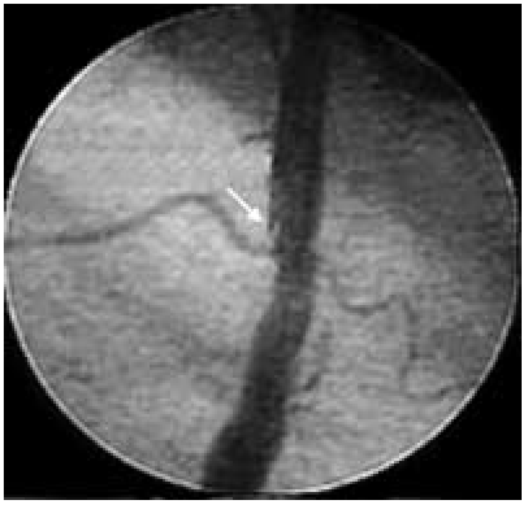
Figure 1. Angiography reveals a dissected flap within the lumen, indicating a ruptured plaque.
Cardiovascmed 07 00349 g001
Figure 2. Angioscopy shows (A). a red thrombus on a plaque (1), blocking a part of the lumen (2). (B). After removal of thrombus and plaque with atherectomy, a white flap is visible (3). (C). Post angioplasty angioscopy can show a dissecting flap (4) within the lumen.
Cardiovascmed 07 00349 g002
Figure 3. The intra-vascular ultrasound cross-section shows an eccentric ruptured plaque with deep ulcus (arrows). A thin flap is still covering parts of the ulceration. (Courtesy of Paul Schoenhagen).
Cardiovascmed 07 00349 g003
Figure 4. The palpogram shows an eccentric plaque with a big calcification. On the left shoulder there is a high strain spot of an otherwise less deformable plaque, probably representing a vulnerable plaque.
Cardiovascmed 07 00349 g004
Figure 5. Optical coherence tomography produces high-resolution, real-time, cross-sectional, or three-dimensional images of tissues at a exceptionally high, histology-like resolution of a eccentric plaque with a thin cap (arrows). (Courtesy of Evelyn Regar).
Cardiovascmed 07 00349 g005
Figure 6. 3D reconstruction technique combining ANGiography and IVUS allowing an exact reconstruction of the vessel shape.
Cardiovascmed 07 00349 g006