Next Article in Journal
Aortic Valve Repair with a Novel Rigid Annuloplasty Ring
Previous Article in Journal
A New Deputy Editor for Cardiovascular Medicine
 
 
Cardiovascular Medicine is published by MDPI from Volume 28 Issue 1 (2025). Previous articles were published by another publisher in Open Access under a CC-BY (or CC-BY-NC-ND) licence, and they are hosted by MDPI on mdpi.com as a courtesy and upon agreement with Editores Medicorum Helveticorum (EMH).
Interesting Images

An Unusual Heart Disease in a Young Old Boy

by
Corinna Leoni Foglia
1,*,
Marcello Di Valentino
2 and
Andrea Menafoglio
2
1
Department of Paediatric Cardiology, San Giovanni Hospital, Bellinzona, Switzerland
2
Department of Cardiology, San Giovanni Hospital, Bellinzona, Switzerland
*
Author to whom correspondence should be addressed.
Cardiovasc. Med. 2018, 21(6), 174; https://doi.org/10.4414/cvm.2018.00570
Submission received: 20 March 2018 / Revised: 20 April 2018 / Accepted: 20 May 2018 / Published: 20 June 2018
A 17-year-old boy was referred for a cardiological consultation because of progressive dyspnoea for 1 year. The boy was known to have a learning disability, progerioid features, muscular dystrophy, lipodystrophy, joint contractures, hyperlipidaemia and liver steatosis (Figure 1).
There were no known cardiomyopathies or neuromuscular diseases in his family.
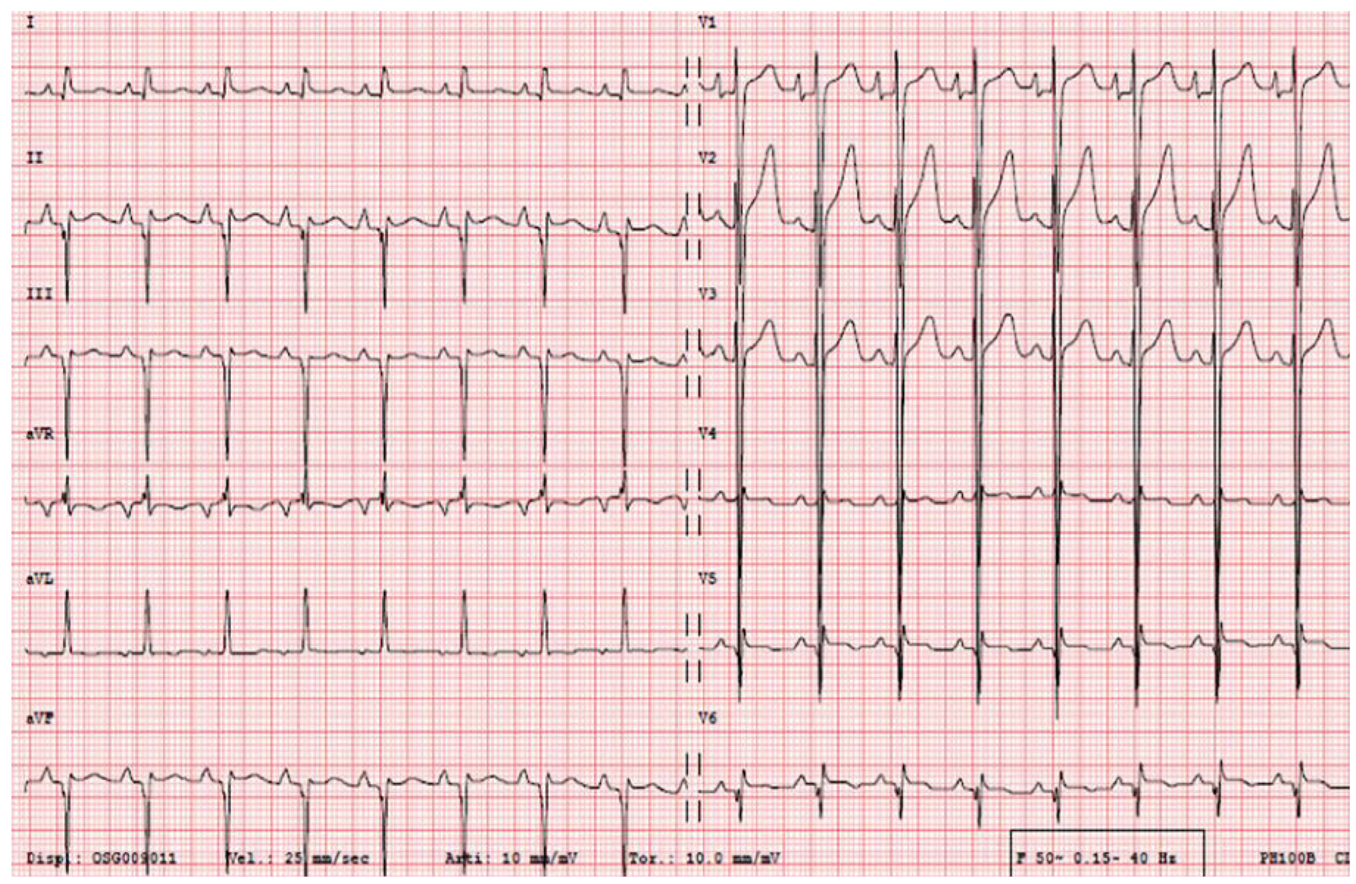
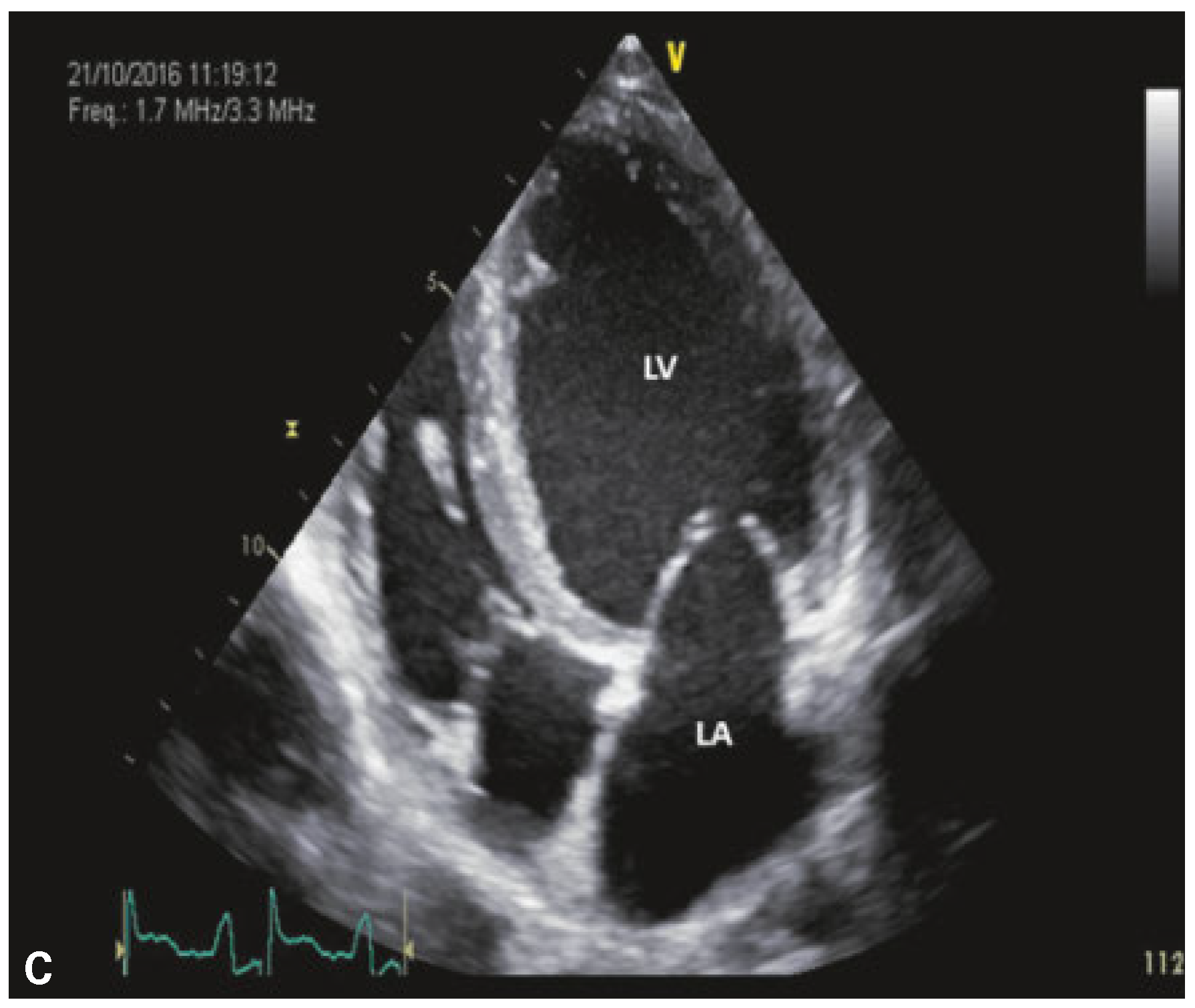
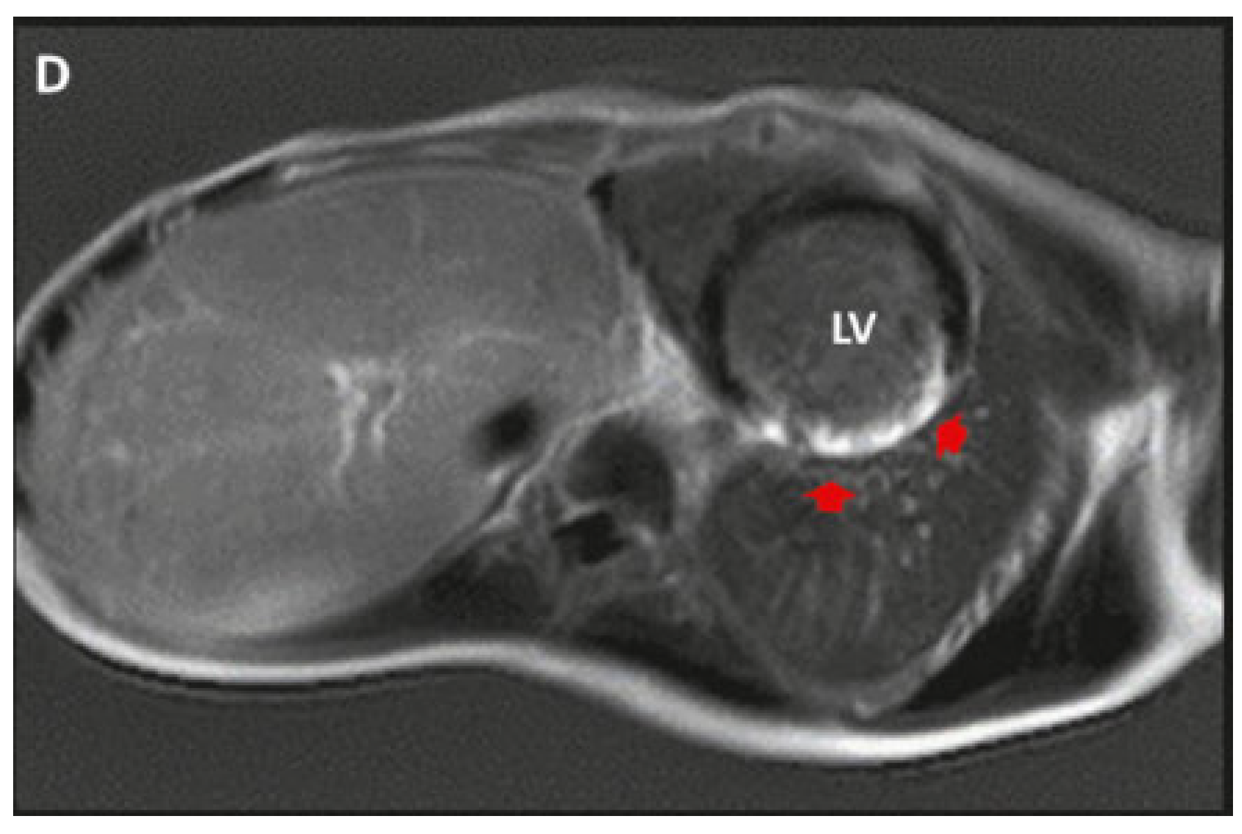
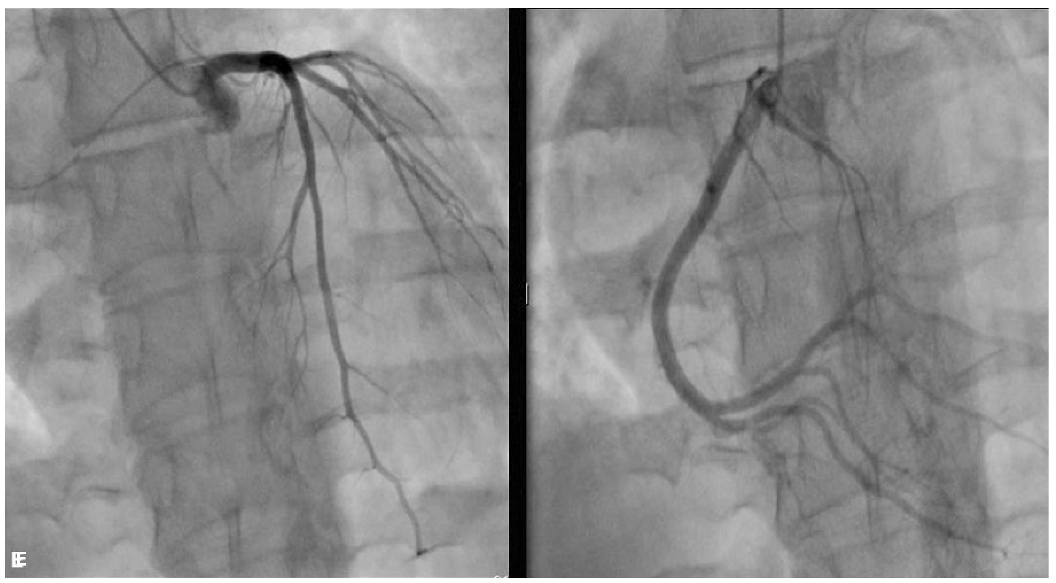
The electrocardiogram (ECG) showed left ventricular hypertrophy and Q-waves in the inferolateral leads (Figure 2). Echocardiography revealed left ventricular dilatation and severe systolic dysfunction (ejection fraction 25%) (Figure 3). On cardiac magnetic resonance imaging (MRI), myocardial transmural late gadolinium enhancement in the inferolateral wall was found (Figure 4). Coronary angiography showed normal coronary arteries (Figure 5). A 24-hour Holter ECG showed about 1000 isolated polymorphic ventricular premature beats. Treatment with a beta-blocker, angiotensin converting-enzyme (ACE) inhibitor and spironolactone was started with symptomatic improvement. Furthermore, we are considering inplantable cardioverter-defibrillator (ICD) implantation as primary prevention for sudden cardiac death.
A genetic test showed a very rare lamin gene mutation: LMNA:NM_005572:exon1:c.C29T:p.T10l.
This rare laminopathy is probably responsible for the whole clinical picture of this young patient.
The lamin gene codes for lamin A/C, which is a structural protein of the nuclear envelope. Laminopathies cause a wide variety of diseases involving skeletal muscles, adipose tissue, peripheral nerves, premature aging and cardiac involvement, including left ventricular dysfunction, atrioventricular block, and atrial and ventricular arrhythmias. The clinical manifestations could involve one or more systems. It has been estimated that LMNA mutations cause up to 10% of familial dilated cardiomyopathies. The cardiac involvement is progressive, with poor prognosis [1,2,3].
Typically, in the LMNA cardiomyopathy, a midventricular septal fibrosis is described on cardiac MRI. In our patient, despite normal coronary arteries, an inferolateral transmural scar was documented, similar to the pattern described in Duchenne and Becker muscular dystrophy [4].
In a young patient with dilated cardiomyopathy, particularly if multisystemic manifestations are present, it is important to suspect a laminopathy, because of the poor prognosis of this disease. In the presence of clinical manifestations that raise suspicion of a laminopathy, a cardiological assessment should be considered.

Conflicts of Interest

No financial support and no other potential conflict of interest relevant to this article was reported.

References

  1. Charron, P.; Arbustini, E.; Bonne, G. What should the cardiologist know about lamin disease. Arrhythm Electrophysiol Rev. 2012, 1, 22–28. [Google Scholar] [CrossRef] [PubMed]
  2. Kumar, S.; Baldinger, S.; Grandjbakhch, E.; Maury, P.; Sellal, J.M.; Androulakis, A.F.; et al. Long-term arrhythmic and nonarrhythmic outcomes of Lamin A/C mutation carriers. J Am Coll Cardiol. 2016, 68, 229–307. [Google Scholar] [CrossRef] [PubMed]
  3. van Rijsingen, I.A.; Arbustini, E.; Elliot, P.; Mogensen, J.; Hermans-van Ast, J.F.; van der Kooi, A.J.; et al. Risk factors for malignant ventricular arrhythmias in Lamin A/C mutation carriers. J Am Coll Cardiol. 2012, 59, 493–500. [Google Scholar] [CrossRef] [PubMed]
  4. Holmström, M.; Kivistö, S.; Heliö, T.; Jurkko, R.; Kaartinen, M.; Antila, M.; et al. Late gadolinium enhanced cardiovascular magnetic resonance of lamin A/C gene mutation related dilated cardiomyopathy. J Cardiovasc Magn Reson. 2011, 13, 30. [Google Scholar] [CrossRef] [PubMed]
Figure 1. Clinical appearance of the patient: note the progerioid features, the muscular dystrophy, the lipodystrophy and the joint contractures. The picture of the patient is published with his consent and the consent of his parents.
Cardiovascmed 21 00174 g001
Figure 2. ECG showing sinus rhythm, Q-waves in the inferolateral leads and signs of left ventricular hypertrophy.
Cardiovascmed 21 00174 g002
Figure 3. Echocardiography four chamber view showing left ventricular (LV) dilatation. LA: left atrium.
Cardiovascmed 21 00174 g003
Figure 4. Cardiac magnetic resonance imaging with myocardial transmural late gadolinium enhancement in the inferolateral wall (red arrowheads).
Cardiovascmed 21 00174 g004
Figure 5. Coronary angiography showing normal coronary arteries.
Cardiovascmed 21 00174 g005