Next Article in Journal
Tricuspid Regurgitation: Assessment and New Frontiers
Previous Article in Journal
Der Leiter des Universitären Herzzentrums Zürich, Professor Thomas F. Lüscher, Folgt Einem Ruf Nach London—Und Bleibt Doch Ein Bisschen in Zürich
 
 
Cardiovascular Medicine is published by MDPI from Volume 28 Issue 1 (2025). Previous articles were published by another publisher in Open Access under a CC-BY (or CC-BY-NC-ND) licence, and they are hosted by MDPI on mdpi.com as a courtesy and upon agreement with Editores Medicorum Helveticorum (EMH).
Interesting Images

Incomplete Stent Apposition Resolves One Year After Implantation

by
Florian Basile Stierlin
,
Stéphane Cook
* and
Mathieu Stadelmann
Department of Cardiology, University & Hospital Fribourg, CH-1708 Fribourg, Switzerland
*
Author to whom correspondence should be addressed.
Cardiovasc. Med. 2017, 20(9), 216; https://doi.org/10.4414/cvm.2017.00515
Submission received: 20 June 2017 / Revised: 20 July 2017 / Accepted: 20 August 2017 / Published: 20 September 2017

Abstract

Incomplete stent apposition (ISA) is proven to increase thrombotic risk, particularly late stent thrombosis of a drug-eluting stent. Less is known about ISA and bioresorbable scaffold (BRS). We describe the coronary angiogram and optical coherence tomography (OCT) changes at one year in a patient with ISA of Magmaris BRS, in the context of coronary artery disease without acute coronary syndrome. In this particular case, the OCT demonstrated a very good result with complete stent resorption, suggesting that the rapid BRS kinetics resorption could lessen the risk of complications attributed to ISA.

Case report

We report the case of a 61-year-old man known for hypercholesterolaemia, a history of smoking and positive family history, who presented with progressive dyspnoea without oppressive chest pain. Stress echocardiography showing inferior basal akinesia extending to the septum during maximal effort prompted a coronary angiogram.
The coronary angiogram demonstrated two significant lesions in the right coronary artery. The first situated was on the proximal portion (visually estimated to be 70% occlusion) and the second on the distal portion (visually estimated to be 70–90%) (Figure 1a). We first treated the proximal stenosis. We performed an angioplasty (NC EMERGE 3 × 12 mm—Boston Scientific©) at a maximal pressure of 12 atm. for 50 s and implanted a bioresorbable scaffold (BRS) (Magmaris 3.5 × 15 mm—Biotronik®) with an inflation of 12 atm. for 30 s.
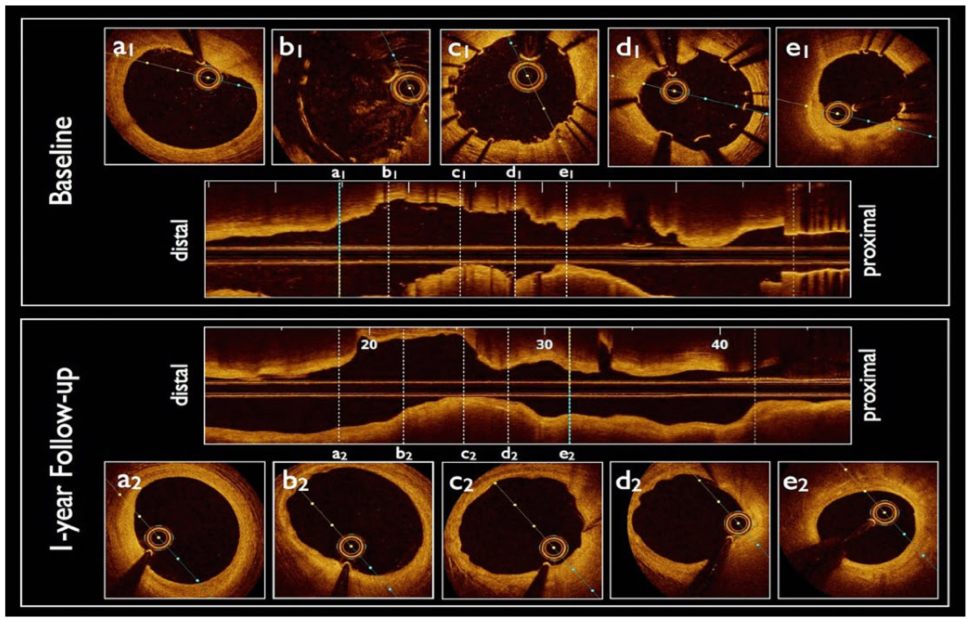
An optical coherence tomography (OCT) revealed a 330–720 µm incomplete stent apposition (ISA) of the BRS, which was treated with two further balloon inflations (NC EMERGE 3.5 × 15 mm—Boston scientific®) at 14 atm. for 23 s (Figure 2a1, b1, c1, d1, e1,). Despite these inflations, the BRS ISA, measured as a maximum of 570 µm, persisted (Figure 2d1). Because of this unsatisfactory OCT result, the distal lesion was treated with a conventional metallic everolimus drug-eluting stent (2.75 × 18 mm Xience Alpine—Abbott®). The final angiographic end-result was satisfactory. The patient was prescribed aspirin 100 mg/d and prasugrel 10 mg/d for 12 months.
Thirteen months later, facing a persistent dyspnea, a new coronary angiogram was performed. The examination confirmed an excellent result post-stenting of the BVS on the right proximal coronary artery despite the initial ISA (panel b, Figure 1). An OCT (Dragon Fly Optis—St. Jude®) confirmed the absence of stenosis and showed a complete dissolution of the BVS initially implanted (panels a2, b2, c3, d2 and e2, Figure 2). The 1-year follow-up OCT also revealed presence of struts residues with peristrut low intensity areas (PSLIA) and evaginations (panels b2, c2, d2, Figure 2) corresponding with the vessel wall areas where the BVS was initially well-apposed. On the contrary, the areas where te BVS was initially mal-apposed showed an intact artery wall and complete BRS resorption (panel d2 and e2,. Figure 2).

Discussion

ISA is defined as an absence of contact between at least one stent strut and the intimal surface of the artery not overlying a side branch, and can be acute (at the time of implantation) or late [1]. The diagnosis is made by means of intravascular imaging, such as OCT. ISA reduces strut coverage [2,3], which enhances the risk of stent thrombosis, particularly late stent thrombosis, as already described, for drug-eluting stents [4].
During the drug-eluting stent era, four distinct mechanisms of ISA were reported: (a) incomplete apposition of the stent in the vicinity of a severely calcified lesion with localised expansion of the platform (lever principle); (b) gradual recoil of a weak stent platform due to coronary pulsatility (chronic recoil); (c) dissolution of a former jailed thrombus or soft plaque (jailed thrombus); (d) positive remodelling of the coronary artery due to a hypersensitivity reaction, with subsequent ISA.
In this case, and despite good lesion preparation and optimal sizing, the Magmaris BRS had a non-trivial ISA that directly followed the proximal RCA bend (superior knee). The reason for ISA in this case probably reflects the weak radial strength of the BRS platform in the middle of the RCA bend. On the basis of the study by Onuma and colleagues [5], it is considered that magnesium alloy has a 5- to 6-fold lower tensile strength than cobalt chromium. Postdilatation did not affect the ISA. An aggressive postdilatation with larger balloons would not have improved the radial strength of the BRS and would have entailed a significant risk of strut rupture. Owing to rapid BRS resorption kinetics (within the 1-year period of dual antiplatelet therapy), the operator estimated that the risk of stent thrombosis of the Magmaris was lower with the residual ISA than in the case of rupture (and collapse) of the scaffold. Aggressive postdilatation has been promoted by Cuculi and colleagues [6]. The Luzern team presented an increased rate of stent thrombosis due to Magmaris rupture and collapse at the 2017 SSC (Swiss Cardiology Society) meeting held in Baden. In the case of Magmaris implantation, our opinion is that aggressive postdilatation is not mandatory. This case set a good example, since the 13-month OCT follow-up demonstrated that the Magmaris BRS was completely resorbed, and this patient did not experience any clinical event under “strong” dual antiplatelet therapy.

Disclosure Statement

No financial support and no other potential conflict of interest relevant to this article was reported.

References

  1. Lindsey, J.B.; Marso, S.P. Incomplete stent apposition: Should we appose or oppose? JACC Cardiovasc Interv. 2010, 3, 495–497. [Google Scholar] [CrossRef] [PubMed]
  2. Foin, N.; Gutiérrez-Chico, J.L.; Nakatani, S.; Torii, R.; Bourantas, C.V.; Sen, S.; et al. Incomplete stent apposition causes high shear flow disturbances and delay in neointimal coverage as a function of strut to wall detachment distance: Implications for the management of incomplete stent apposition. Circ Cardiovasc Interv. 2014, 7, 180–9. [Google Scholar] [CrossRef] [PubMed]
  3. Gomez-Lara, J.; Radu, M.; Brugaletta, S.; Farooq, V.; Diletti, R.; Onuma, Y.; et al. Serial analysis of the malapposed and uncovered struts of the new generation of everolimus-eluting bioresorbable scaffold with optical coherence tomography. JACC Cardiovasc Interv. 2011, 4, 992–1001. [Google Scholar] [CrossRef] [PubMed]
  4. Cook, S.; Wenaweser, P.; Togni, M.; Billinger, M.; Morger, C.; Seiler, C.; et al. Incomplete stent apposition and very late stent thrombosis after drug-eluting stent implantation. Circulation. 2007, 115, 2426–2434. [Google Scholar] [CrossRef] [PubMed]
  5. Onuma, Y.; Serruys, P.W. Rather Thick, Yet Antithrombogenic: Is the Magmaris Scaffold a New Hope for Bioresorbable Coronary Scaffold? Circ Cardiovasc Interv. 2017, 10. [Google Scholar] [CrossRef] [PubMed]
  6. Puricel, S.; Cuculi, F.; Weissner, M.; Schmermund, A.; Jamshidi, P.; Nyffenegger, T.; et al. Bioresorbable Coronary Scaffold Thrombosis: Multicenter Comprehensive Analysis of Clinical Presentation, Mechanisms, and Predictors. J Am Coll Cardiol. 2016, 67, 921–931. [Google Scholar] [CrossRef] [PubMed]
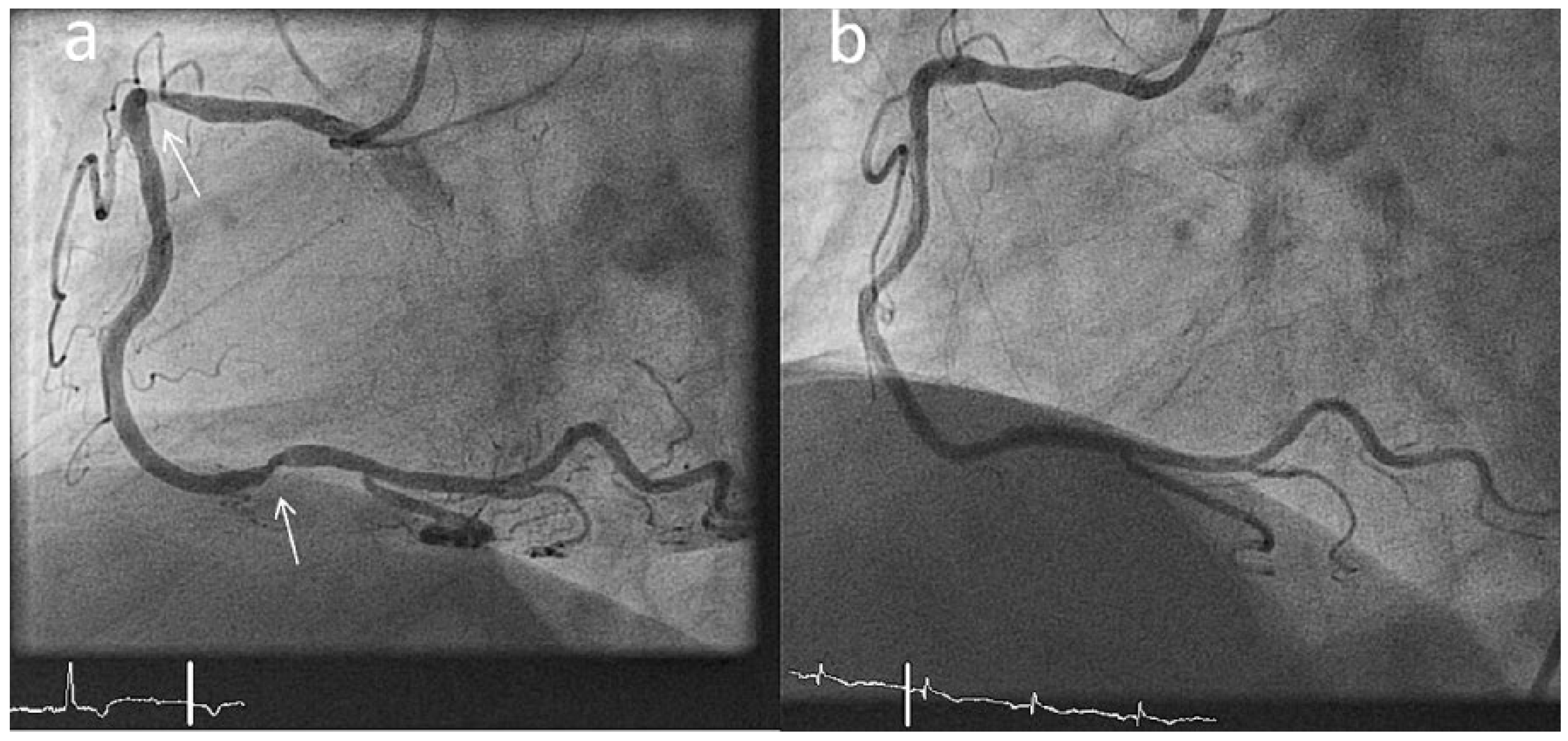
Figure 1. Right coronary angiogram showing stenosis on the proximal and distal portion of the artery (panel a). One-year follow-up right coronary angiogram confirming absence of restenosis of the vessel (panel b).
Cardiovascmed 20 00216 g001
Figure 2. Optical coherence tomography (OCT) of the right coronary artery during the first coronary angiogram (baseline) and one year after (1-year follow-up). Incomplete stent implantation after balloon inflations (panels a1, b1, c1, d1, e1). One-year follow-up OCT showing peri-strut low intensity areas and endothelial evaginations where the BRS was well-apposed (panels b2, c2, d2) and normal endothelial wall where the incomplete stent apposition existed (panels a2, e2).
Cardiovascmed 20 00216 g002