Next Article in Journal
Going to High Altitude with Heart Disease
Previous Article in Journal
Data Sharing: A New Editorial Initiative of the International Committee of Medical Journal Editors
 
 
Cardiovascular Medicine is published by MDPI from Volume 28 Issue 1 (2025). Previous articles were published by another publisher in Open Access under a CC-BY (or CC-BY-NC-ND) licence, and they are hosted by MDPI on mdpi.com as a courtesy and upon agreement with Editores Medicorum Helveticorum (EMH).
Interesting Images

Myxoma of the Pulmonary Valve

by
Olivier Roux
1,
Francine Tinguely
2,
Jean-Luc Barras
3,
René Prêtre
3 and
Jean-Jacques Goy
1,*
1
Division of Cardiology, University and Hospital, Fribourg, Switzerland
2
Clinique Cecil, Lausanne, Switzerland
3
University Hospital CHUV, Lausanne, Switzerland
*
Author to whom correspondence should be addressed.
Cardiovasc. Med. 2017, 20(5), 136; https://doi.org/10.4414/cvm.2017.00474
Submission received: 17 February 2017 / Revised: 17 March 2017 / Accepted: 17 April 2017 / Published: 17 May 2017
An 16-year-old male patient with a family history of hypertrophic obstructive cardiomyopathy was referred for screening echocardiography. He was asymptomatic with no medical history, took no medication and exercised regularly by playing football. Clinical status and 12-lead ECG were entirely normal. Echocaradiography showed a normal left ventricle anatomy with normal function. On transthoracic echocardiography, an 8 × 10 mm mass located in the right ventricular infundibulum, without haemodynamic obstruction, was incidentally found (Figure 1A,B). The differential diagnosis was fibroelastoma, organised thrombus, lipoma, rhabdomyoma, fibroma, or myxoma. Echocardiographic findings were confirmed on a computed tomography scan (Figure 1C,D). The mass had the same density as the septal muscle. The patient underwent surgical removal, which confirmed the presence of the mass partially attached to the pulmonary valve (Figure 1E,F). Pathophysiological analysis revealed a cardiac myxoma with a typical histopathological appearance: this lesion was composed of uniform spindle- and stellate-shaped cells with abundant eosinophilic cytoplasm, indistinct cell borders, oval and small nuclei with open chromatin. The cells were dispersed in a relatively myxoid stroma in which sparse capillarysized blood vessels were seen. The margin was not well-defined and invaded the non-atrophic cardiomyocytes on which it lay (Figure 1G). The tumour cells were diffusely positive for SMA (smooth muscle actin – Figure 1H), and for CD34 (Figure 1J). The outcome was uneventful and transthoracic echocardiography at day 30 and year 1 was normal.
This is an extremely rare case of a myxoma of the right-sided cavities, especially with involvement of the pulmonary valve and right ventricular outflow tract.

Conflicts of Interest

No financial support and no other potential conflict of interest relevant to this article was reported.

References

  1. Gribaa, R.; Slim, M.; Kortas, C.; Kacem, S.; Ben Salem, H.; Ouali, S.; et al. Right ventricular myxoma obstructing the right ventricular outflow tract: a case report. J Med Case Rep. 2014, 8, 435. [Google Scholar] [CrossRef] [PubMed]
  2. Rao, P.A.; Nagendra Prakash, S.N.; Vasudev, S.; Girish, M.; Srinivas, A.; Guru Prasad, H.P.; et al. A rare case of right ventricular myxoma causing recurrent stroke. Indian Heart J. 2016, 68, 97–101. [Google Scholar] [CrossRef] [PubMed][Green Version]
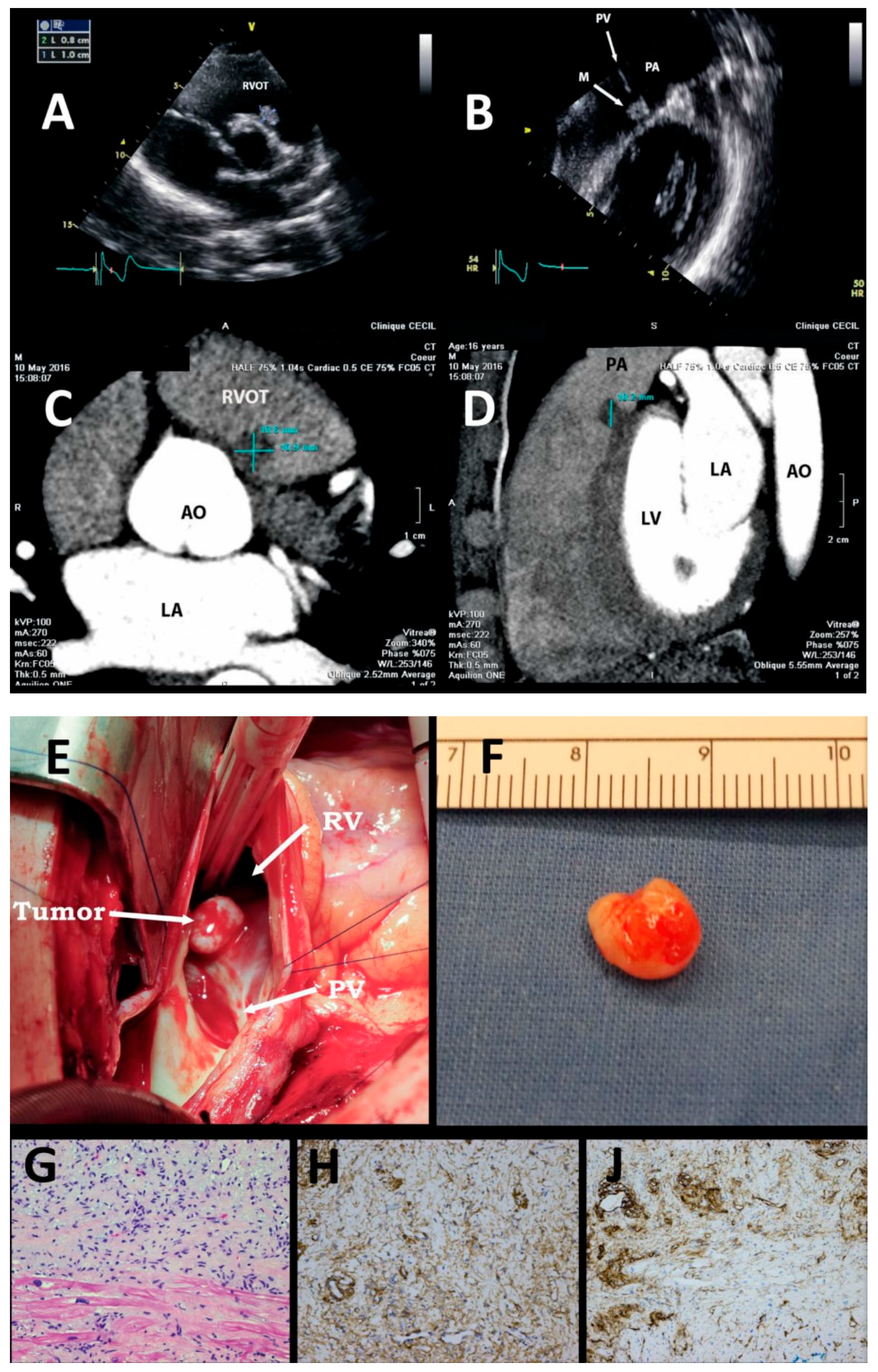
Figure 1. The mass with different imaging methods and views : by transthoracic echocardiography (Figure 1A,B), by CT scan transverse view (Figure 1C) and sagittal view (Figure 1D), surgical view of the mass, partially attached to the PV in the RVOT (Figure 1E), the surgical specimen (Figure 1F), and histopathology of the mass (Figure 1G, H, J). AO = aorta; LA = left atrium; LV = left ventricle; M = mass; PA = pulmonary artery; PV = pulmonary valve; RVOT = right ventricle outflow tract.
Cardiovascmed 20 00136 g001