Next Article in Journal
Pericarditis in a Swiss Regional Hospital
Previous Article in Journal
The Evolution of Surgical Valves
 
 
Cardiovascular Medicine is published by MDPI from Volume 28 Issue 1 (2025). Previous articles were published by another publisher in Open Access under a CC-BY (or CC-BY-NC-ND) licence, and they are hosted by MDPI on mdpi.com as a courtesy and upon agreement with Editores Medicorum Helveticorum (EMH).
Case Report

Double Trouble—A Case of Atrial Fibrillation and Pulmonary Embolism

by
Valerian Valitona
*,
Nicolas Brugger
,
Denis Graf
,
Stéphane Cook
and
Diego Arroyo
1
Department of Cardiology, University and Hospital Fribourg, Fribourg, Switzerland
2
Department of Cardiology, Bern University Hospital, Bern, Switzerland
*
Author to whom correspondence should be addressed.
Cardiovasc. Med. 2017, 20(12), 310; https://doi.org/10.4414/cvm.2017.00531
Submission received: 20 September 2017 / Revised: 20 October 2017 / Accepted: 20 November 2017 / Published: 20 December 2017

Abstract

Atrial fibrillation is a cause of left atrial thrombus leading to cardioembolic stroke, which can be effectively prevented with oral anticoagulation. Right atrial appendage thrombus is a rare complication of atrial fibrillation that can also lead to cardioembolic pulmonary embolism. We present the case of a 71-year-old male with atrial fibrillation, thrombus in the right atrial appendage and pulmonary embolism.

Case report

A 71-year-old male with permanent atrial fibrillation and recently diagnosed stage IIA colorectal adenocarcinoma was admitted to the hospital electively for colorectal surgery. His usual treatment included acenocoumarol, verapamil, and oxazepam. There were no symptoms related to atrial fibrillation (European Heart Rhythm Association score I), the ventricular rate was well controlled with verapamil, and the patient had never undergone cardioversion nor catheter ablation. He was scheduled for subtotal colectomy with ileo-colostomy. The anticoagulation regimen was switched to therapeutic enoxaparin 60 mg subcutaneously twice daily during the perioperative period. The immediate postoperative period was uneventful but the patient developed Proteus vulgaris abdominal sepsis on day 13. An exploratory laparotomy showed peritonitis without any perforation or anastomotic leak. He fully recovered with extensive peritoneal lavage and a course of intravenous antibiotic therapy. During the abdominal sepsis workup, a thoracic and abdominal computed tomography angiography was performed, which showed an incidental hypodense lesion of 17×30 mm attached to the upper lateral portion of the right atrium, as well as a right segmental posterior and lateral basal pulmonary embolism (Figure 1). The patient was asymptomatic from the pulmonary embolism.
During the perioperative period, the patient was insufficiently anticoagulated with enoxaparin 60 mg subcu-taneously twice daily, with an anti-Xa activity between 0.18 and 0.22 UAXA/ml (target: 0.5–1.0 UAXA/ml).
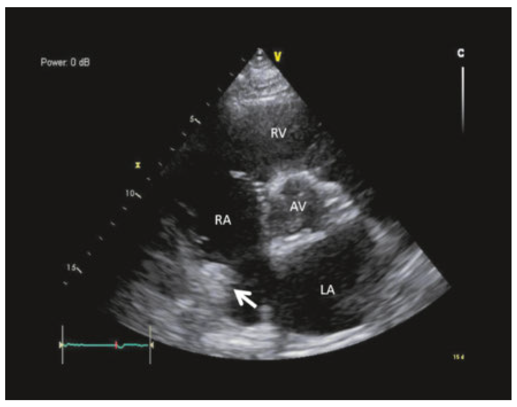
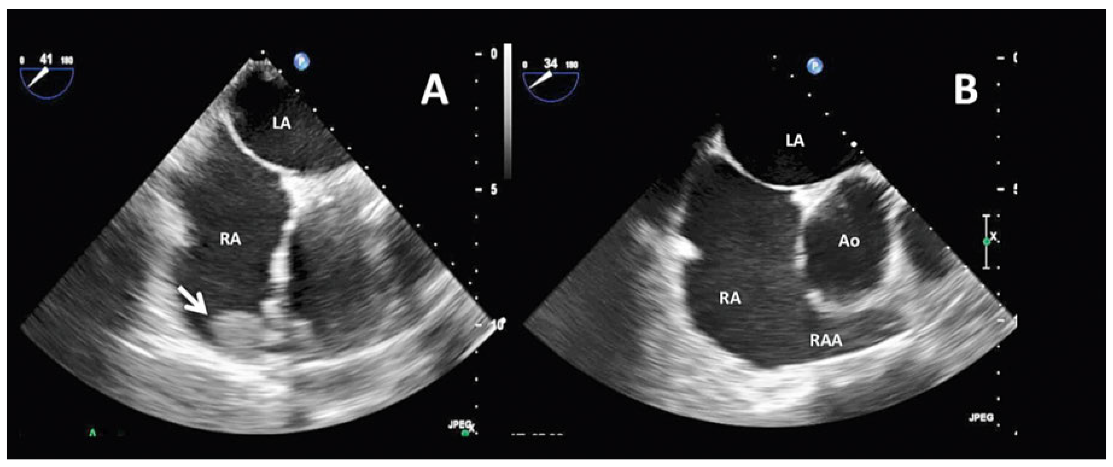
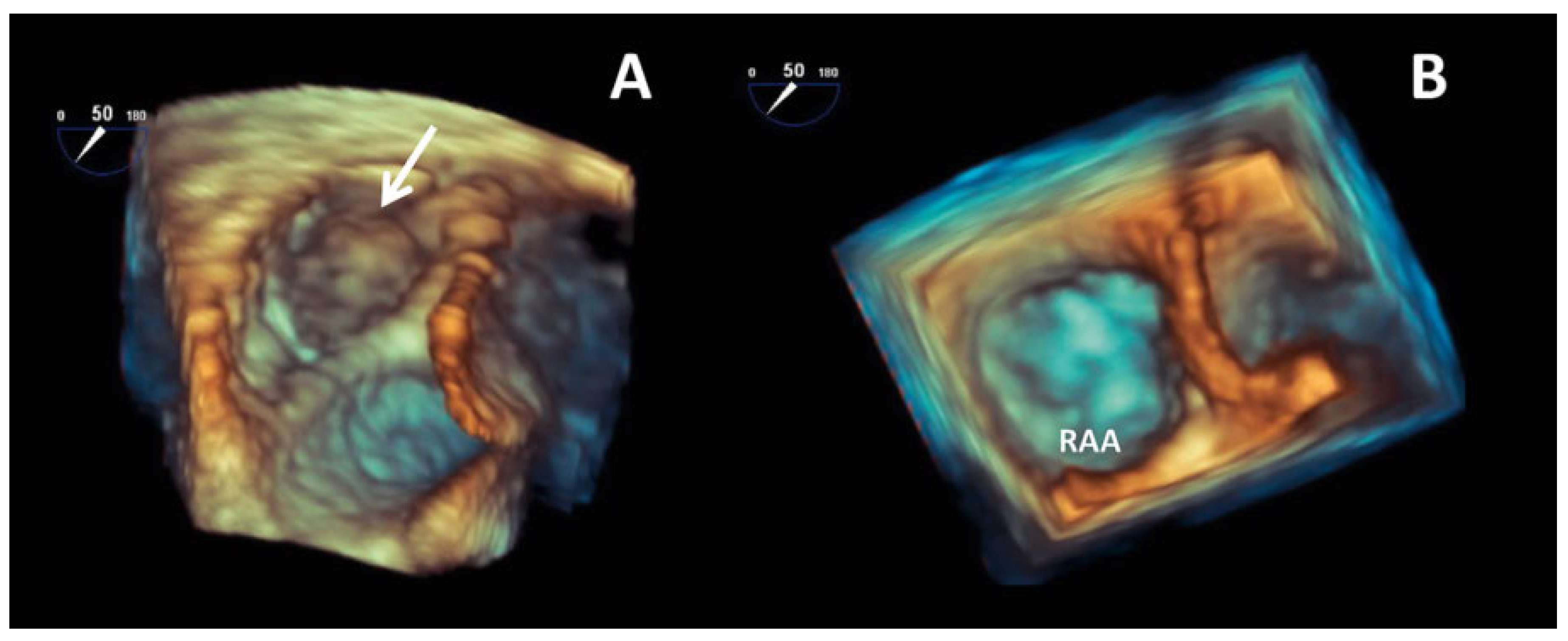
The workup was completed with transthoracic echocardiography, which revealed a moderately dilated, hypertrophic left ventricle with preserved systolic function, bi-atrial dilatation, moderate mitral regurgitation and an nonspecific thickening of the right atrium (Figure 2). Transoesophageal echocardiography (TEE) with 3D reconstruction confirmed a right atrial appendage mass which was suspected to be a thrombus (Figure 3A and Figure 4A). The left atrium and left atrial appendage were thrombus free.
The enoxaparin dose was increased to 80 mg subcutaneously twice daily as the previous dose was below the effective therapeutic range. The suspected thrombus and pulmonary embolism were considered to be due to the insufficiently anticoagulated atrial fibrillation, and decision was made to resume acenocoumarol with target INRs of 2–3. The right atrial appendage mass had disappeared at 3-month follow-up (Figure 3B and Figure 4B).
Discussion
Whereas the pathophysiology of left atrial appendage thrombus and its role in cardioembolic strokes is well defined and studied, data on right atrial appendage thrombus are scarce [1]. This could be explained, in part, by the less severe consequences of a small thrombus in the lung circulation compared to that of a cardioembolic stroke. The prevalence of right atrial appendage thrombus in atrial fibrillation patients varies in the literature from 0.7 to 5.8% [2,3,4]. In a large study, Cresti et al. showed that among 805 patients with atrial fibrillation or flutter who underwent TEE-guided cardioversion, the incidence of right atrial thrombus was 0.75% (6 of 805 patients) compared with 10.3% (83 of 805 patients) for the left atrial appendage [4].
In atrial fibrillation patients, the right and left atrial appendages are larger and the emptying velocities lower compared with those in sinus rhythm [2]. The morphology and the inner structure of the right atrium seem to be less favourable for thrombus formation because of a better blood flow compared with the left atrium [5]. The smaller amount of pectinate muscle in the right atrium and its dendritic structure may contribute to a decreased blood stagnation compared with the left atrium.
The location, risk-factors and the fact that the right atrial appendage mass disappeared under effective anticoagulation, point towards right atrial appendage thrombus rather than any other cause. Several different factors, such as cancer, the perioperative period and the subtherapeutic anticoagulation, in addition to atrial fibrillation may have contributed to right atrial appendix thrombus formation and pulmonary embolism.
The relationship between pulmonary embolism and atrial fibrillation is complex and bi-causal. The increased right ventricular afterload seen in some pulmonary embolism patients could trigger atrial fibrillation. On the other hand, as was more likely the case here, atrial fibrillation can lead to right atrial appendage thrombus formation and pulmonary embolism.
Some patient characteristics are simply shared factors such as advanced age, heart failure, obesity and inflammatory states [6]. Free-floating large right atrial thrombi generally have a poor prognosis, with reported mortalities of up to 17% [7]. Depending on patient characteristics and specific contraindications, treatment options include thrombolysis, and surgical or percutaneous mechanical embolectomy.
This case highlights the possibility of right atrial appendage thrombus in atrial fibrillation and its implication in cardioembolic pulmonary embolism. The right atrial appendage should be screened for thrombus, particularly if TEE is performed in the workup of a patient with a suspected cardioembolic event or for any reason in a patient with atrial fibrillation.

Conflicts of Interest

No financial support and no other potential conflict of interest relevant to this article was reported.

References

  1. Manning, W.J.; Silverman, D.I.; Waksmonski, C.A.; Oettgen, P.; Douglas, P.S. Prevalence of residual left atrial thrombi among patients with acute thromboembolism and newly recognized atrial fibrillation. Arch. Intern. Med. 1995, 155, 2193–2198. https://www.ncbi.nlm.nih.gov/pubmed/7487241. [CrossRef] [PubMed]
  2. De Divitiis, M.; Omran, H.; Rabahieh, R.; Rang, B.; Illien, S.; Schimpf, R.; et al. Right atrial appendage thrombosis in atrial fibrillation: its frequency and its clinical predictors. Am. J. Cardiol. 1999, 84, 1023–1028. https://www.ncbi.nlm.nih.gov/pubmed/10569657. [CrossRef] [PubMed]
  3. Manning, W.J.; Silverman, D.I.; Keighley, C.S.; Oettgen, P.; Douglas, P.S. Transesophageal echocardiographically facilitated early cardioversion from atrial fibrillation using short-term anticoagulation: final results of a prospective 4.5-year study. J. Am. Coll. Cardiol. 1995, 25, 1354–1361. https://www.ncbi.nlm.nih.gov/pubmed/7722133. [CrossRef] [PubMed]
  4. Cresti, A.; García-Fernández, M.A.; Miracapillo, G.; Picchi, A.; Cesareo, F.; Guerrini, F.; et al. Frequency and significance of right atrial appendage thrombi in patients with persistent atrial fibrillation or atrial flutter. J. Am. Soc. Echocardiogr. 2014, 27, 1200–1207. https://www.ncbi.nlm.nih.gov/pubmed/25240491. [CrossRef] [PubMed]
  5. Shinoda, K.; Hayashi, S.; Fukuoka, D.; Torii, R.; Watanabe, T.; Nakano, T. Structural Comparison between the Right and Left Atrial Appendages Using Multidetector Computed Tomography. Biomed. Res. Int. 2016, 2016, 6492183. https://www.ncbi.nlm.nih.gov/pubmed/27900330. [CrossRef] [PubMed]
  6. Bikdeli, B.; Abouziki, M.D.; Lip, G.Y.H. Erratum: Pulmonary Embolism and Atrial Fibrillation: Two Sides of the Same Coin? A Systematic Review. Semin Thromb Hemost. 2017. ISSN 1098-9064.: https://www.ncbi.nlm.nih.gov/pubmed/28427092. https://www.ncbi.nlm.nih.gov/pubmed/28427092.
  7. Barrios, D.; Rosa-Salazar, V.; Morillo, R.; Nieto, R.; Fernández, S.; Zamorano, J.L.; et al. Prognostic Significance of Right Heart Thrombi in Patients With Acute Symptomatic Pulmonary Embolism: Systematic Review and Meta-analysis. Chest. 2017, 151, 409–416. https://www.ncbi.nlm.nih.gov/pubmed/27746202. [CrossRef]
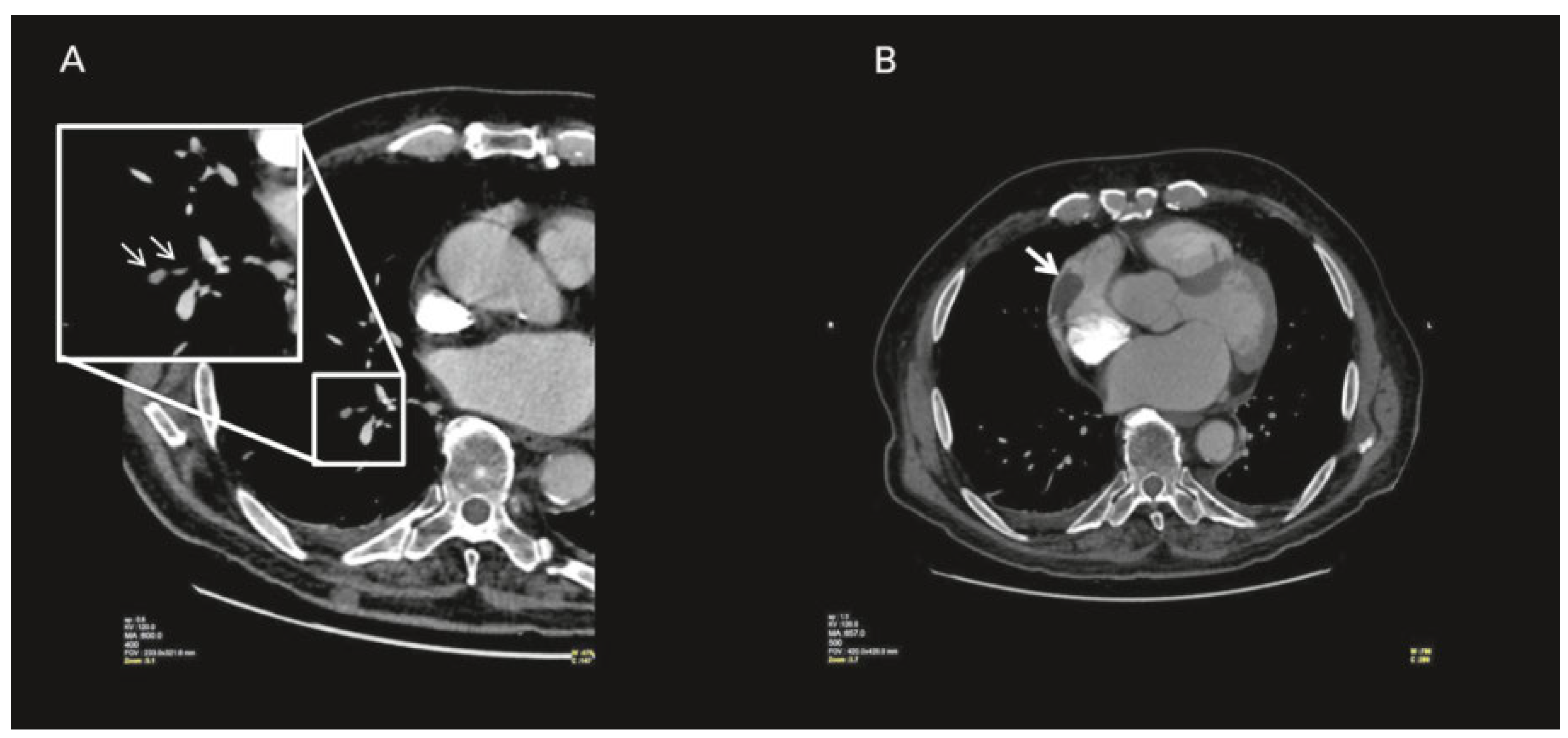
Figure 1. Thoracic computed tomography with contrast; (A) white arrow point to right segmental pulmonary embolus; (B) white arrow points at thrombus in the right atrium.
Cardiovascmed 20 00310 g001
Figure 2. Transthoracic echocardiography parasternal short-axis view: white arrow point to thrombus in the right atrium.
Cardiovascmed 20 00310 g002
Figure 3. 2-Dimension transoesophageal echocardiography, mid-oesophageal 40° view; (A) white arrow points at thrombus at time of presentation; (B) 3 months later after anticoagulation.
Cardiovascmed 20 00310 g003
Figure 4. 3-Dimensional transoesophageal echocardiography; (A) white arrow points at thrombus in right atrial appendage; (B) 3 months later after anticoagulation.
Cardiovascmed 20 00310 g004