Case Report
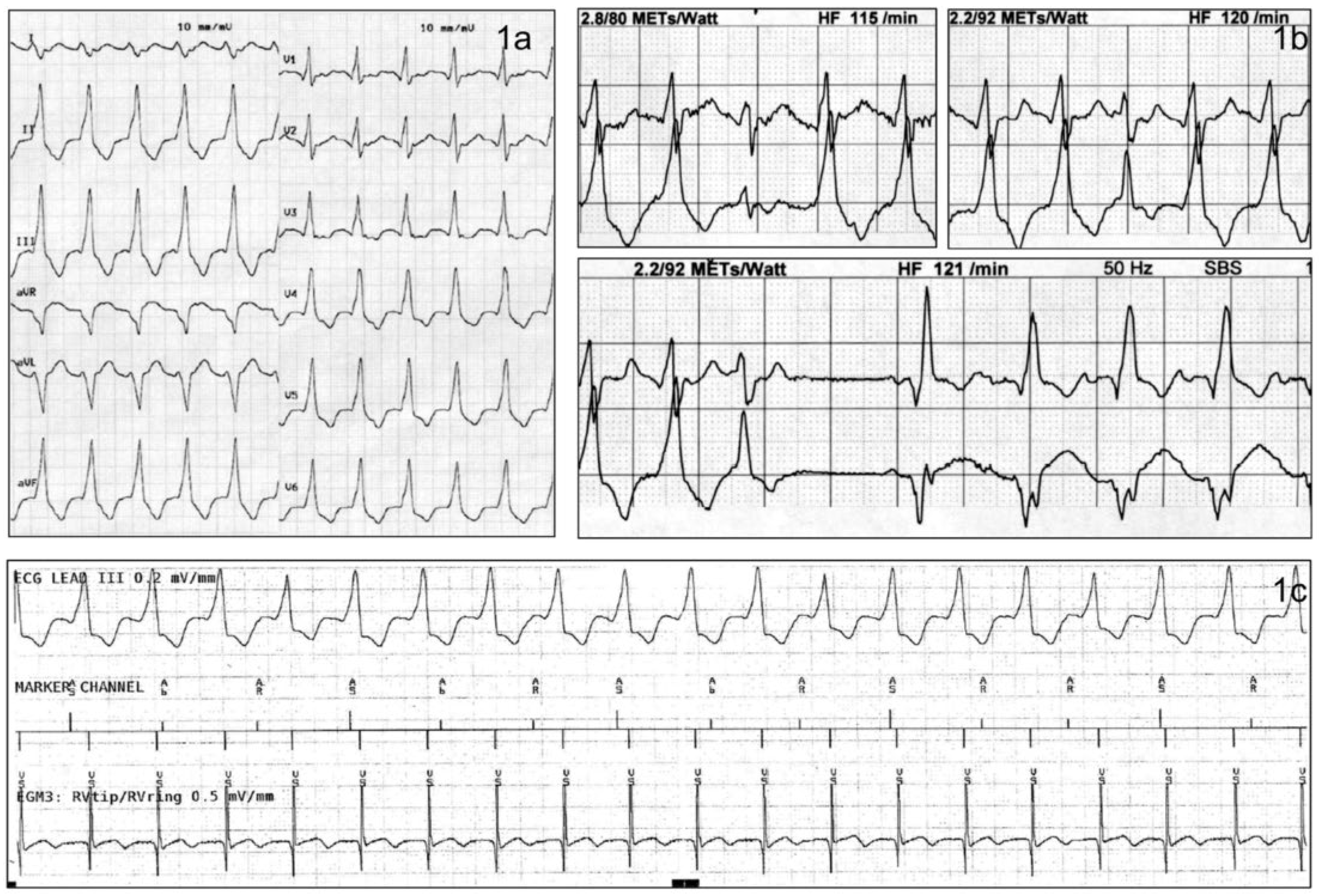
A 47-year-old male patient with ischaemic cardiomyopathy and severe left ventricular dysfunction (left ventricular ejection fraction 18%) presented for a regular follow-up in our outpatient heart failure clinic. In 2004, a dual-chamber implantable cardioverter-defibrillator (ICD-Medtronic® Evera XT DR™, Medtronic plc, Dublin, Ireland) was implanted for primary prevention of sudden cardiac death. A ventricular tachycardia (VT) monitor zone was programmed from 150– 170 bpm, a VT1-zone from 171–202 bpm and a ventricular fibrillation zone from 203 bpm. At presentation, the patient reported a slightly reduced physical exercise capacity over the last 6 months, with recurrent palpitations. Physical examination revealed no signs of fluid overload. The resting 12-lead ECG demonstrated a broad complex tachycardia with a heart rate of 105 bpm (
Figure 1a). QRS duration was 176 ms with an inferior axis and positive concordance in the chest leads. To evaluate exercise capacity, a bicycle stress test was performed. The ECG at the beginning of the investigation was unchanged compared with the one described above. The heart rate increased from 105 to 121 bpm, when suddenly the QRS morphology changed (
Figure 1b) without clinical impact (no additional dyspnoea, angina or dizziness). The patient continued for another 2 minutes on the treadmill.
Discussion
In retrospect, the broad complex tachycardia at the beginning was a slow VT. The proofs of a VT are the presence of atrioventricular dissociation, fusion beats and capture beats (fig. 1b). A change in QRS morphology or a shift of the QRS axis compared with sinus rhythm, as seen in our example, may be helpful as well. Additionally, positive or negative concordance (entirely positive or negative QRS complexes in the chest leads) is another sign indicative of a VT. Device interrogation confirmed the presence of a VT (V>A; fig. 1c). Interestingly, there was no retrograde atrial activation during VT (shown by atrioventricular dissociation), which at the present cycle length of the VT is most likely due to missing ventriculoatrial conduction and not a result of atrioventricular (AV) node electrical refractory phase. In contrast, the atrial overstimulation due to the accelerating sinus node demonstrated the presence of antegrade conduction properties of the AV node. This discrepancy between ante- and retrograde AV node conduction properties is not rarely observed during electrophysiological testing.
Presence of a slow VT is a rare, but important finding in patients suffering from advanced heart failure. Typically, the majority of these patients remain largely asymptomatic. Indeed, a German study investigating patients with heart failure and primary prevention ICD showed a prevalence of slow VTs of 6%, with all of the patients being free of clinical symptoms [
1].
Antitachycardia pacing or overdrive stimulation is a successful way to terminate an ongoing ventricular tachyarrhythmia without delivery of a shock. During antitachycardia pacing, short bursts of rapid ventricular pacing (typically 8–12 beats) are delivered at a rate slightly faster than the rate of the detected tachycardia (empirically 85–88% of the tachycardia cycle length).
Conclusions
In our case, during the exercise stress test the slow VT was terminated owing to the accelerating sinus rhythm, which terminated the slow ventricular tachycardia by a mechanism similar to that of overdrive pacing.
To the best of our knowledge, this is the first documented case of a slow ventricular tachycardia terminated by intrinsic sinus tachycardia during an exercise stress test.
Conflicts of Interest
Dr. Breitenstein has received speaker fees from BMS/Pfizer and Medtronic as well as educational grants from Biosense Webster, Biotronik and Actelion. Dr. Steffel has received consultant and/or speaker fees from Amgen, Astra-Zeneca, Atricure, Bayer, Biosense Webster, Biotronik, Boehringer-Ingelheim, Boston Scientific, Bristol-Myers Squibb, Cook Medical, Daiichi Sankyo, Medtronic, Novartis, Pfizer, Sanofi-Aventis, Sorin, St. Jude Medical / Abbott and Zoll. He reports ownership of CorXL. Dr. Steffel has received grant support through his institution from Bayer Healthcare, Biosense Webster, Biotronik, Boston Scientific, Daiichi Sankyo, Medtronic, and St. Jude Medical / Abott.
References
- Lusebrink, U.; Duncker, D.; Hess, M.; Heinrichs, I.; Gardiwal, A.; Oswald, H.; et al. Clinical relevance of slow ventricular tachycardia in heart failure patients with primary prophylactic implantable cardioverter defibrillator indication. Europace. 2013, 15, 820–826. [Google Scholar] [CrossRef] [PubMed][Green Version]
© 2025 by the authors. Licensee MDPI, Basel, Switzerland. This article is an open access article distributed under the terms and conditions of the Creative Commons Attribution (CC BY) license (https://creativecommons.org/licenses/by/4.0/).


