Next Article in Journal
Topsy-Turvy Ventricular Tachycardia
Previous Article in Journal
An Unusual Case of Upper Rate Behaviour
 
 
Cardiovascular Medicine is published by MDPI from Volume 28 Issue 1 (2025). Previous articles were published by another publisher in Open Access under a CC-BY (or CC-BY-NC-ND) licence, and they are hosted by MDPI on mdpi.com as a courtesy and upon agreement with Editores Medicorum Helveticorum (EMH).
Case Report

Cardiac Sarcoidosis with Coeliac Disease

by
Stéphane Fournier
1,
Julien Regamey
1,
Samuel Rotman
2,
Etienne Pruvot
1 and
Roger Hullin
1,*
1
Department of Cardiology, University Hospital Centre Vaudois (CHUV), Lausanne, Switzerland
2
Institute of Pathology, University Hospital Centre Vaudois (CHUV), Lausanne, Switzerland
*
Author to whom correspondence should be addressed.
Cardiovasc. Med. 2016, 19(4), 128; https://doi.org/10.4414/cvm.2016.00412
Submission received: 20 January 2016 / Revised: 20 February 2016 / Accepted: 20 March 2016 / Published: 20 April 2016

Abstract

This case of cardiac sarcoidosis started with manifestation of a complete atrioventricular block in a 42-year-old female without other cardiac dysfunction. Two years later, the patient presented with acute heart failure symptoms at the Emergency Department. Echocardiography at admission showed thinning and hyperdensity of the basal interventricular septum, which is a rare but typical echocardiographic sign of cardiac sarcoidosis. Endomyocardial biopsy, positron emission tomography–computed to mography and transbronchial biopsy confirmed the clinical suspicion of sarcoidosis. Of note, the patient also had coeliac disease, which can occur conjointly with sarcoidosis. After 5 months of immunosuppressive treatment with methylprednisolone and azathioprine the burden of inflammatory lesions was significantly reduced and the patient had improved to New York Heart Association class I.

Case report

The long history of the 44-year-old woman included two uneventful pregnancies, dust-mite-induced asthma treated by occasional inhalation of β2adrenergic receptor agonists, and a suspicion of coeliac disease based on an intestinal biopsy obtained 2 weeks before admission with acute heart failure. In 2013, she had presented with complete atrioventricular (AV) block with a ventricular escape rhythm at 40 bpm treated with implantation of a double -chamber pacemaker; cardiac magnetic resonance imaging (MRI) and Lyme serology were negative at that time. Between 2013 and 2015, her New York Heart Association (NYHA) class increased from I to II and left ventricular ejection fraction (LVEF) decreased from normal to 40% with global hypokinesia. In April 2015, she was admitted to the Emergency Wards for palpitations, reduced exercise tolerance, tiredness and prolonged recovery from exercise. Clinical examination revealed bilateral ankle oedema and a positive hepatojugular reflux. The laboratory results showed elevation of N-terminal pro-brain-type natriuretic peptide (NT-proBNP) (2018 ng/L), D-Dimers (2151 ng/mL), high sensitive troponin (16 ng/L) and creatinine (93 μmol/L). The echocardiogram showed a non-dilated left ventricle (LV) with global hypokinesia, a LVEF of 35%, and basal thinning of the interventricular septum (Figure 1); the ejection fraction (EF) of the nondilated right ventricle (RV) was severely reduc ed. A pacemaker interrogation demonstrated a ventricular stimulation rate of 98.9% and therefore pacemaker-induced dysfunction initially entered into the differential diagnosis. Computed tomography (CT) angiography in the Emergency Department excluded pulmonary embolism but showed bilateral hilar adenopathy. The ECG revealed recurrent unsustained polymorphic ventricular tachycardia (VT), for which low-dose β-blocker therapy was started. In the following night, a haemo dynamically tolerated sustained VT was successfully treated with 300 mg intravenous amiodarone; thereafter the patient was given an oral loading dose of 10 g amiodarone with subsequent decrease in the incidence of VT episodes.
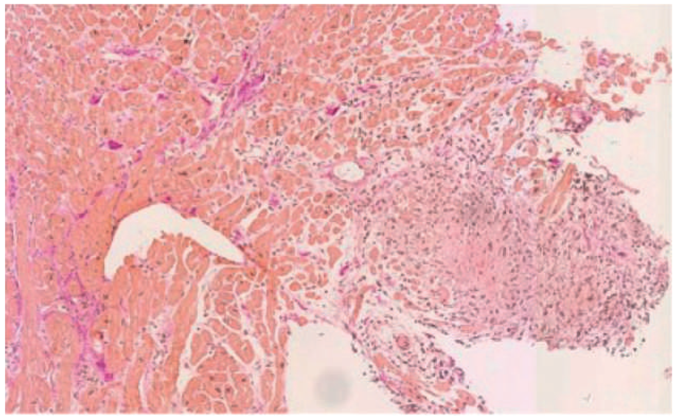
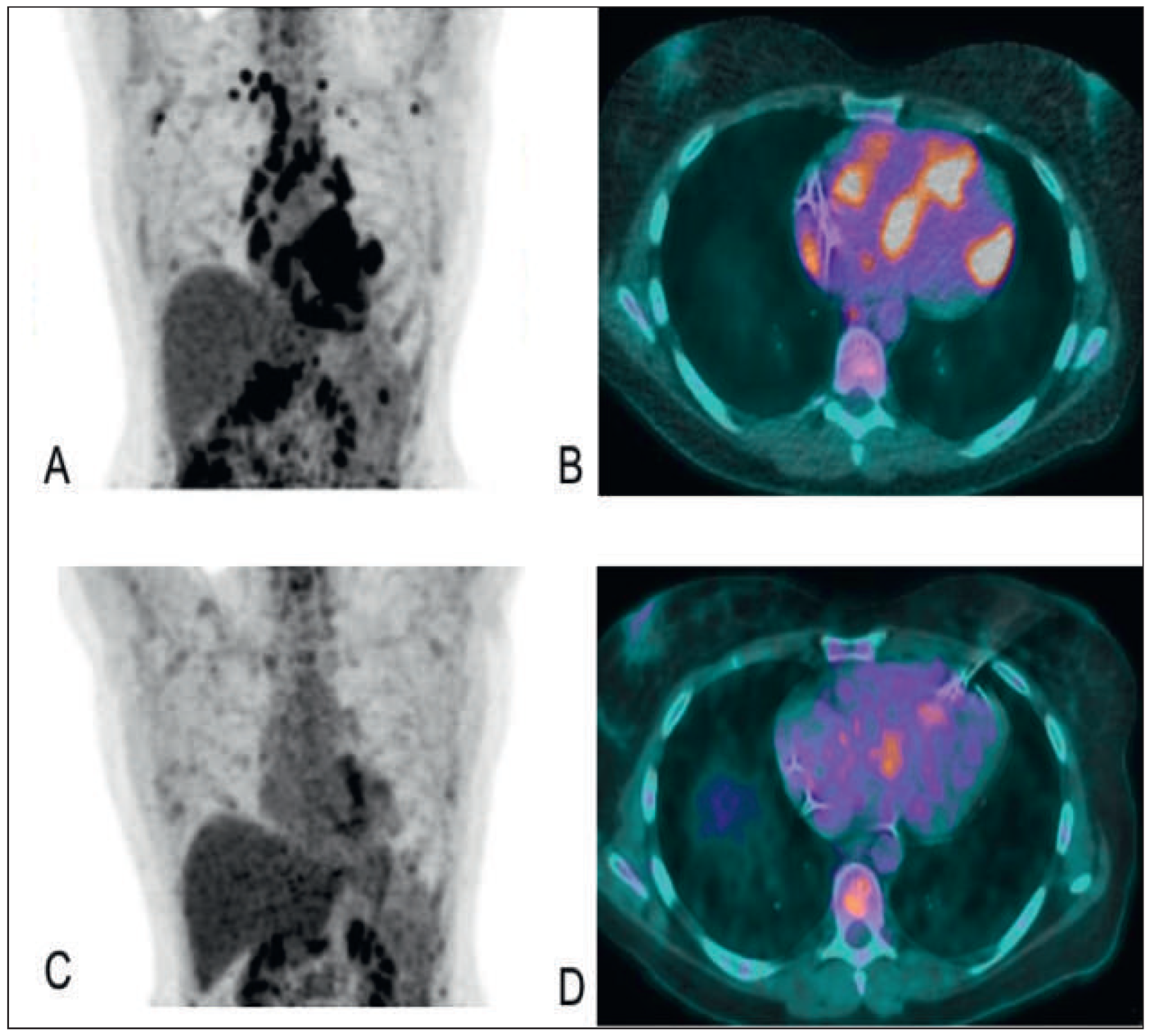
Work-up was negative for metabolic, endocrinological, rheumatological and infectious disorders, C-reactive protein and erythrocyte sedimentation rate were normal. However, titres of antitransglutaminase and antigliadine IgA antibodies were elevated (84 U/L and 31 U/L, respectively) in accordance with the coeliac disease diagnosed in a duodenal biopsy obtained 10 days before admission. Late gadolinium sequences of cardiac MRI showed subepicardial enhancement of anterior, septal and inferior segments and a mid-ventricular sign in the inferior septum. The coronaryangiog ram was normal, but haemodynamic measurements revealed a low cardiac index (1.5 L/min/m2). Endomyocardial biopsies of the RV septum revealed nonnecrosing granulomatous myocarditis (Figure 2) and ruled out cardiac desmosomal disease (right ventricular dysplasia). 18fluorodeoxygenase (FDG) PET-CT showed multiple hypermetabolic lesions especially in the heart, the lung, the spleen and multiple lymph nodes in the axillary, supraclavicular, media stinal, hilar and retroperitoneal positions (Figure 3A,B). Broncho alveolar lavage showed a mild alveolar lymphocytosis; transbronchial biopsies demonstrated granulomatous lymphadenitis. After exclusion of other aetiologies associated with non-necrotising granulomas (tuberculosis, Churg-Strauss vascultis), the diagnosis of systemic sarcoidosis combined with celiac disease was established. In fact, both disease entities are linked to class II haplotype HLA-DR3, DQ2, and B8 expression, which result in an increased susceptibility to both diseases [1].
Corticosteroid treatment was initiated as 1 mg methylprednisolone/kg body weight and after 3 weeks of cortico steroid treatment the patient received a first dose of 50 mg methotrexate aiming at a more rapid reduction of corticosteroid treatment. The next day the patient consulted urgently for palpitation and dizziness starting a few hours previously. The ECG showed sustained VT of inferior origin with a frequency of 170 bpm, which spontaneously converted to sinus rhythm at the Emergency Wards. Only 3 h later a VT with a frequency of 141 bpm originating from the right ventricular outflow tract occurred, for which reason the patient received an additional 6 g of amiodarone loading (800 mg/d) and a maximum dose of carvedilol was given. Despite of disappearance of sustained VT thereafter, her DDD pacemaker was upgraded to a cardiac resynchronisation therapy-defibrillator (CRT-D) because of the underlying severe cardiac pathology. As methotrexate was suspected to promote ventricular dysrhythmias [2], treatment with azathioprine was started with a consecutive taper of cortico steroid therapy to 0.5 mg/kg body weight. At 3-month follow-up, an echocardiogram showed a moderate LVEF improvement from 35 to 41% and at 5-month follow-up, the patient was in functional NYHA class I without any clinical signs of systemic sarcoidosis. A 18FDG-PET-CT showed significant reduction/disappearance of the previously described lesions (Figure 3C,D).

Cardiac sarcoidosis

Sarcoidosis is a multisystem granulomatous disease of unknown cause often seen in young adults. Age-adjusted incidence rates in the United States are 35.5/ 100 000 for African-Americans and 10.9/100 000 for whites with an overall prevalence of 20/100 000. Cardiac manifestation is present in 20–27% of sarcoidosis patients in the United States but up to 58% in Japan [3].

ECG in sarcoidosis

Almost 70% of patients with cardiac sarcoidosis have ECG abnormalities. Most common are first-degree, second-degree or complete AV block (23–30%). Complete AV block tends to present at a younger age than idiopathic heart block. Unsustained VT is the second most common manifestation of cardiac sarcoidosis, and VT in combination with complete AV block accounts for almost two thirds of sudden deaths.

Echocardiography in sarcoidosis

Usually, dilated cardiomyopathy with regional wall motion abnormalities due to scattered granulomas are observed. Whereas there is initial wall thickening, wall thinning due to scarring and fibrosis occurs eventually. Thinning of the basal anterior septum in a young patient with dilated cardiomyopathy is rare but highly suggestive of sarcoidosis [3] (Table 1).

Endomyocardial biopsy in sarcoidosis

Endomyocardial biopsy is the gold standard for diagnosis of cardiac sarcoidosis. However, its sensitivity is low, as shown by Ardehali et al., who reported that only 7 out of 28 patients with presumed cardiac sarcoidosis had positive biopsy results [4]. In accordance, in 50–80% of patients with extracardiac sarcoidosis and clinical signs of cardiac manifestation biopsies are negative (Table 1).

PET in sarcoidosis

Tissue affected by sarcoidosis shows increased uptake of 18FDG, which correlates well with histological disease activity. PET-based perfusion images also depict the extent of fibrogranulomatous replacement in the whole heart. Overall, PET sensitivity is reported to be as high as 82–100% [3] (Table 1).

Cardiac magnetic resonance imaging

Cardiac MRI visualises infiltration in late gadoliniumenhanced images and local inflammation with oedema in T2-weighted images, in addition to functional and morphological characterisation. A cardiac MRI study from The Netherlands demonstrated in 58 patients with cardiac sarcoidosis a sensitivity of 100%, a specificity of 78%, and positive predictive value and negative predictive values of 55 and 100%, respectively. Since rare cases of congestive heart failure due to right ventricular dysplasia may mimic cardiac sarcoidosis, MRI should be always performed to rule out right ventricular dysplasia if cardiac sarcoidosis is suspected (Table 1).
Table 1. Revised criteria for diagnosing cardiac sarcoidosis [5].
Cardiac sarcoidosis is confirmed
I. Histological diagnosis group: when myocardial biopsy specimens demonstrate noncaseating epithelioid cell granulomas with histological or clinical diagnosis of extracardiac sarcoidosis.
II. Clinical diagnosis group: although myocardial biopsy specimens do not demonstrate noncaseating epitheloid cell granulomas, if extracardiac sarcoidosis is diagnosed histologically or clinically and satisfies the following conditions and more than one in six basic diagnostic criteria:
II.1. More than two of four major criteria are satisfied:
II.1.1. Advanced atrioventricular block.
II.1.2. Basal thinning of the interventricular septum.
II.1.3. Positive cardiac 67Ga uptake.
II.1.4. Depressed LVEF (<50%).
II.2. One in four major criteria and more than two in five minor criteria are satisfied:
II.2.1. Abnormal ECG findings: ventricular arrhythmias (ventricular tachycardia, multifocal or frequent premature ventricular contractions), complete right bundle-branch block, axis deviation or abnormal Q-wave.
II.2.2. Abnormal echocardiography: regional abnormal wall motion or morphological abnormality (ventricular aneurysm, wall thickening).
II.2.3. Nuclear medicine: perfusion defect detected by 201Tl myocardial scintigraphy or 99Tc myocardial scintigraphy.
II.2.4. Gadolinium-enhanced MRI: delayed enhancement of myocardium.
II.2.5. Endomyocardial biopsy: interstitial fibrosis or monocyte infiltration over moderate grade.

Management

Immunosuppressive therapy

Corticosteroids are the main therapy for cardiac sarcoidosis despite lack of confirmatory evidence. In one large retrospective study of 95 patients with cardiac sarcoidosis, patients treated with corticosteroids had a 5-year survival rate of 75% as compared with 10% in untreated patients [3]. The standard recommendation is to start with high doses (1 mg methylprednisolone/kg bodyweight and day) followed by progressive tapering. However, the starting dose is debated since reports show similar long-term survival when patients are started with prednisolone ≤40 mg/d, maintained for a minimum of 6 months, with progressive tapering to 5–15 mg/d over the following 12 months. If prolonged high-dose corticosteroid treatment is necessary for control of disease activity, implementation of supplementary methotrexate, ciclosporin, cyclophosphamide, as well as azathioprine treatment may be discussed in order to decrease the dose of corticosteroids. Methotrexate is the most studied alternative to steroids but its cardiac efficacy is not established.

Cardiac pharmacological therapy

Heart failure treatment even when caused by cardiac sarcoidosis remains based on recommendations in guidelines. In particular, angiotensin converting-enzyme inhibitors or angiotensin receptor blockers are recommended in all patients with impaired systolic function.

Arrhythmias

No prospective trials have tested the efficacy of β-blockers or amiodarone treatment. The secondary effects of amiodarone treatment are of concern; in particular, pulmonary fibrosis may contribute to further deterioration of lung function in patients with pulmonary sarcoidosis.

Implantable cardioverter defibrillator

Cardiac arrest or sudden cardiac death (SCD) may be the first manifestation of cardiac sarcoidosis, therefore implantation of an implantable cardiac defibrillator (ICD) should be strongly considered once the diagnosis of cardiac sarcoidosis is established. However, evidence from prospective trials is lacking. Therefore, attention should be given to symptoms such as syncope, heart failure status, LV function and spontaneous or induced ventricular arrhythmias to make individualised decisions about primary prevention of SCD [3].

Radiofrequency ablation

The effectiveness of radiofrequency ablation in prevention of recurrent arrhythmias in patients with cardiac sarcoidosis is limited. In a single-centre experience, ablation therapy was effective in controlling arrhythmias in only two of eight patients with recurrent monomorphic VT [3].

Disclosure statement

No financial support and no other potential conflict of interest relevant to this article was reported.

References

  1. Reggoug, S.; Benelbarhdadi, I.; Essamri, W.; Ajana, F.Z.; Afifi, R.; Benazzouz, M.; et al. Celiac disease associated with sarcoidosis. Gastroenterol Clin Biol. 2009, 33, 430–432. [Google Scholar] [CrossRef] [PubMed]
  2. Perez-Verdia, A.; Angulo, F.; Hardwicke, F.L.; Nugent, K.M. Acute cardiac toxicity associated with high-dose intravenous methotrexate therapy: case report and review of the literature. Pharmacotherapy 2005, 25, 1271–1276. [Google Scholar] [CrossRef] [PubMed]
  3. Golwala, H.; Dernaika, T. Atrial fibrillation as the initial clinical manifestation of cardiac sarcoidosis: a case report and review of the literature. J Cardiovasc Med (Hagerstown). 2015, 16 (Suppl. S2), S104–S112. [Google Scholar] [CrossRef] [PubMed]
  4. Ardehali, H.; Howard, D.L.; Hariri, A.; Qasim, A.; Hare, J.M.; Baughman, K.L.; et al. A positive endomyocardial biopsy result for sarcoid is associated with poor prognosis in patients with initially unexplained cardiomyopathy. Am Heart J. 2005, 150, 459–463. [Google Scholar] [CrossRef] [PubMed]
  5. Diagnostic standard and guidelines for sarcoidosis. Jpn J Sarcoidosis Granulomatous Disorders. 2007, 27, 89–102.
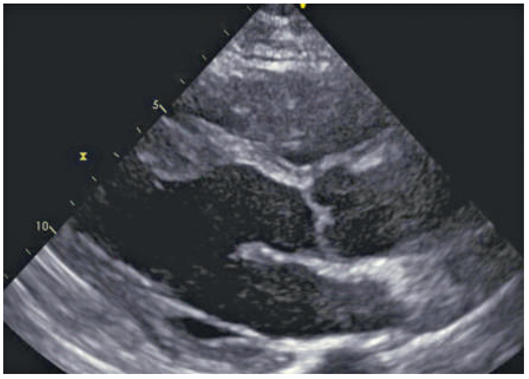
Figure 1. Echocardiogram showing basal thinning of the basal interventricular septum.
Cardiovascmed 19 00128 g001
Figure 2. Non-necrotising granuloma with perigranulomatous mononuclear infiltrate with rare eosinophilic cells.
Cardiovascmed 19 00128 g002
Figure 3. (A) Baseline PET-CT showing multiple metabolic lesions of the heart, spleen and several pulmonary nodules. (B) Baseline PET-CT showing multiple metabolic lesions of the heart in the interventricular septum and the free left and right ventricular wall. (C,D) Same PET-CT at 5 months follow-up showing significant reduction/disappearance of the previously described lesions.
Cardiovascmed 19 00128 g003