Next Article in Journal
Leadless Cardiac Stimulation: Ready to Take Centre Stage?
Previous Article in Journal
A Rare Electrocardiographic Presentation of Acute Ischemic Heart Disease
 
 
Cardiovascular Medicine is published by MDPI from Volume 28 Issue 1 (2025). Previous articles were published by another publisher in Open Access under a CC-BY (or CC-BY-NC-ND) licence, and they are hosted by MDPI on mdpi.com as a courtesy and upon agreement with Editores Medicorum Helveticorum (EMH).
Case Report

Flexible Pusher Cable for Percutaneous Closure of an Atrial Septal Defect

by
Sabine Pallivathukal
,
Jean-Pierre Pfammatter
and
Bernhard Meier
*
Universitätsklinik für Kardiologie, Inselspital, Bern, Switzerland
*
Author to whom correspondence should be addressed.
Cardiovasc. Med. 2016, 19(3), 95; https://doi.org/10.4414/cvm.2016.00398
Submission received: 16 December 2015 / Revised: 16 January 2016 / Accepted: 16 February 2016 / Published: 16 March 2016

Abstract

The case report describes a new flexible pusher cable (HyperionFlex-Pusher) intended for safer percutaneous atrial septal defect device closure.

Introduction

Transcatheter closure of an atrial septal defect (ASD) with different occluder systems yields excellent long term results [1,2,3,4,5,6]. Embolisation of occluders occurs in about 2% [7]. Transcatheter retrieval of embolised devices has a success rate from 50% to 75% and cardiac surgery may be required [7,8]. We describe a new delivery system (HyperionFlexPusher, Comed, Bolsward, Holland) with a flexible pusher cable providing the opportunity to assess whether the device is in a safe anatomical position before detachment in order to reduce the risk of occluder embolisation. This position is close to the final one aher detachment. Retrieval of the device for repositioning is still possible at that point by retracting the device with the core wire of the pusher cable into the long delivery sheath.

Case Report

An 8-mm central secundum atrial septal defect without signs of multifenestration or signs of atrial septal aneurysm was detected in the transthoracic echocardiography (TTE) of a five-year-old girl. There were satisfying rims and mild signs of volume overload of the right ventricle, but no signs of pulmonary arterial hypertension.

Implantation Procedure

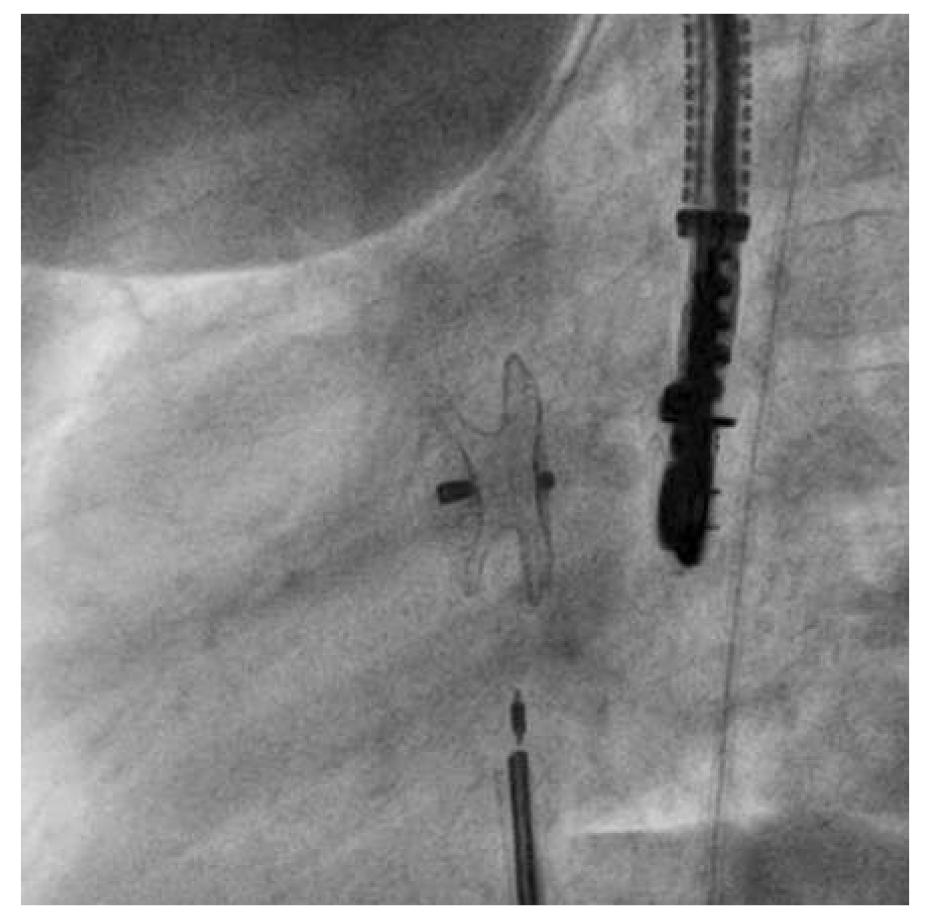
The procedure was conducted under general anaesthesia and fluoroscopic guidance combined with transoesophageal echocardiography monitoring. Aher tracheal intubation, venous access was gained through the right femoral vein with a 6 French CheckFlow introducer set and the patient was treated with 1 000 units of intravenous unfractionated heparin and 900 mg of intravenous cefuroxime. The atrial defect was passed under fluoroscopic guidance with a 4 French multipurpose catheter. A stiff guidewire was placed in the leh superior pulmonary vein. A 14-mm Hyperion Atrial Septal occluder (Comed) was chosen on the basis of the findings of echocardiography and balloon sizing (24-mm Amplatzer sizing balloon, St. Jude Medical, Plymouth, USA). We decided to close the defect with 14-mm Hyperion Atrial Septal Occluder (Comed). The 6 French introducer sheath was exchanged for a 10 French delivery Hyperion sheath (Comed) and advanced into the leh atrium. The preassembled Hyperion occluder was loaded and the leh atrial disc of the occluder was opened. Aherwards, the delivery sheath and the disc were retracted as a unit until the disc hugged the septum. Subsequently, the right atrial disc was opened in the right atrium. At this stage, the HyperionFlexPusher cable allowed for retraction of its stiff sleeve with the device remaining attached to the screw at the end of the core wire. This relieves the distortion by relaxing the tension of the system. The device position achieved came close to the final position with the precise orientation to the atrial septum (Figure 1, video 1). At this point, the device could still be retrieved and redeployed or replaced. The device was then released by counterclockwise torquing the core wire (Figure 2, video 2). Ideally, the stiff sleeve should be readvanced at least partially before that to avoid entangling of the flexible core wire during torquing. The patient was discharged on the following day aher a TTE and chest X-ray had confirmed good device position. Acetylsalicylic acid (100 mg per day) was recommended for 6 months.

Follow Up

The subsequent course was uneventful. The TTE at 4 months postimplantation showed a well-seated device without residual shunt and neither interference with atrioventricular valves nor thrombus.

Discussion

A flurry of ASD closure devices have been developed since the first report approximately 40 years ago [1,2,3]. The described pusher modification [9,10] facilitates the placement under anatomically difficult conditions and potentially reduces the risk for device embolisation.

Disclosure Statement

Bernhard Meier has received research grants to the institution by St. Jude Medical and speaker fees by Comed and St. Jude Medical.

References

  1. King, T.; Mills, N. Nonoperative closure of atrial septal defects. Surgery. 1974, 75, 383–8. [Google Scholar] [PubMed]
  2. King, T.; Thompson, S.L.; Steiner, C.; Mills, N.L. Secundum atrial septal defect: Nonoperative closure during cardiac catheterization. JAMA. 1976, 235, 2506–9. [Google Scholar] [CrossRef] [PubMed]
  3. Lock, J.; Cockerham, J.T.; Keane, J.F.; Finley, J.P.; Wakely P.E., Jr.; Fellows, K.E. Transcatheter umbrella closure of congenital heart defects. Circulation. 1987, 75, 593–9. [Google Scholar] [CrossRef] [PubMed]
  4. Roymanee, S.; Promphan, W.; Tonklang, N.; Wongwaitaweewong, K. Comparison of the Occlutech ® Figulla ® Septal Occluder and Amplatzer ® Septal Occluder for atrial septal defect device closure. Pediatr Cardiol. 2015, 36, 935–41. [Google Scholar] [CrossRef] [PubMed]
  5. Akagi, T. Current concept of transcatheter closure of atrial septal defect in adults. J Cardiol. 2015, 65, 17–25. [Google Scholar] [CrossRef] [PubMed]
  6. Bissessor, N. Current perspectives in percutaneous atrial septal defect closure devices. Medical Devices: Evidence and Research 2015, 8, 297–303. [Google Scholar] [CrossRef] [PubMed]
  7. Chessa, M.; Carminati, M.; Butera, G.; Bini, R.M.; Drago, M.; Rosti, L.; et al. Early and late complications associated with transcatheter occlusion of secundum atrial defect. J Am Coll Cardiol. 2002, 39, 1061–5. [Google Scholar] [CrossRef] [PubMed]
  8. Goel, P.; Kapoor, A.; Batra, A.; Khanna, R. Transcatheter retrieval of embolized Amplatzer septal occluder. Tex Heart Inst J. 2012, 39, 653–6. [Google Scholar] [PubMed]
  9. Meier, B. Catheter-based atrial shunt occlusion, when the going gets even tougher: editorial comment to use of a straight, side-hole (SSH), delivery sheath for improved delivery of Amplatzer ASD occluder. Cathet Cardiovasc Interv. 2007, 69, 21–2. [Google Scholar] [CrossRef] [PubMed]
  10. Freixa, X. The Amplatzer™ Cardiac Plug 2 for leh atrial appendage occlusion: novel features and first-in-man experience. EuroInter-vention. 2013, 8, 1094–8. [Google Scholar] [CrossRef] [PubMed]
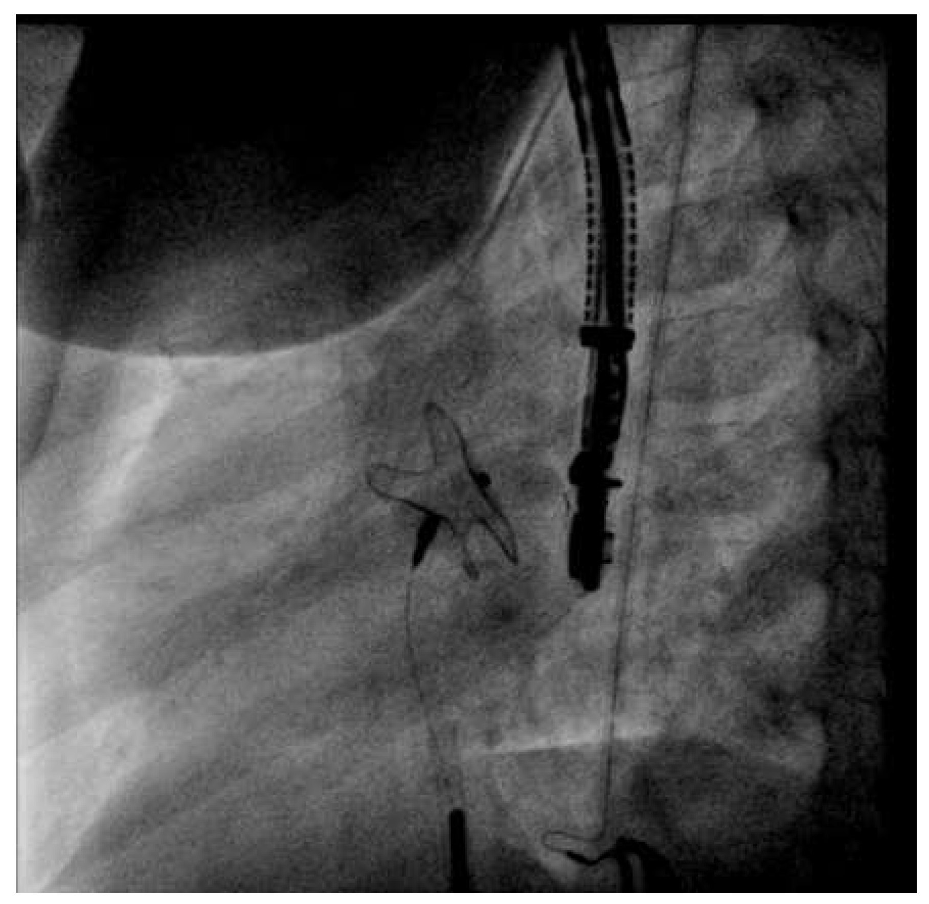
Figure 1. Video 1. HyperionFlexPusher Cable with 14-mm Hyperion Occluder in position before releasing (left anterior oblique 72° / caudal 12° projection).
Cardiovascmed 19 00095 g001
Figure 2. Video 2. After releasing the device (left anterior oblique 72° / caudal 12° projection).
Cardiovascmed 19 00095 g002