Next Article in Journal
A Preliminary Study on the Introduction of Peer-Assisted Learning in Auscultation Courses
Previous Article in Journal
Leadless Cardiac Stimulation: Ready to Take Centre Stage?
 
 
Cardiovascular Medicine is published by MDPI from Volume 28 Issue 1 (2025). Previous articles were published by another publisher in Open Access under a CC-BY (or CC-BY-NC-ND) licence, and they are hosted by MDPI on mdpi.com as a courtesy and upon agreement with Editores Medicorum Helveticorum (EMH).
Case Report

Symptomatic Polymorphic Ventricular Tachycardia in a Young Woman

by
André dos Santos Rocha
1,*,
Zahurul Alam Bhuyian
2,
Florence Fellman
2,
Jürg Schlaepfer
4,
Michèle Bovy
3 and
Alain Delabays
1,4
1
Department of Internal Medicine, Ensemble Hospitalier de la Côte, Morges, Switzerland
2
Division of Genetics, Centre Hospitalier Universitaire Vaudois, Lausanne, Switzerland
3
Department of Anaesthesiology, Ensemble Hospitalier de la Côte, Morges, Switzerland
4
Division of Cardiology, Centre Hospitalier Universitaire Vaudois, Lausanne, Switzerland
*
Author to whom correspondence should be addressed.
Cardiovasc. Med. 2016, 19(3), 90; https://doi.org/10.4414/cvm.2016.00395
Submission received: 16 December 2015 / Revised: 16 January 2016 / Accepted: 16 February 2016 / Published: 16 March 2016

Abstract

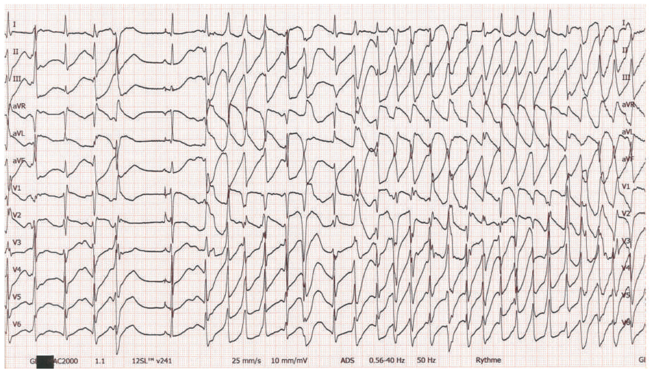
A 32-year-old woman presented to the emergency department with fainting, headaches, nausea and fatigue, associated with episodes of syncope. She reported a medical history of fainting, dizziness and vertigo of unknown origin since her childhood. During the last 2 weeks, she had recurrent episodes of chest pain, accompanied by palpitations and dyspnoea. On admission, she complained of intense oppressive chest pain. Her ECG is shown in Figure 1.

Question

What is the underlying arrhythmia causing the symptoms of this young woman? What are the possible mechanisms of this arrhythmia?

Clinical History

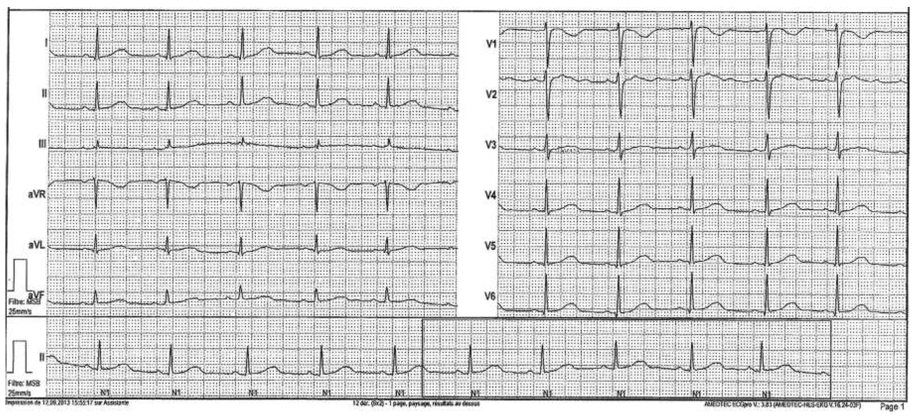
We report the case of a 32-year-old woman who presented with a long-standing medical history of fainting, dizziness and vertigo of unknown origin. She mentioned several episodes of syncope since her childhood with a clear worsening of symptoms for the last couple of years, when vertigo, headaches, nausea and fatigue began to impinge on her daily life activities. She got a broad evaluation by her general practitioner (GP), with no plausible cause found on radiology, laboratory testing or ECG (Figure 2).
Figure 1. Twelve-lead ECG of the patient on admission.
Cardiovascmed 19 00090 g001
Figure 2. Twelve-lead ECG of the patient 2 years before the emergency department admission.
Cardiovascmed 19 00090 g002
Under the diagnoses of migraine and depressive disorder, the patient was given antidepressive, anti-emetic and analgesic treatment with a poor symptom control. Aher an 18-month period of treatment with daily sertraline and occasional paracetamol and domperidone, with no symptom improvement, her GP decided to change the antidepressive medication to fluoxetine.
Aher this modification, in addition to the customary vertigo and fainting episodes, she started to have brief episodes of chest pain accompanied by palpitations, which is why she came to the emergency department. On admission, the patient exhibited intense oppressive chest pain, fluctuating over time, with mild dyspnoea and palpitations. ECG monitoring was initiated, revealing frequent periods of polymorphic ventricular tachycardia (Figure 1) in relationship to the symptoms. Electrical stability was then achieved by intravenous administration of magnesium sulphate, potassium chloride and calcium, revealing the baseline sinus rhythm, as shown in Figure 3.

Workup and Evolution

An impressive 780-millisecond QT interval was found (Figure 3) and potential QT-elongating drugs, namely fluoxetine and domperidone, were promptly stopped. Simultaneously, the patient received propranolol 10 mg.
The patient was kept under close surveillance for some days in the intensive care unit, while the QT interval progressively diminished on magnesium replacement and beta-blocker therapy, firstly with propranolol 10 mg four times a day and subsequently with nadolol 80 mg once a day. Even though domperidone was stopped, the patient did not experience nausea or vertigo and was completely asymptomatic aher the cessation of tachycardia. Echocardiography demonstrated slightly reduced leh ventricular function (ejection fraction calculated at 50%), associated with anterior, septal and lateral hypokinesia. These findings might probably have been linked to the repeated dysrhythmic events as well as with the repolarisation problems. Once the beta-blocker treatment was ongoing, treadmill stress testing was performed. No symptoms or arrhythmia were elicited during the test.
Under the aforementioned treatment for a 7-day period, the patient was asymptomatic but still had an elongated QT interval of 510 ms. Even though the patient had taken at least two different drugs promoting QT elongation, the anamnestic data were suggestive of a lifelong problem (recurrent syncope since childhood, previous to the therapy). This particular feature prompted us to consider the possibility of a congenital long QT syndrome (LQTS).
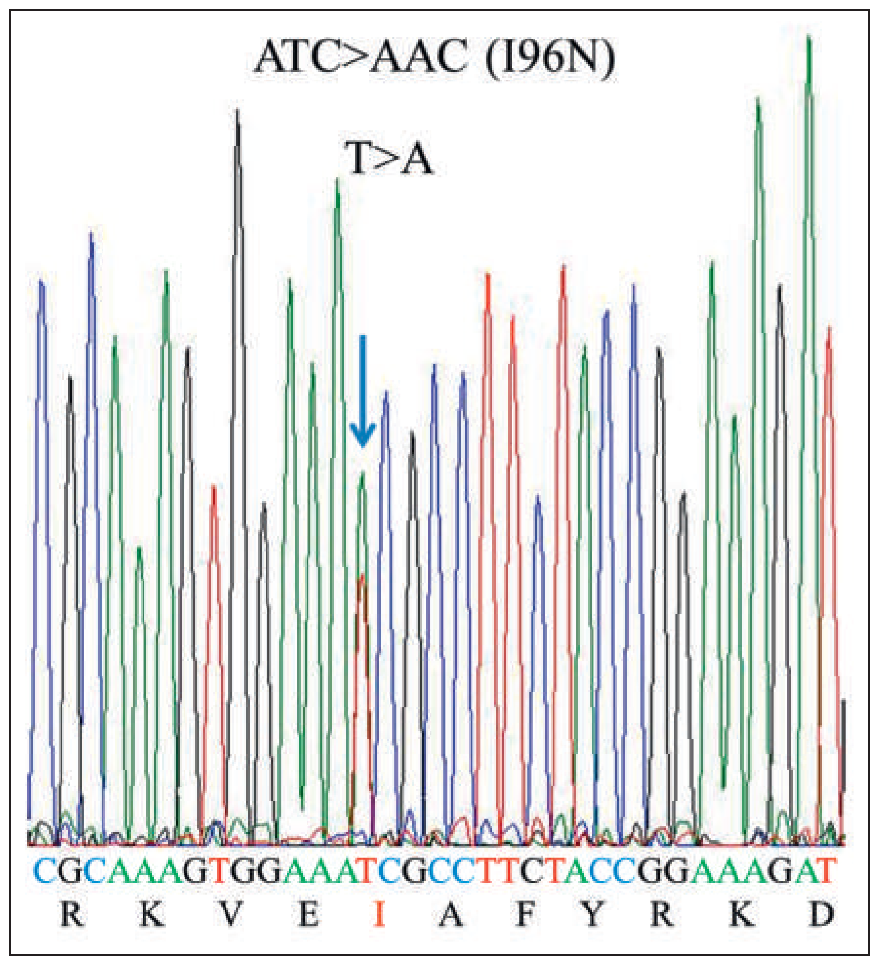
The patient underwent genetic testing. From the three genes most frequently associated with LQTS, we searched for possible mutations in the KCNQ1 and KCNH2 genes, with polymerase chain-reaction (PCR) testing and bidirectional sequencing, as the T-wave pattern and drug-induced history pointed to either KCNQ1 or KCNH2 genes, respectively. The results showed the presence, in the heterozygous state, of the c.287T>A mutation (p.I96N) in the KCNH2 gene. The p. I96N mutation was considered as pathological, especially as the mutation that causes nonsynonymous substitution of the amino acid I96 (p.I96T) present in this patient (Figure 4) has been described in association with LQTS [1].
The index case is the first child of nonconsanguineous parents. The family history is negative for syncopal events or sudden cardiac death, except repeated syncope events in a sister of the paternal grand-father, who died at 80 years old from stroke. The patient has many paternal and maternal uncles, aunts and cousins without any cardiac symptoms. The patient’s sister is healthy with a normal QTc interval on her ECG. She too underwent genetic analysis, which showed no mutation in the KCNH2 gene. As other relatives do not live in Switzerland, family information was given to organise genetic testing abroad.
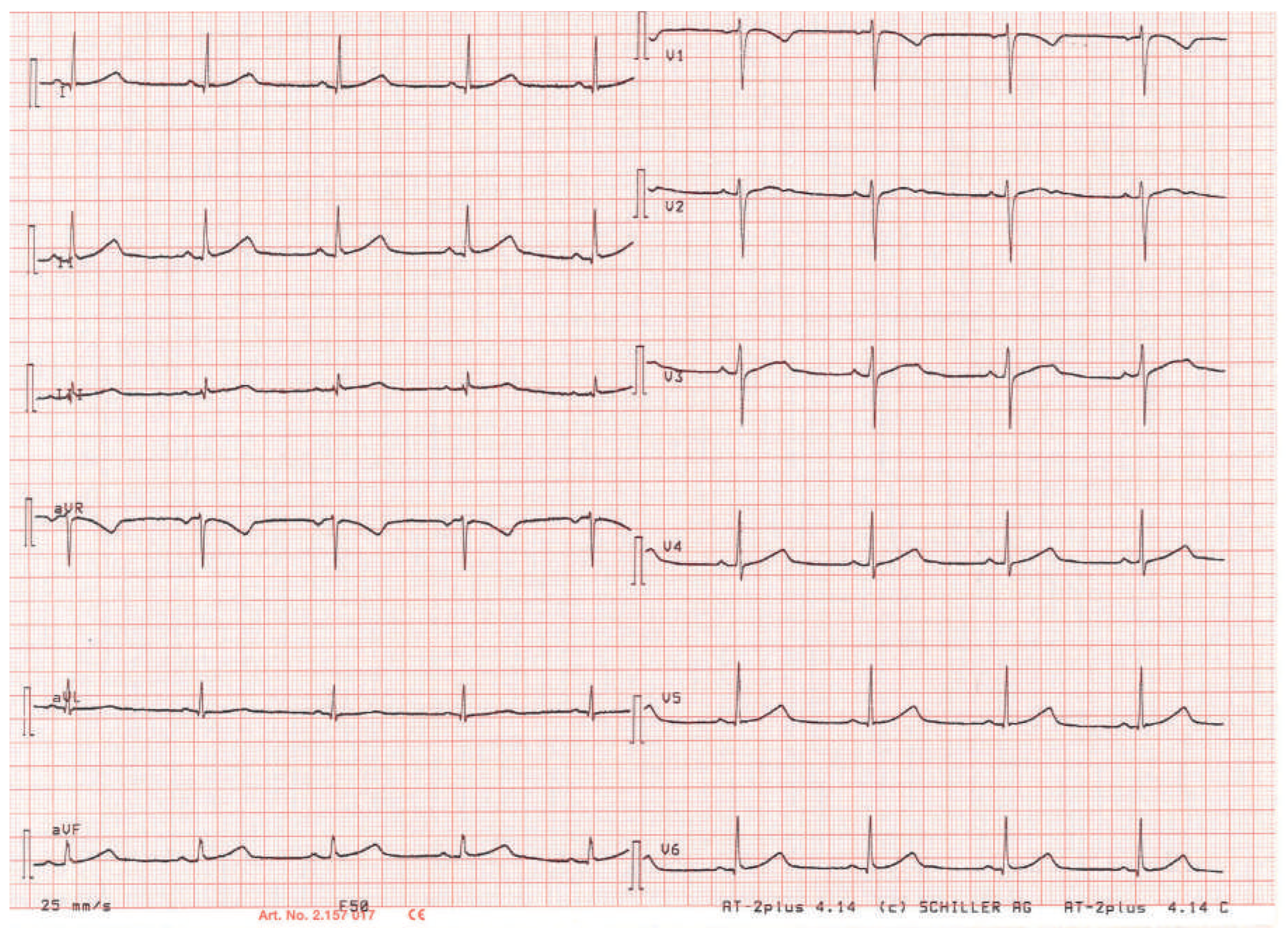
The patient was discharged aher implantation of a continuous a loop recorder (Reveal®), under beta-blocker treatment with nadolol 80 mg once daily. One month later, she was completely asymptomatic with a QTc interval of 510 ms, as shown in Figure 5.

Discussion

The patient’s clinical presentation was very typical of cardiac syncope, as a long-lasting history of fainting and dizziness, associated with oppressive chest pain, dyspnoea and palpitations. Nevertheless, the broad differential diagnosis of syncope does not usually allow early identification of a condition such as a congenital arrhythmia. Syncope may be caused by peripheral circulatory inadequacy (vasovagal or postural hypotension), cardiac disorders (rhythmic, ischaemic, valvular and malformation conditions), respiratory and pulmonary diseases, cerebral disorders and, finally, it may be of primary psychic origin [2]. Recurrently, patients are misdiagnosed as having psychiatric conditions without sufficient investigation of somatic causes, like the patient in this report.
Some key points in this clinical history are particularly thought-provoking and could have permitted an earlier diagnosis. As a first observation, any patient with recurrent syncope should have a broad cardiocirculatory and neurological examination before the symptoms are attributed to a psychiatric cause. Apparently, the diagnosis of depression was made on the simple finding of fatigue and depressed mood. The patient denied having decreased interest or pleasure, weight or appetite change, change in sleep, activity or concentration. Likewise, she had neither guilt/worthlessness feelings nor thoughts of death or suicide [3]. At this point, one may argue that the institution of antidepressive treatment with sertraline was probably too hasty, before other somatic causes were excluded. Additionally, another crucial moment on the workup was her clear worsening when fluoxetine and domperidone were introduced, two different QT-elongating drugs. Her cardiac arrhythmia was then noticeably unmasked, since a perplexing 780-millisecond QT interval degenerated into a polymorphic ventricular tachycardia (Torsade de pointes).
As soon as the LQTS diagnosis was suspected, fluoxetine and domperidone were stopped. A particular pharmacological property of one of these drugs complicated the management of this situation, since the half-life of fluoxetine and its metabolites can be as long as 16 days [4]. Therefore, the reduction of the patient’s QT interval took many days and prolonged cardiac monitoring was necessary because of the high risk of arrhythmia recurrence.
A beta-blockers is the first-line treatment for LQTS [5]. It dramatically diminishes event rates, from 0.97 to 0.31 events per patient per year. This efficacy is attributable to the prominent involvement of adrenergic stimulation in the pathogenesis [6,7]. Nadolol was chosen because not all beta-blockers are equal in the management of LQTS. A comparative study showed a significantly lower event-free survival for symptomatic patients receiving metoprolol compared with propranolol/nadolol [8]. For patients who have frequent syncope despite maximal doses of beta-blocker, an implantable cardioverter defibrillator is recommended [10]. However, recent data questioned this attitude. A systematic review in the Cochrane Database showed a lack of evidence to recommend the use of an implantable cardioverter defibrillator to prevent sudden cardiac death in people with cardiac ion channalopathies [9].
The reported prevalence of congenital LQTS is approximately 1/2 000 in the general population [11]. As in this report, the vast majority of LQTS patients presents with syncope, and rarely as a cardiac arrest (1–3%) [12]. Moreover, from those who become symptomatic, a first cardiac event is found in 90% before the age of 40 [13]. There are more than 15 LQTS subtypes [14], based on different affected genes and ion channels, but three of them correspond to more than 90% of the cases, LQTS subtype 1 (KCNQ1 gene), LQTS subtype 2 (KCNH2 gene) and LQTS subtype 3 (SCN5A gene) [15]. Our patient was found to have LQTS subtype 2, based on genetic testing. However, some distinctive features of the three types can arise from simple anamnesis, as specific triggers of symptoms are known in each subtype. In LQTS subtype 1, for example, cardiac events occur during physical exertion, typically during swimming [6], whereas in subtype 2, cardiac events are ohen associated with emotional stress, auditory stimulation and the postpartum period [6,16,17]. Distinctively, in subtype 3, symptoms are most ohen observed during rest or at night [6,18].
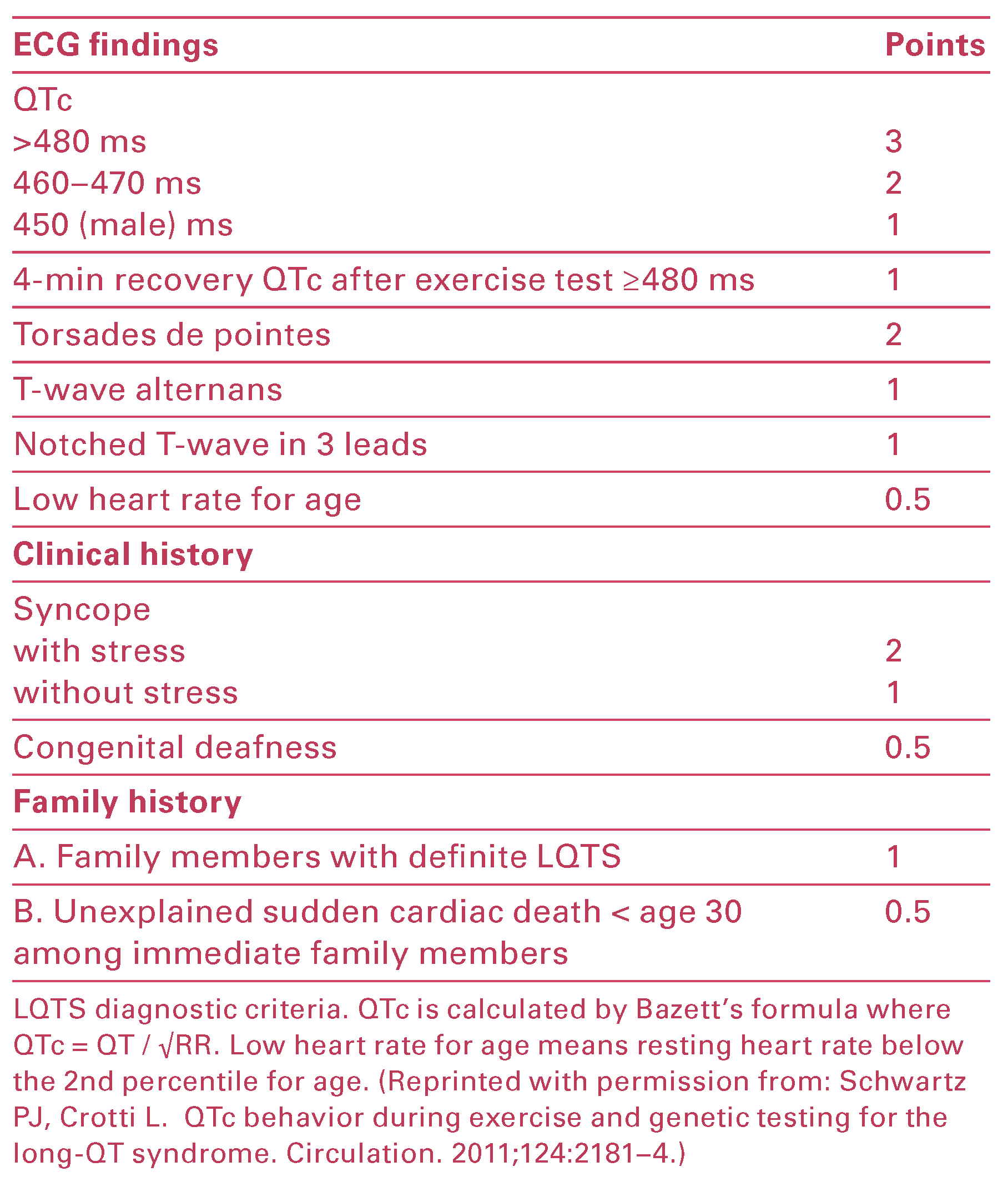
The diagnosis can be eased by use of a scoring system (updated Schwartz Score) (Table 1) which includes ECG, symptoms and family history [19]. Patients with a Schwartz Score ≥3.5, without a secondary cause for QT prolongation, are diagnosed as having LQTS [5].
Some important clues are present on the ECG which must not be neglected. The elongated QT interval in LQTS is the cornerstone of diagnosis; however, an expert eye on the ST-T morphology may help to differentiate the three LQTS subtypes [20]. As shown by a multinational registry analysis, ST-T-wave patterns can be used to identify subtype 1 and subtype 2 LQTS [21]. In LQTS subtype 1, T-waves appeared broad-based, peaked and asymmetrical. In LQTS subtype 2, T-waves were typically bifid and of low amplitude. Additionally, it was concluded that the patterns associated with each subtype were similar, regardless of the specific mutation, in different individuals and families. Remarkably, our patient presenting with a braod-based, peaked double-notched T-wave turned out to have a LQTS sub type 2. Such a large Himalayan T-wave was described in association with KCNQ1 mutation (LQTS subtype 1) [22]. To the best of our knowledge, this is the first work reporting such a wide and tall T-wave associated with KCNH2 mutation.
Concerning the patterns of other arrhythmogenic syncopes, T-wave inversion in leads V1–V3 accompanied by the epsilon wave indicates arrhythmogenic right ventricular cardiomyopathy [23]. Persistent ST elevations in leads V1–V3 with a right bundle-branch block and of ten a prolongation of the PR interval should raise the suspicion of Brugada syndrome [24].
Accordingly to the clinical history of our patient, type 2 LQTS was rather plausible as most drugs that cause LQTS do so by blocking the potassium rectifying current via the KCNH2 gene [25,26,27]. By this means, there is a relation between KCNH2 mutations and drug duced LQTS.
The QT prolongation on the ECG reflects the longer du ration of repolarisation, orchestrated by mutated sodium, potassium and calcium channels or their auxiliary proteins [28]. The mutations in their respective genes in approximately 85% of the cases are inherited from one of the parents and in the remaining 15% are de novo mutations [29]. Additionally, even in genotypepositive cases of LQTS, approximately half of them have no lifetime symptoms and 10–50% show no apparent QT prolongation [30–32]. Concerning risk assessment, even patients with a normal QTc interval (<440 ms), still show a >10-fold risk of fatal arrhythmia [33]. Thus, genetic counselling is advised for relatives.
As a concluding note, clinicians should be aware that syncope is only a symptom and most ohen it represents only one manifestation of an underlying disease. While many fainting experiences are trivial, sometimes they are the manifestation of severe illnesses and sporadically the syncope ends up in sudden death [3]. With this in mind, it is of prime importance to extensively exclude somatic causes of syncope before attributing it to a primary psychiatric origin. Regarding arrhythmogenic syncope, it should not be excluded on the basis of a normal ECG, especially in likely scenarios. In these situations, the clinician should actively search for the arrhythmia through a detailed family history of malaise or syncope, a resting and a stress ECG, and Holter monitoring or a prolonged recording, given the brief and transient nature of some of these episodes. In particular situations, a loop recorder can be implanted, or electrophysiological testing or a drug challenge to reveal a Brugada pattern can be used. As illustrated by this report, psychogenic syncope, although not rare, should stay a diagnosis of exclusion, for the benefit of the patient.

Disclosure Statement

No financial support and no other potential conflict of interest relevant to this article was reported.

References

  1. Harmon, K.G.; Asif, I.M.; Klossner, D.; Drezner, J.A. Incidence of sudden cardiac death in national collegiate athletic association athletes. Circulation 2011, 123, 1594–600. [Google Scholar] [CrossRef]
  2. Maron, B.J.; Doerer, J.J.; Haas, T.S.; Tierney, D.M.; Mueller, F.O. Sudden deaths in young competitive athletes: analysis of 1866 deaths in the United States, 1980–2006. Circulation 2009, 119, 1085–92. [Google Scholar] [CrossRef]
  3. Marijon, E.; Tafflet, M.; Celermajer, D.S.; Dumas, F.; Perier, M.C.; Mustafic, H.; et al. Sports-related sudden death in the general population. Circulation 2011, 124, 672–81. [Google Scholar] [CrossRef]
  4. Harmon, K.G.; Asif, I.M.; Klossner, D.; Drezner, J.A. Incidence of sudden cardiac death in National Collegiate Athletic Association athletes. Circulation. 2011, 123, 1594–600. [Google Scholar] [PubMed]
  5. Maron, B.J.; Thompson, P.D.; Ackerman, M.J.; Balady, G.; Berger, S.; Cohen, D.; et al. American Heart Association Council on Nutrition, Physical Activity, and Metabolism. Recommendations and considerations related to preparticipation screening for cardiovascular abnormalities in competitive athletes: 2007 update: a scientific statement from the American Heart Association Council on Nutrition, Physical Activity, and Metabolism: endorsed by the American College of Cardiology Foundation. Circulation. 2007, 115, 1643–455. [Google Scholar]
  6. Corrado, D.; Pelliccia, A.; Bjørnstad, H.H.; Vanhees, L.; Biffi, A.; Borjesson, M.; et al. Cardiovascular pre-participation screening of young competitive athletes for prevention of sudden death: proposal for a common European protocol. Consensus Statement of the Study Group of Sport Cardiology of the Working Group of Cardiac Rehabilitation and Exercise Physiology and the Working Group of Myocardial and Pericardial Diseases of the European Society of Cardiology. Eur Heart J 2005, 26, 516–524. [Google Scholar]
  7. Bille, K.; Figueiras, D.; Schamasch, P.; Kappenberger, L.; Brenner, J.I.; Meijboom, F.J.; et al. Sudden cardiac death in athletes: the Lausanne Recommendations. Eur J Cardiovasc Prev Rehabil. 2006, 13, 859–75. [Google Scholar]
  8. Dvorak, J.; Grimm, K.; Schmied, C.; Junge, A. Development and implementation of a standardized precompetition medical assessment of international elite football players--2006 FIFA World Cup Germany. Clin J Sport Med. 2009, 19, 316–21. [Google Scholar] [CrossRef] [PubMed]
  9. Pelliccia, A.; Fagard, R.; Bjørnstad, H.H.; Anastassakis, A.; Arbustini, E.; Assanelli, D.; et al. Recommendations for competitive sports participation in athletes with cardiovascular disease: a consensus document from the Study Group of Sports Cardiology of the Working Group of Cardiac Rehabilitation and Exercise Physiology and the Working Group of Myocardial and Pericardial Diseases of the European Society of Cardiology. Eur Heart J. 2005, 26, 1422–45. [Google Scholar] [PubMed]
  10. Drezner, J.A.; Rogers, K.J. Sudden cardiac arrest in intercollegiate athletes: detailed analysis and outcomes of resuscitation in nine cases. Heart Rhythm. 2006, 3, 755–759. [Google Scholar] [CrossRef]
  11. Corrado, D.; Basso, C.; Schiavon, M.; Thiene, G. Screening for hypertrophic cardiomyopathy in young athletes. N Engl J Med. 1998, 339, 364–369. [Google Scholar] [CrossRef] [PubMed]
  12. Corrado, D.; Basso, C.; Pavei, A.; Michieli, P.; Schiavon, M.; Thiene, G. Trends in Sudden Cardiovascular Death in Young Competitive Athletes After Implemention of a Preparticipation Screening Program. JAMA 2006, 296, 1593–1601. [Google Scholar] [CrossRef] [PubMed]
  13. Basso, C.; Corrado, D.; Marcus, F.I.; Nava, A.; Thiene, G. Arrhythmogenic right ventricular cardiomyopathy. Lancet 2009, 373, 1289–300. [Google Scholar] [CrossRef]
  14. Corrado, D.; Pelliccia, A.; Heidbuchel, H.; Sharma, S.; Link, M.; Basso Cet, a.l. Recommendations for interpretation of 12-lead electrocardiogram in the athlete. Eur Heart J 2010, 31, 243–59. [Google Scholar] [CrossRef]
  15. Baggish, A.L.; Hutter, A.M., Jr.; Wang, F.; Yared, K.; Weiner, R.B.; Kupperman, E.; et al. Cardiovascular screening in college athletes with and without electrocardiography: A cross-sectional study. Ann Intern Med. 2010, 152, 269–75. [Google Scholar] [CrossRef]
  16. Riding, N.R.; Salah, O.; Sharma, S.; Carré, F.; George, K.P.; Farooq, A.; et al. ECG and morphologic adaptations in Arabic athletes: are the European Society of Cardiology’s recommendations for the interpretation of the 12-lead ECG appropriate for this ethnicity? Br J Sports Med 2014, 48, 1138–43. [Google Scholar] [CrossRef]
  17. Papadakis, M.; Carre, F.; Kervio, G.; Rawlins, J.; Panoulas, V.F.; Chandra, N.; et al. The prevalence, distribution, and clinical outcomes of electrocardiographic repolarization patterns in male athletes of African/Afro-Caribbean origin. Eur Heart J 2011, 32, 2304–13. [Google Scholar] [CrossRef]
  18. Magalski, A.; McCoy, M.; Zabel, M.; Magee, L.M.; Goeke, J.; Main, M.L.; et al. Cardiovascular screening with electrocardiography and echocardiography in collegiate athletes. Am J Med 2011, 124, 511–8. [Google Scholar] [CrossRef] [PubMed]
  19. Uberoi, A.; Stein, R.; Perez, M.V.; Freeman, J.; Wheeler, M.; Dewey, F.; et al. Interpretation of the electrocardiogram of young athletes. Circulation 2011, 124, 746–57. [Google Scholar] [CrossRef]
  20. Drezner, J.A.; Ackerman, M.J.; Anderson, J.; Ashley, E.; Asplund, C.A.; Baggish, A.L.; et al. Electrocardiographic interpretation in athletes: the ‘Seattle Criteria’. Br J Sports Med 2013, 47, 122–124. [Google Scholar] [CrossRef]
  21. Drezner, J.A.; Fischbach, P.; Froelicher, V.; Marek, J.; Pelliccia, A.; Prutkin, J.M.; et al. Normal electrocardiographic findings: recognising physiological adaptations in athletes. Br J Sports Med 2013, 47, 125–136. [Google Scholar] [CrossRef] [PubMed]
  22. Drezner, J.A.; Ackerman, M.J.; Cannon, B.C.; Corrado, D.; Heidbuchel, H.; Prutkin, J.M.; et al. Abnormal electrocardiographic findings in athletes: recognising changes suggestive of primary electrical disease. Br J Sports Med 2013, 47, 153–167. [Google Scholar] [CrossRef] [PubMed]
  23. Drezner, J.A.; Ashley, E.; Baggish, A.L.; Börjesson, M.; Corrado, D.; Owens, D.S.; et al. Abnormal electrocardiographic findings in athletes: recognising changes suggestive of cardiomyopathy. Br J Sports Med 2013, 47, 137–152. [Google Scholar] [CrossRef] [PubMed]
  24. Brosnan, M.; La Gerche, A.; Kalman, J.; Lo, W.; Fallon, K.; MacIsaac, A.; et al. The Seattle Criteria increase the specificity of preparticipation ECG screening among elite athletes. Br J Sports Med 2014, 48, 1144–50. [Google Scholar] [CrossRef]
  25. Riding, N.R.; Sheikh, N.; Adamuz, C.; Watt, V.; Farooq, A.; Whyte, G.P.; et al. Comparison of three current sets of electrocardiographic interpretation criteria for use in screening athletes. Heart. 2015, 101, 384–90. [Google Scholar] [CrossRef]
  26. Pickham, D.; Zarafshar, S.; Sani, D.; Kumar, N.; Froelicher, V. Comparison of three ECG criteria for athlete pre-participation screening. J Electrocardiol. 2014, 47, 769–74. [Google Scholar] [CrossRef]
  27. Sheikh, N.; Papadakis, M.; Ghani, S.; Zaidi, A.; Gati, S.; Adami, P.E. Comparison of electrocardiographic criteria for the detection of cardiac abnormalities in elite black and white athletes. Circulation. 2014, 129, 1637–49. [Google Scholar] [CrossRef]
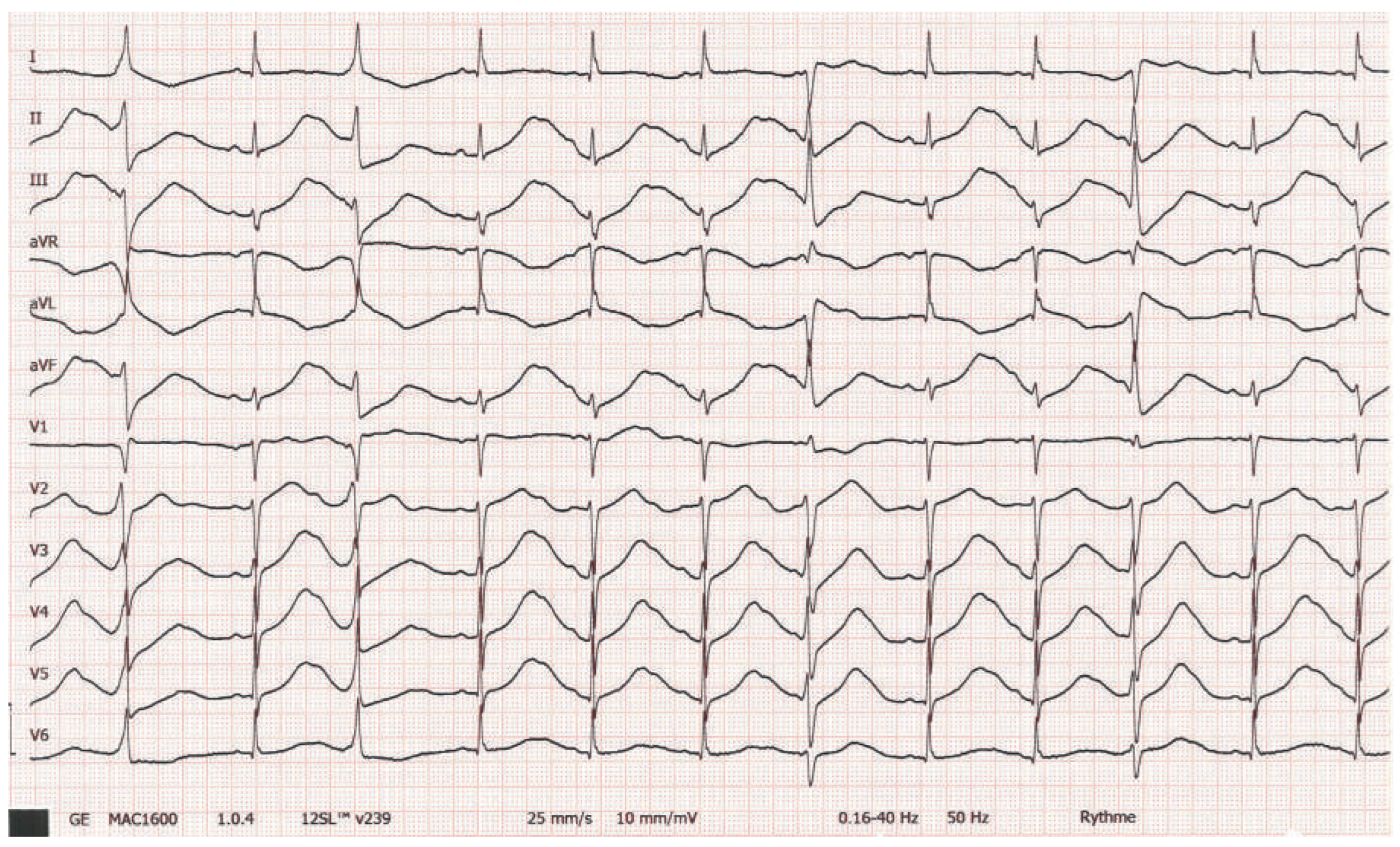
Figure 3. Twelve-lead ECG of the patient after ventricular tachycardia cessation.
Cardiovascmed 19 00090 g003
Figure 4. PCR of the KCNH2 gene, showing a missense mutation of the amino acid I96 (c.287T>A).
Cardiovascmed 19 00090 g004
Figure 5. Twelve-lead ECG of the patient on beta-blocker treatment, after weaning off QT-elongating drugs.
Cardiovascmed 19 00090 g005
Table 1. Schwartz Score (updated in 2011).
Cardiovascmed 19 00090 i001