Next Article in Journal
Symptomatic Polymorphic Ventricular Tachycardia in a Young Woman
Previous Article in Journal
Flexible Pusher Cable for Percutaneous Closure of an Atrial Septal Defect
 
 
Cardiovascular Medicine is published by MDPI from Volume 28 Issue 1 (2025). Previous articles were published by another publisher in Open Access under a CC-BY (or CC-BY-NC-ND) licence, and they are hosted by MDPI on mdpi.com as a courtesy and upon agreement with Editores Medicorum Helveticorum (EMH).
Editorial

Leadless Cardiac Stimulation: Ready to Take Centre Stage?

by
Nicolas B. Dayal
and
Haran Burri
University Hospital of Geneva, Geneva, Switzerland
Cardiovasc. Med. 2016, 19(3), 83; https://doi.org/10.4414/cvm.2016.00397
Submission received: 16 December 2015 / Revised: 16 January 2016 / Accepted: 16 February 2016 / Published: 16 March 2016

Abstract

Over the past few years, the field of cardiac stimulation has witnessed the accelerated development of leadless cardiac pacing, a technology first conceived almost 50 years ago. Currently, two fully intracardiac devices allow single-chamber right-ventricular stimulation: the Nanostim™ and Micra™leadles pacemakers. Both are still undergoing long-term study, but only the latter device is commercially available in Switzerland. The field of cardiac resynchronisation therapy is also exploring leadless technology, with the development of the Wireless-CRT system, although its state of development is less advanced. These technologies, though still in relatively early stages, represent initial steps in a revolution in cardiac stimulation.

Introduction

The advent of cardiac stimulation devices has revolu tionised the treatment of various cardiac disorders, from syncope to arrhythmias and, more recently, in the field of heart failure. During the twentieth century, several milestones were reached leading to the current state of cardiac stimulation, from the first successful use of an external electrical stimulation device in 1932 by Albert S. Hyman [1] to the first implantable epicar dial pacemaker by Rune Elmquist and Ake Senning in 1958 [2]. In parallel, interest in development of trans venous lead systems grew, with the first implantation of a temporary transvenous pacing lead in 1958 by Sey mour Furman [3], and, finally, the appearance of the first implantable permanent pacemaker systems with transvenous leads in 1962 [4].
Currently, most cardiac implantable electronic devices consist of a pulse generator implanted in the sub cutaneous or submuscular tissue, connected to one or more transvenous leads implanted on the endocardial surface of the right chambers or the coronary sinus. The surgical implantation procedure, though a fre quently performed intervention in most centres, can have complications in almost 10% of cases [5,6]. These include haematoma (2–3%) and infection (approxi mately 1%) at the pocket site, as well as cardiac perfora tion (0.4–0.6%), lead dislodgment (2–3%) and pneumo thorax (around 2%) following placement of the transvenous leads. Further lead related concerns aher device implantation include fracture, insulation de fects, connector problems and infection. Despite great advances in pacemaker and lead technologies, these issues are far from being resolved and may even be growing in importance owing to the increasing num ber of devices and leads per device implanted. Indeed, the morbidity associated with device related complica tions remains high, and these complications must be balanced against the potential benefit in this popula tion of ohen very elderly patients.
Since early on, research has investigated the possibility of dispensing with the leads and external generators altogether as one way potentially to eliminate these issues. The feasibility of intracardiac stimulation devices was demonstrated from as early as 1970, with several reports of successful implantation and short term function in canine models [7,8]. Though these devices showed great promise, it was not until more re cently that the research community and industry have accelerated the development of human leadless pace makers. At first, the concept of leadless devices still required the implantation of an extracardiac genera tor that would transmit wirelessly to an intracardiac receiver, either by ultrasound energy [9,10,11] or mag netic field induction energy [12]. However, since 2012, several fully intracardiac devices have been developed and later commercialised for pacing the right ventricle. Leadless cardiac pacing is also being developed in the field of cardiac resynchronisation therapy (CRT). Al though the results of CRT therapy have been impres sive in carefully selected populations, it is still marred by several major issues, such as a relatively high rate of nonresponders and leh ventricular (LV) lead failure. LV endocardial pacing offers the theoretical advantage of a wider selection of pacing sites compared with the highly variable anatomy of the coronary sinus branches, and possibly a greater haemodynamic effect.Until recently, this required a transvenous approach with passage through the atrial or ventricular septum, but with a high rate of systemic thromboembolism [13]. Thus, wireless leh ventricular endocardial pacing is also being developed in order to address these issues.

Characteristics of the Different Leadless Pacing Systems

Right Ventricular Pacing Devices

The first fully intracardiac leadless pacing system to be developed was the NanostimTM Leadless Cardiac Pace maker System (St Jude Medical, Sylmar, CA, USA), intro duced in 2012 and obtaining the CE mark in 2013. This device was followed by the MicraTM Transcatheter Pac ing System (Medtronic, Minneapolis, MN, USA), which obtained CE certification recently in 2015. Both devices remain investigational in the United States.
These devices incorporate the battery and electronic circuit as well as bipolar sensing and pacing electrodes in a small capsule shaped format that is designed to be implanted directly in the right ventricle (RV) (Figure 1). In terms of function, both pacemakers operate as single chamber VVI devices with optional rate adaptive pacing, and both use a bipolar sensing and pacing configuration. They possess steroid eluting cathode tips to ensure similar maturation compared with standard transvenous devices.
Key differences between both devices are highlighted in Table 1. The MicraTM has a shorter, wider profile, thus needing a larger gauge delivery sheath (27 F or 0.9 cm). The device is fixated with the help of four self expand ing nitinol tines that are deployed with retraction of the delivery sheath and anchor the device to the myo cardium. In contrast, the NanostimTM possesses a pri mary screw in helix with maximal penetration in the myocardium of 1.3 mm and a secondary mechanism of angled nylon tines. Electrodes in the MicraTM consist of a steroid eluting cathode tip and a titanium ring. In the NanostimTM, a steroid eluting disk in the centre of the helix acts as the cathode, and the anode, located more than 10 mm away, consists of the non coated part of the titanium case. Further differences include the modality of rate adaptive pacing: whereas the MicraTM uses a three axis accelerometer (with filters to distin guish cardiac motion from patient acceleration), the NanostimTM relies on a central venous temperature sensor.
The telemetry connection with the MicraTM is estab lished via radio frequency with a standard Medtronic model 2090 Programmer and programming head. With the NanostimTM, the system requires two way conduction with the help of an external module that acts as the link between a model 3650 Merlin™ Patient Care System Programmer and standard ECG skin electrodes placed on the subject’s torso. Signals are transmitted to and from the device via subliminal 250 kHz pulses applied to the skin electrodes that do not interfere with pacemaker function.

Left Ventricular Pacing System

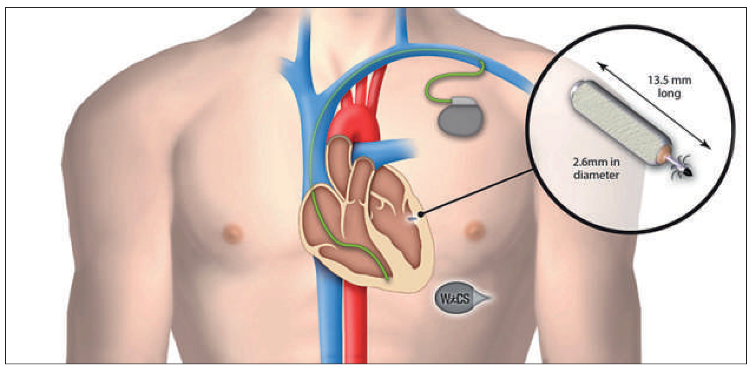
The ultrasound based WiCS™ system (Wireless Cardiac Stimulation; EBR Systems, Sunnyvale, CA, USA) has been developed to offer leadless LV endocardial pacing with the use of ultrasound transmission and has recently been granted CE mark approval. The system consists of a subcutaneous pulse generator communi cating via ultrasound with a receiver electrode (13.5 × 2.6 mm) implanted in the leh ventricle (Figure 2). The re ceiver converts the ultrasound pulses produced by the generator from acoustic to electrical energy, thus pacing the LV. A conventional pacing device (pace maker, CRT device or implantable cardiac defibrillator) is necessary in order to synchronise LV pacing with RV pacing, using a programmed 3 ms delay.

Implantation Procedures of Leadless Devices

Right Ventricular Pacing Systems

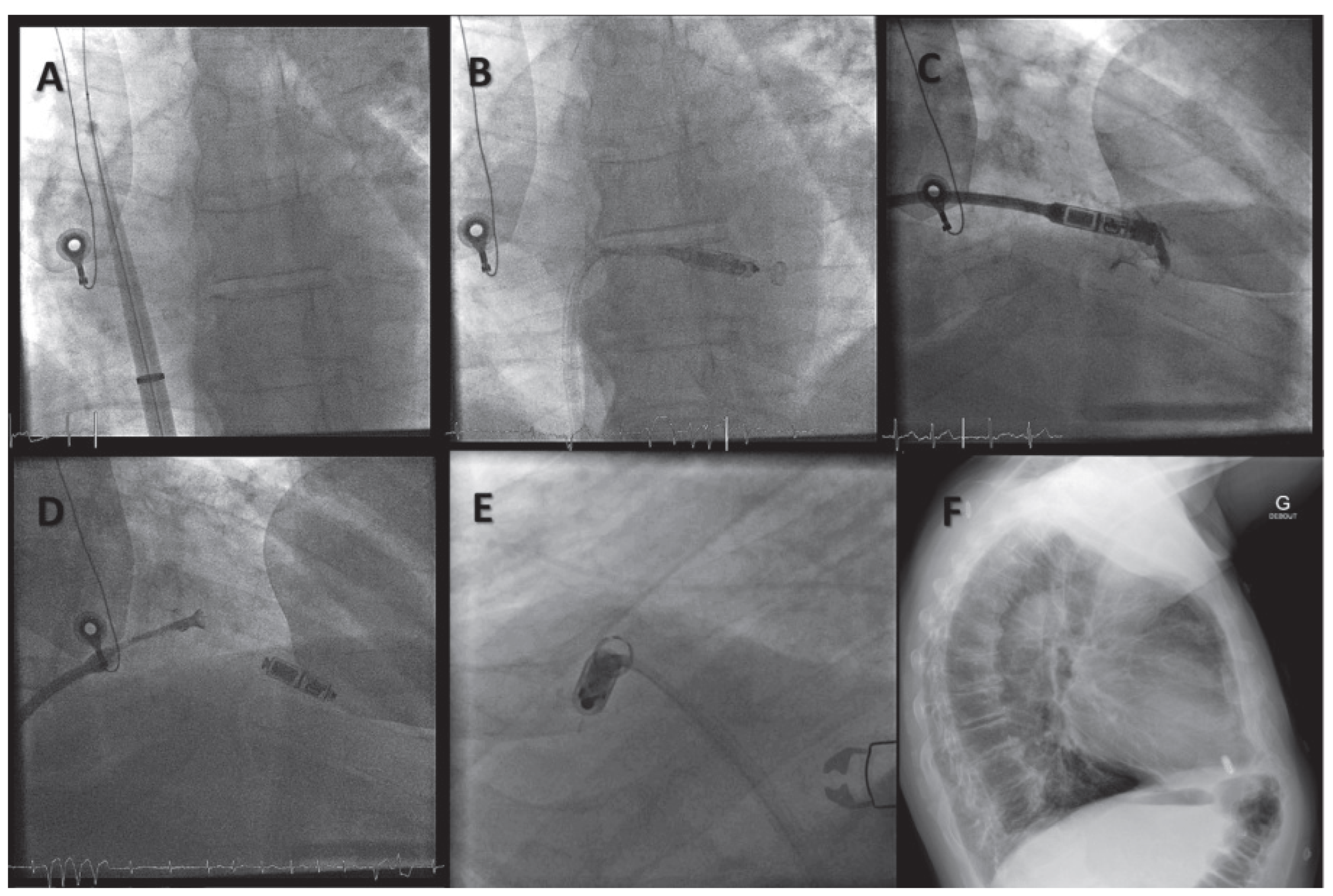
Both devices can be implanted under local anaesthesia and fluoroscopic guidance. The delivery systems con tain a deflectable tip that is directed through the right atrium and into the RV via the tricuspid valve. Ideally, the devices are directed to the apical or apico septal region. Once in position, the implantation of the Nanostim™ LCP device requires retraction of an ex tendable protective sleeve, exposing the helix, which is then rotated 1.25 turns, enabling fixation [14]. The Micra™ implantation also requires retraction of a sleeve, thus deploying the four tines, of which at least two should hook into the myocardium for a successful deployment. Once fixated, both devices are undocked, but remain tethered to the delivery system, allowing evaluation of stability and pacing performance, and easy repositioning if necessary. Before final release, the operator performs a “pull and hold” under fluoros copy, then cuts the tethers, freeing the device. Figure 3 illustrates the implantation procedure of the Micra™ TPS system. Apart from formal training by the manu facturers of the different devices, operators undertak ing implantation of leadless devices should be experi enced in femoral access as well as cardiac stimulation. It is a significant asset to have experience with electro physiological procedures, as steering of the catheter re lates more to this domain than to manipulation of pac ing leads. Furthermore, the physician should ideally be competent in lead extraction, as he or she will be more proficient with large bore femoral access as well as device retrieval in case of need. Device implantation should ideally be undertaken in an environment with high quality fluoroscopy such as an electrophysiology or catheterisation laboratory, especially with the Mi cra™ system as correct visualisation of the four tines is mandatory for adequate device placement.

Wireless Cardiac Resynchronisation Therapy System

The leadless receiver is implanted in the LV endocar dium via a retrograde aortic approach, and anchored with the help of three self expanding nitinol tines. The optimal position of the pulse generator in the WiCS™ system is chosen with use of echocardiography in order to find an appropriate acoustic window. Usually this window is found lateral to the leh parasternal re gion in the fourth to sixth intercostal spaces, the fihh intercostal ohen being the most appropriate [15].

Current Scientific Evidence Regarding Leadless Pacing Technology

Micra™ Transcatheter Pacing System

Aher initial feasibility studies performed in animals [16], early results of the Micra Transcatheter Pacing Study have been published recently. This ongoing safety and efficacy study is a worldwide, prospective, multi centre single arm study that has recruited 744 patients with a class I or II indication for single chamber ventric ular pacing, of whom 725 underwent an implantation at tempt. Preliminary results once the first 60 patients had completed 3 months follow up were published in 2015 [17] and, more recently, a planned interim analysis of the first 300 patients completing 6 months of follow up was published [18]. Implantation was successful in 719 pa tients (99.2%), with a mean procedure time of 34.8 ± 24.1 min, and fluoroscopy time of 8.9 ± 16.6 min, higher than the usual fluoroscopy time (<5 minutes) for a single chamber transvenous pacemaker. Among the first 140 patients, optimal device position was achieved on the first attempt in 59% of patients, and 18.9% required more than two deployments.
With regards to pacemaker efficacy, 98.3% of patients reached the primary efficacy endpoint consisting of a capture threshold <2.0 V at 6 months, without signifi cant (>1.5 V) increase since implantation (p <0.001). Mean pacing threshold value was 0.54 V at 0.24 ms, mean R wave amplitude 15.3 mV and mean impedance was 627 Ω. Based on these values, device longevity was estimated at 12.5 years, with 94% lasting over 10 years. The efficacy of the device was further highlighted by the inclusion, approved by the US Food and Drug Ad ministration (FDA), of pacemaker dependant patients aher analysis of the first 25 patients [19].
In terms of safety endpoints, the overall rate of major procedure or system related adverse effects at 6 months was 3.45%, corresponding to 28 complications in 25 patients, 4 of whom had unsuccessful implants. Traumatic cardiac injury was reported in 11 cases (1.5%) with 3 cardiac perforations (0.41%) and 8 pericardial effusions (1.1%), higher than usually reported in trans venous pacemaker implantations. Other complica tions included 5 access site complications (0.69%), 2 thromboembolic events (0.28%), 2 pacing issues (ele vated thresholds, 0.28%) and 8 other complications (1.1%). Compared with a historical cohort of 2 667 trans venous implants, and aher adjustment for differences in population, the Micra patients had a lower risk of major complications (hazard ratio 0.46, confidence in terval 0.28–0.74, p = 0.006), although these post-hoc re sults must be interpreted with caution. Concerning the risk of cardiac perforation specifically, mechanical as sessment of the Micra™ delivery system in cadavers pointed to an acceptable margin between the force ex erted at the tip of the catheter and the force necessary to perforate the RV [20], although these are not clinical data and perforation of other structures (vena cava, right atrium, etc.) has not been evaluated. The possibil ity of mechanical trauma linked to the delivery system is also highlighted by the presence of transient atrioven tricular block or right bundle branch block in some patients. Overall, however, the implant procedure was well tolerated in the Micra TPS study, with a mean time to discharge of 1 day in the analysis of the first 140 pa tients [17].
The system has been available for limited market re lease in Switzerland since June 2015.

Nanostim™ Leadless Cardiac Pacemaker

The LEADLESS study [14] was a multicentre, single arm European study evaluating the safety and efficacy of the Nanostim™ LCP device in 33 patients. Similarly to the Micra TPS study, patients were included if they had an indication for single chamber pacing, consisting of: (1) permanent atrial fibrillation with atrioventricular block or slow ventricular response; (2) normal sinus rhythm with second or third degree atrioventricular block and a low level of physical activity or short expected life span; or (3) sinus bradycardia with in frequent pauses or unexplained syncope with electro physiological findings. The initial results have been de scribed in previous reviews [21,22], and showed a 97% rate of successful implantation, 70% requiring only one deployment of the device, and two serious adverse device endpoints (one tamponade and one placement in the leh ventricle via a patent foramen ovale). Retro spective 1 year analysis of the LEADLESS trial [23] showed an absence of further complications and stable pacemaker performance values (mean pacing thres hold at a 0.4 ms pulse width of 0.43 ± 0.30 V; R wave amplitude of 10.3 ± 2.2 mV; and impedance 627 ± 209 Ω). More recently, interim results of the LEADLESS II study, a prospective, nonrandomised, multicentre study aimed at obtaining FDA approval for the Nanostim™ device, have been published [24]. The primary analysis concerned the 6 month safety and efficacy results of the first 300 patients, but the study also published the outcomes of all 526 patients enrolled to date. Inclusion and exclusion criteria were similar for the LEADLESS trial, although pacemaker dependant patients were no longer excluded. The results showed a Nanostim™ im plantation success rate of 95.8%, with a mean proce dure time of 28.6 ± 17.8 minutes, and fluoroscopy time of 13.9 ± 9.1 minutes, once again significantly longer than for a transvenous VVI pacemaker. Similarly to the LEADLESS trial, 70.2% of devices were successfully im planted aher initial deployment. Mean hospital stay was comparable to that in the Micra TPS study (1.1 ± 1.7 days). Regarding pacemaker efficacy at 6 months, 90% of patients reached the combined primary efficacy endpoint of (1) acceptable pacing threshold (≤2.0 V at 0.4 msec) and (2) an acceptable sensing amplitude (R wave ≥5.0 mV, or a value equal to or greater than the value at implantation). In the total cohort, the mean values at 12 months were: R wave amplitude 9.2 ± 2.9 mV and pacing capture threshold (at 0.4 msec) 0.58 ± 0.31 V. According to results at the 6 month follow up, battery longevity could be estimated to be 15 ± 6.7 years.
With regards to safety, the rate of device related seri ous adverse events was 6.7% over 6 months, with 1.3% cardiac perforation, 1.7% device dislodgement, 1.3% el evated pacing thresholds necessitating device retrieval and replacement, and 1.3% vascular complications. This study also pointed to a learning curve, as in most medical procedures, with an almost halved rate of complications aher 10 procedures (3.6 vs 6.8%), al though this difference was not statistically significant. Of note, another long term safety trial is ongoing in Europe (The LEADLESS Observational Study; NCT02051972).

Wireless Cardiac Resynchronisation Therapy System

Initial evaluation of the WiCS™ device was performed in the WISE CRT study [15], which was a multicentre European prospective observational study evaluating the feasibility and safety of the system. It aimed to enrol 100 patients with indications for CRT therapy and with either failed coronary sinus lead implanta tion, clinical nonresponse aher conventional CRT im plantation or necessity to upgrade to a CRT system. The study was interrupted prematurely aher just 17 patient enrolments, because of a high rate (three patients or 18%) of procedural serious pericardial effusions linked to manipulation of the delivery catheter or guidewire, with one fatality. Of the 17 patients, 1 additional subject had a failed implantation as a result of inadequate pacing thresholds. From an efficacy point of view, the system showed promising results in the few patients followed up for 6 months.
Following revision of the delivery system, the WiCS system is currently under evaluation in the SELECT LV nonrandomised trial [25]. Early results from 35 patients continue to show promising results in terms of effi cacy, with a shortening in mean QRS duration from 174 to 137 ms at 1 month, an improvement of LV ejection fraction from 27 to 33.7% at 6 months and reduced ven tricular volumes. Importantly, patients also showed an improvement in their New York Heart Association (NYHA) class from 2.6 to 1.8 at 6 months. With regards to safety, there were no cases of tamponade with the revised delivery system. Nevertheless, rates of compli cations were not negligible, with 4 of the 35 patients suffering device or procedure related events within 24 hours (one groin fistula necessitating surgery, one groin pseudoaneurysm treated conservatively, one electrode embolisation to the lower leg and one case of ventricular fibrillation with aborted implantation). At 1 month, there were 10 further complications, 7 related to the device and 3 concerning infection.

Remaining Questions

Leadless Right Ventricular Pacing

To date, no data exist regarding the actual battery longevity of leadless pacemakers, the only indications coming from theoretical calculations or estimates based on early results of the aforementioned studies. Moreover, much of the data was retrieved from non pacemaker dependant patents, such as in the LEAD LESS trial. Even in the LEADLESS II trial, mean rates of ventricular pacing were 51.6% at 12 months. Never theless, these estimates point toward similar device longevity to single chamber conventional pacemakers. In the case of the Nanostim™, the high efficiency of the unit results in a current drain of approximately 1 µA, around six times less than a transvenous unit from the same manufacturer [22]. As illustrated in Table 1, the projected device longevity of the Micra™ at the Inter national Organization for Standardization (ISO) values is substantially less than the Nanostim™, but thanks to energy saving algorithms such as capture manage ment, the projected values at real world settings are improved.
Furthermore, the optimal strategy once a leadless de vice reaches the end of its service is unknown. Device abandonment with placement of a new device has yet to be tested comprehensively. Removal of a chronically implanted leadless pacemaker also remains poorly studied in humans, the only data emanating from the LEADLESS II study where seven patients underwent successful retrieval of devices that had been implanted 160 ± 180 days previously. In the Micra TPS study, one patient underwent successful percutaneous removal 17 days aher implantation because of intermittent loss of capture. Even available animal data concern rela tively recently implanted devices (5 months in 10 sheep implanted with a Nanostim™ [26] and 18 months in 4 sheep implanted with a Micra™ [27]). Moreover, the optimal strategy in the event of infection of the lead less pacemaker remains unclear. Data from the LEAD LESS II study showed an uncomplicated percutaneous retrieval of six embolised devices (four in the pulmo nary artery and two in the right femoral vein) rela tively early aher implantation (8.0 ± 6.4 days).
The effectiveness of the rate response algorithms in leadless pacemakers has yet to be studied. In the case of the Micra TPS study, no treadmill data are available yet. For the Nanostim™ device, 1 year retrospective analysis of the LEADLESS trial described an adequate rate response function in the 19 patients out of 31 pro grammed in VVIR at 12 months. Evaluation of rate re sponse will be interesting to follow, especially in the case of the Nanostim™, as the responsiveness of cen tral venous temperature to an increase in activity may be slower than the more standard accelerometer used in the Micra™. The performance of the novel rate re sponse algorithm used by the Micra™ also needs to be evaluated.
Although both devices are labelled “MRI conditional” because of the lack of ferrous components, currently data are lacking. One case of a Micra™ implanted pa tient safely undergoing a 1.5 Tesla (T) magnetic reso nance imaging (MRI) scan of the brain was reported in the Micra TPS study [17]. Theoretical data based on modelling results predict a reduced risk of MRI interac tion with the Micra™ device compared with conven tional devices, with a predicted rise in temperature of less than 0.4° in 99% of cases at 1.5 T and 3 T [28]. More clinical experience is needed before the devices can be considered fully MRI safe.

Leadless Endocardial Left Ventricular Pacing

The efficacy results of the Wise CRT and SELECT LV trials seem to indicate the systems could be a useful alternative to standard CRT systems in cases of nonre sponse or LV lead failure, but data are available for only a short follow up of 6 months. Obviously the main con cern with the system remains the safety of the implan tation procedure, which is not fully addressed, as well as the currently unknown rate of chronic complica tions.
Other concerns are the seemingly short battery life of the device; in the Wise CRT study, projected battery longevity aher 6 months follow up was only 18 months (range 9–42 months). Ease of extraction and/or replace ment of the different devices contained in the system remains unknown. Issues with restricted acoustic windows that limit energy transmission need to be addressed. Finally, the thromboembolic risk linked to the leadless receiver unit in the LV in comparison with standard transvenous LV leads needs to be further studied.

Future Directions

One important issue that will be addressed in the fu ture is the possibility of multichamber leadless pacing, although anchoring the device in a safe and effective manner in the thin walled atrium, and issues with additional energy requirements resulting with com munication between the devices, need to be addressed. Currently the available RV pacing devices only allow single chamber ventricular pacing, thus restricting their application in everyday life, with rates of VVI pacemaker implantation at around 8–25% according to different regions of the world [6,29]. The combination of leadless devices with the subcutaneous defibrillator (Boston Scientific, Marlborough, MA, USA) will allow antitachycardia and antibradycardia pacing, and will be introduced in the near future. Finally, researchers are working on a device which will harvest kinetic en ergy from the heart’s motion for providing power – thereby dispensing of the need for device replacement.

Conclusions

Leadless pacemakers are a milestone in pacing ther apy. They are clearly the preferred pacing modality in a niche group of patients (e.g., those with venous access issues), but they can be considered in other situations where VVI(R) pacing is indicated. Currently, only one system is commercially available in Switzerland, with a limited market release. The implantation procedure requires special skills that need to be acquired prop erly, as complications such as tamponade are a con cern. Other issues are management at battery deple tion, extractability, and cost (which is likely to evolve depending upon the uptake of the technology and the introduction of new models from competitors). The therapy will no doubt continue to evolve, and provide us with more treatment options for our patients.

Dislosure Statement

N.D. has no conflicts of interest to report; H.B. has received speaker fees from Biotronik, Boston Scientific, Medtronic and Sorin and insti tutional fellowship support/research grants from Biotronik, Boston Scientific, Medtronic, Sorin and St Jude Medical.

References

  1. Hyman, A. Resuscitation of the stopped heart by intracardial ther apy – II. Experimental use of an artificial pacemaker. Arch Intern Med. 1932, 50, 283–05. [Google Scholar]
  2. Larsson, B.; Elmqvist, H.; Ryden, L.; Schuller, H. Lessons from the first patient with an implanted pacemaker: 1958–2001. Pacing Clin Electrophysiol. 2003, 26, 114–24. [Google Scholar] [CrossRef]
  3. Furman, S.; Schwedel, J.B. An intracardiac pacemaker for Stokes Adams seizures. N Engl J Med. 1959, 261, 943–8. [Google Scholar] [CrossRef]
  4. Lagergren, H.; Johansson, L. Intracardiac stimulation for complete heart block. Acta Chir Scand. 1963, 125, 562–6. [Google Scholar]
  5. Kirkfeldt, R.E.; Johansen, J.B.; Nohr, E.A.; Jorgensen, O.D.; Nielsen, J.C. Complications aher cardiac implantable electronic device implanta tions: an analysis of a complete, nationwide cohort in Denmark. Eur Heart J. 2014, 35, 1186–94. [Google Scholar] [CrossRef]
  6. Udo, E.O.; Zuithoff, N.P.; van Hemel, N.M.; de Cock, C.C.; Hendriks, T.; Doevendans, P.A.; et al. Incidence and predictors of short and long term complications in pacemaker therapy: the FOLLOWPACE study. Heart Rhythm. 2012, 9, 728–35. [Google Scholar] [CrossRef]
  7. Spickler, J.W.; Rasor, N.S.; Kezdi, P.; Misra, S.N.; Robins, K.E.; LeBoeuf, C. Totally self contained intracardiac pacemaker. J Electrocardiol. 1970, 3, 325–31. [Google Scholar] [CrossRef]
  8. Vardas, P.P.E.; Manios, E.; Parthenakis, F.; Tsagarakis, C. A miniature pacemaker introduced intravenously and implanted endocardi ally. Preliminary findings from an experimental study. Eur J Card Pacing Electrophysiol. 1991, 1, 27–30. [Google Scholar]
  9. Echt, D.S.; Cowan, M.W.; Riley, R.E.; Brisken, A.F. Feasibility and safety of a novel technology for pacing without leads. Heart Rhythm. 2006, 3, 1202–6. [Google Scholar] [CrossRef]
  10. Lee, K.L.; Lau, C.P.; Tse, H.F.; Echt, D.S.; Heaven, D.; Smith, W.; et al. First hu man demonstration of cardiac stimulation with transcutaneous ultrasound energy delivery: implications for wireless pacing with implantable devices. J Am Coll Cardiol. 2007, 50, 877–83. [Google Scholar] [CrossRef]
  11. Lee, K.L.; Tse, H.F.; Echt, D.S.; Lau, C.P. Temporary leadless pacing in heart failure patients with ultrasound mediated stimulation energy and effects on the acoustic window. Heart Rhythm. 2009, 6, 742–8. [Google Scholar] [CrossRef]
  12. Wieneke, H.; Konorza, T.; Erbel, R.; Kisker, E. Leadless pacing of the heart using induction technology: a feasibility study. Pacing Clin Electrophysiol. 2009, 32, 177–83. [Google Scholar] [CrossRef]
  13. Rademakers, L.M.; van Gelder, B.M.; Scheffer, M.G.; Bracke, F.A. Mid term follow up of thromboembolic complications in leh ventricular endocardial cardiac resynchronization therapy. Heart Rhythm. 2014, 11, 609–13. [Google Scholar] [CrossRef]
  14. Reddy, V.Y.; Knops, R.E.; Sperzel, J.; Miller, M.A.; Petru, J.; Simon, J.; et al. Permanent leadless cardiac pacing: results of the LEADLESS trial. Circulation. 2014, 129, 1466–71. [Google Scholar] [CrossRef]
  15. Auricchio, A.; Delnoy, P.P.; Butter, C.; Brachmann, J.; Van Erven, L.; Spitzer, S.; et al. Feasibility, safety, and short term outcome of leadless ultrasound based endocardial leh ventricular resynchronization in heart failure patients: results of the wireless stimulation endo cardially for CRT (WiSE CRT) study. Europace. 2014, 16, 681–8. [Google Scholar] [CrossRef]
  16. Bonner, M.; Eggen, M.; Haddad, T.; Sheldon, T.; Williams, E. Early Perfor mance and Safety of the Micra Transcatheter Pacemaker in Pigs. Pacing Clin Electrophysiol. 2015. [Google Scholar] [CrossRef]
  17. Ritter, P.; Duray, G.Z.; Steinwender, C.; Soejima, K.; Omar, R.; Mont, L.; et al. Early performance of a miniaturized leadless cardiac pacemaker: the Micra Transcatheter Pacing Study. Eur Heart J. 2015, 36, 2510–9. [Google Scholar] [CrossRef]
  18. Reynolds, D.; Duray, G.Z.; Omar, R.; Soejima, K.; Neuzil, P.; Zhang, S.; et al. A Leadless Intracardiac Transcatheter Pacing System. N Engl J Med. 2015. [CrossRef]
  19. Mont, L.; et al. Micra Transcatheter Pacing System: early perfor mance assessment to demonstrate safety for pacemaker depend ent patients. 2015, 17 (Suppl. 3): (abstr) 1311. Europace. 2015, 17 (Suppl. 3):(abstr 1311).
  20. Bonner, M.; et al. Micra delivery system perforation risk assess ment. Europace. 2015, 17 (Suppl. 3), abstr 1312. [Google Scholar]
  21. Sperzel, J.; Burri, H.; Gras, D.; Tjong, F.V.; Knops, R.E.; Hindricks, G.; et al. State of the art of leadless pacing. Europace. 2015. [Google Scholar] [CrossRef]
  22. Miller, M.A.; Neuzil, P.; Dukkipati, S.R.; Reddy, V.Y. Leadless Cardiac Pace makers: Back to the Future. J Am Coll Cardiol. 2015, 66, 1179–89. [Google Scholar] [CrossRef]
  23. Knops, R.E.; Tjong, F.V.; Neuzil, P.; Sperzel, J.; Miller, M.A.; Petru, J.; et al. Chronic performance of a leadless cardiac pacemaker: 1 year fol low up of the LEADLESS trial. J Am Coll Cardiol. 2015, 65, 1497–504. [Google Scholar] [CrossRef]
  24. Reddy, V.Y.; Exner, D.V.; Cantillon, D.J.; Doshi, R.; Bunch, T.J.; Tomassoni, G.F.; et al. Percutaneous Implantation of an Entirely Intracardiac Lead less Pacemaker. N Engl J Med. 2015, 373, 1125–35. [Google Scholar] [CrossRef]
  25. Reddy, V.Y. Wireless LV endocardial stimulation for CRT: Primary results of the Safety and Performance of Electrodes implanted in the Leh Ventricle (SELECT LV) Study. Presented at HRS 2015, Late Breaking clinical trials. 2015.
  26. Sperzel, J.K.A.; Ligon, D.; Zaltsberg, S. Feasibility, efficacy and safety of percutaneous retrieval of a leadless cardiac pacemaker in an in vivo bovine model. Europace. 2013, 15, 859. [Google Scholar]
  27. Bonner, M.N.N.; Byrd, C.; Schaerf, R.; Goode, L. Extraction of the Micra transcatheter pacemaker system (abstract). Heart Rhythm. 2014, 11, S342. [Google Scholar]
  28. Soejima, K. RF Heating of Micra: Can Patients with the Trans catheter Pacemaker Safely Undergo MRI? Heart Rhythm. 2015, 12, abstrAB06–5. [Google Scholar]
  29. Crossley, G.H.; Chen, J.; Choucair, W.; Cohen, T.J.; Gohn, D.C.; Johnson, W.B.; et al. Clinical benefits of remote versus transtelephonic monitor ing of implanted pacemakers. J Am Coll Cardiol. 2009, 54, 2012–9. [Google Scholar] [CrossRef]
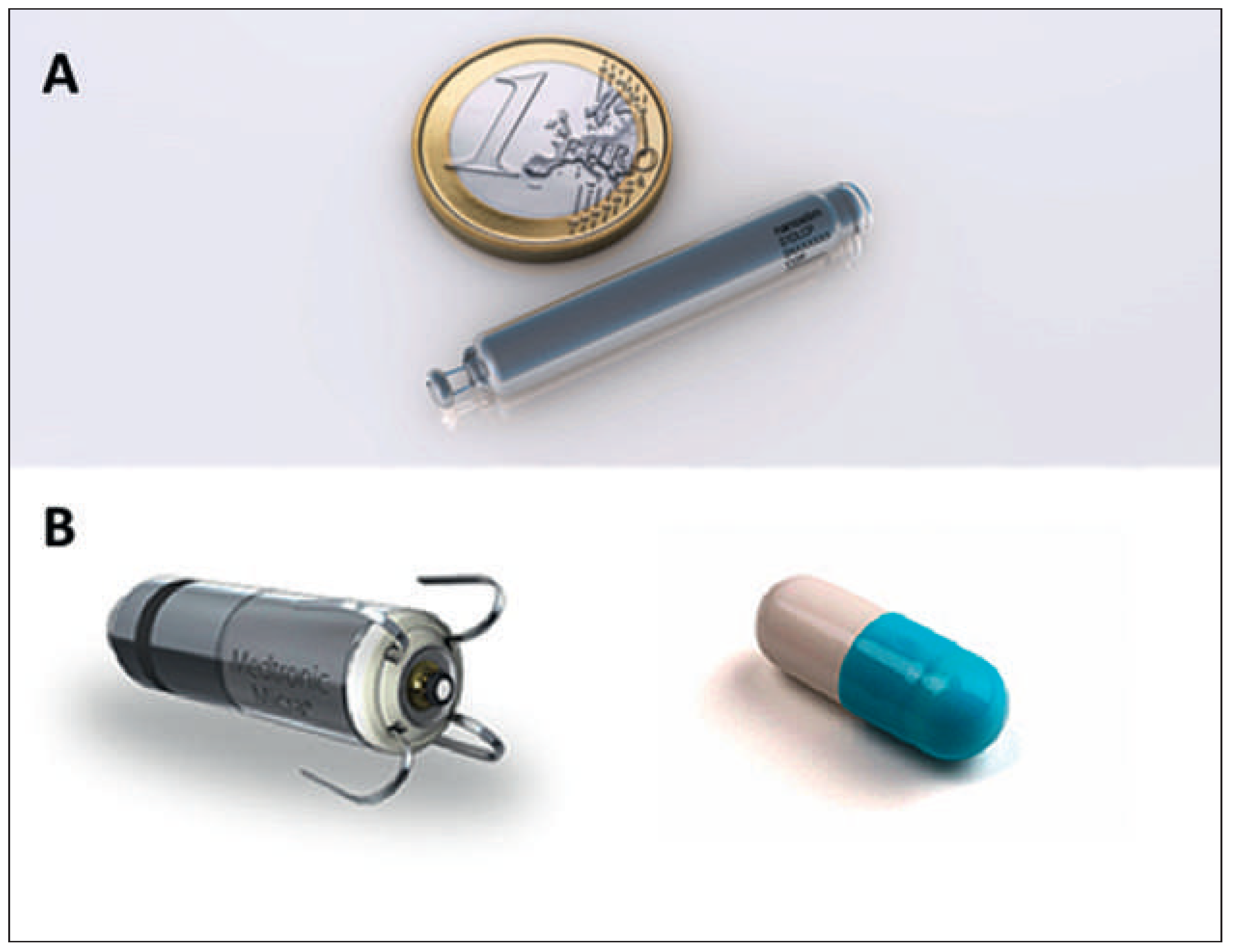
Figure 1. A. The NanostimTM Leadless Cardiac Pacemaker. (St Jude Medical, Sylmar, CA, USA). Courtesy of St Jude Medical. B. The MicraTM Transcatheter Pacing System (Medtronic, Minneapolis, MN, USA). Courtesy of Medtronic.
Cardiovascmed 19 00083 g001
Figure 2. The WiCS™ system (Wireless Cardiac Stimulation; EBR Systems, Sunnyvale, CA, USA), consisting of a standard right ventricular pacemaker/defibrillator and a subcutaneous pulse generator communicating via ultrasound with a receiver electrode (magnified) implanted in the left ventricle. Courtesy of Cambridge Consultants. Image courtesy of EBR Systems.
Cardiovascmed 19 00083 g002
Figure 3. Implantation of a Micra™ system. A. The delivery system is introduced via the femoral vein into the right atrium via a super-stiff guidewire. B. The Micra™ unit is introduced into the deflectable delivery system and positioned across the tricuspid valve into the right ventricle. C. The apical septum is targeted using contrast injection. D. The delivery sheath is withdrawn, thus deploying the tines into the ventricular wall. E. A “pull and hold” test is performed, with viewing in multiple planes to verify that at least two of four tines are fixated, and the tether is then cut to free the device. F. Lateral chest X-ray of the implanted system.
Cardiovascmed 19 00083 g003
Table 1. Key features of the Nanostim™ Leadless Cardiac Pacemaker (LCP) and Micra™.
Cardiovascmed 19 00083 i001