Next Article in Journal
EKG-Interpretation bei Sportlern–Die «Seattle-Kriterien»
Previous Article in Journal
A Preliminary Study on the Introduction of Peer-Assisted Learning in Auscultation Courses
 
 
Cardiovascular Medicine is published by MDPI from Volume 28 Issue 1 (2025). Previous articles were published by another publisher in Open Access under a CC-BY (or CC-BY-NC-ND) licence, and they are hosted by MDPI on mdpi.com as a courtesy and upon agreement with Editores Medicorum Helveticorum (EMH).
Review

The Impact of Fractional Flow Reserve and iFr on Current PCI Strategies

by
Stephane Fournier
and
Olivier Muller
*
Department of Cardiology, University Hospital Centre (CHUV), Lausanne, Switzerland
*
Author to whom correspondence should be addressed.
Cardiovasc. Med. 2016, 19(3), 67; https://doi.org/10.4414/cvm.2016.00390
Submission received: 16 December 2015 / Revised: 16 January 2016 / Accepted: 16 February 2016 / Published: 16 March 2016

Abstract

Fractional flow reserve (FFR) measurement has become the gold standard for assessing myocardial ischaemia and has been recommended in both American and European guidelines as an aid to deciding whether or not to perform myocardial revascularisation. Indeed, large clinical trials have demonstrated that an evaluation of stenoses guided by FFR reduced death, nonfatal myocardial infarction and repeat revascularisation (MACE) when compared with an evaluation guided by angiography only and that percutaneous coronary intervention (PCI) with optimal medical treatment was superior to optimal medical treatment alone to reduce MACE in the presence of a pathological FFR. To measure an FFR, the use of a pharmacological vasodilator (usually adenosine) is necessary. To avoid adenosine administration, the instantaneous wave-free ratio (iFR) recently emerged with the identification of a “wave-free period” of resistances similar in both magnitude and variability to those observed during FFR measurement. Nevertheless, iFR overall accuracy is only 80.4% and therefore iFR should not currently be recomended as an alternative to FFR.

Introduction

The decision to revascularise a patient presenting with stable coronary artery disease (SCAD) is based on the identification, extent and location of epicardial coronary stenosis via angiography. However, several recent trials have shown a benefit of revascularisation over medical treatment in terms of the prognosis when epicardial coronary stenosis induces extensive myocardial ischaemia, whatever the severity of coronary artery stenosis identified at angiography [1,2]. Fractional flow reserve (FFR) measurement has become the gold standard for assessing myocardial ischaemia and has been recommended to aid decision making about myocardial revascularisation in the most recent American and European guidelines [3,4].

What is Fractional Flow Reserve?

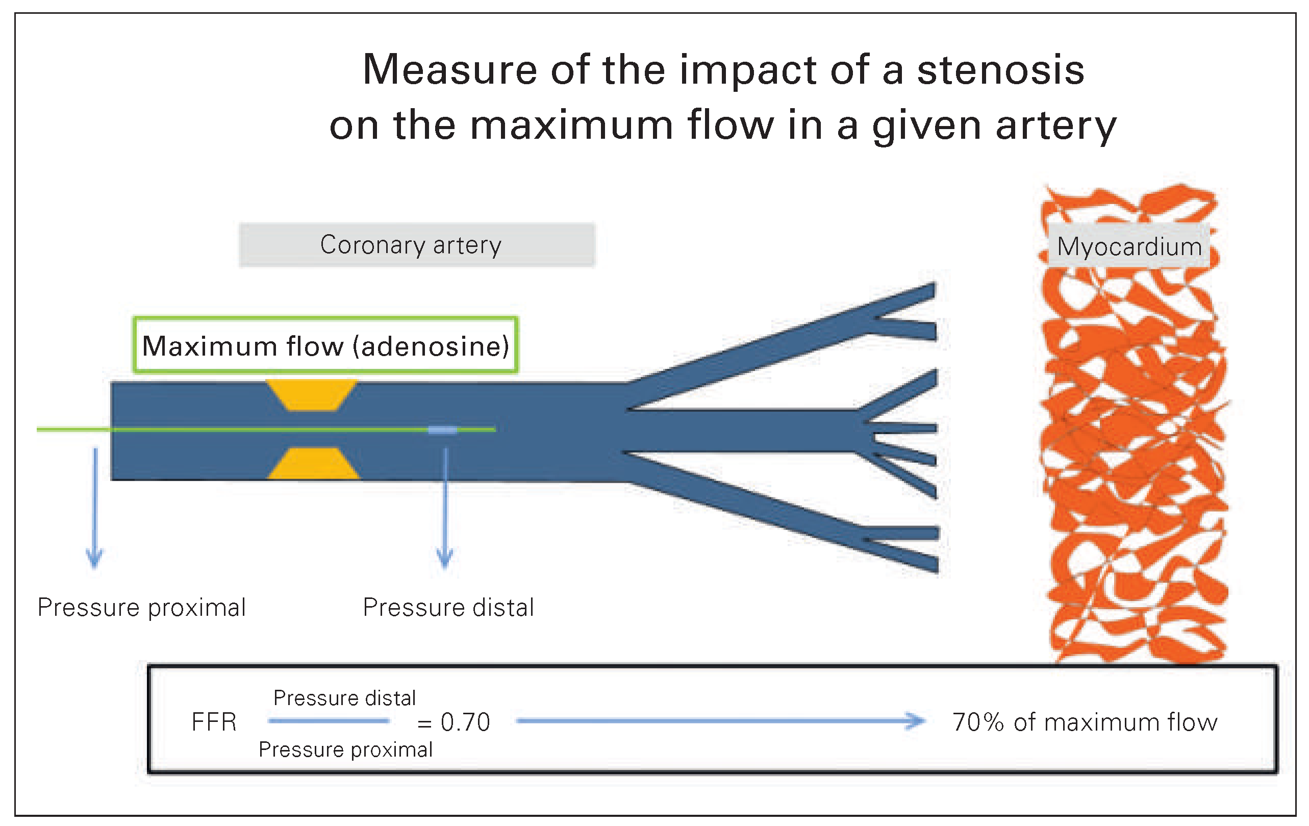
FFR is a technique allowing physicians to assess the impact of epicardial stenosis on the maximum flow in a given coronary artery. In order to measure the FFR, aguide wire must be inserted during coronary angiography to measure pressure distal from the stenosis during maximum hyperaemia. The calculated ratio of pressure distal and proximal to the lesion (FFR) indicates the degree of ischaemia that a stenosis is inducing at maximum vasodilatation. In other words, if FFR is 0.70, this means that maximum myocardial blood flow is only 70% of its normal value. An FFR of 0.70 implies that stenting the focal stenosis responsible for this abnormal ratio would have to improve maximum myocardial blood flow by 43% ([1.0–0.7]/0.7) to reach an FFR of 1.0 – the FFR in a normal coronary artery (Figure 2). An FFR of 0.80 or less identifies ischaemia causing coronary stenoses with an accuracy of more than 90%, and this is the ratio currently used for clinical decision making [5].

Importance of Ischaemia in Stable Coronary Artery Disease Management
Several trials have addressed the question of the benefits of revascularisation over medical treatment in patients presenting with SCAD. In particular, the randomised COURAGE trial, published in 2007, involved 2 287 patients with SCAD [6]. The investigators assigned patients to undergo a percutaneous coronary intervention (PCI) with optimal medical therapy (OMT) or to receive OMT alone. The results demonstrated that PCI did not reduce the risk of death, myocardial infarction, or other major cardiovascular events when added to OMT. The COURAGE trial’s important finding was that revascularisation based on coronary angiography (only one third of patients had proven myocardial ischaemia) in a low risk population had no added value, in terms of prognosis, over OMT. Furthermore, the COUARGE trial’s nuclear substudy showed that the patients with the worst prognosis were those who had extensive myocardial ischaemia, whatever the treatment assigned (Figure 1) [1].
Figure 2. Principle of fractional flow reserve and its relation to myocardial ischaemia (see text).
Cardiovascmed 19 00067 g002

The Value of Coronary Angiography in Detecting Myocardial Ischaemia

Coronary angiography is the gold standard method for detecting epicardial coronary artery stenosis. However, it has several limitations in the detection of myo cardial ischaemia, especially in intermediate stenosis, as it cannot account for all aspects of severity.
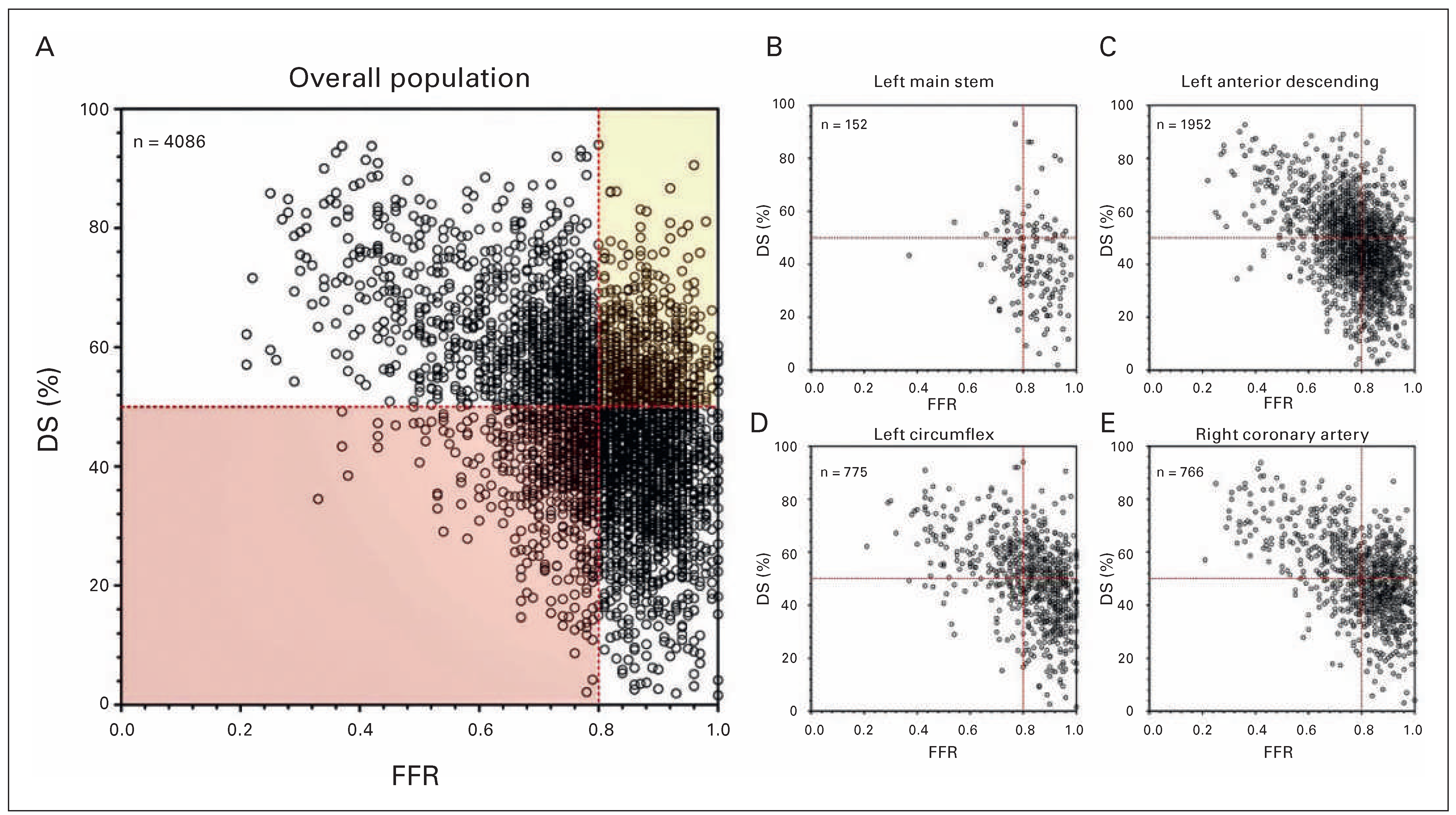
A recent study addressed this issue in a large and unselected patient population; it examined the potential clinical and physiological significance of the discord ance between the severity of coronary artery disease seen on an angiogram and the FFR (Figure 3) [7]. The study compared a sample population of 4 086 patients with at least one stenosis of intermediate angiographic severity. The diagnostic accuracy of a stenosis of ≥50% of the normal vessel diameter predicting an FFR ≤0.80 was 0.64 (95% confidence interval [CI] 0.56–0.72). This study found that one third of a large patient population showed a discordance in the diagnosis of severity thresholds between an angiogram showing stenosis ≥50% diameter and FFR ≤0.8.

What is the Prognosis of Patients Revascularised on the Basis of FFR Measurement?

In the FAME trial, 1 055 patients with stable angina and multivessel coronary artery disease were randomised to undergo PCI guided by angiography alone or PCI guided by angiography and FFR. The primary endpoint was a composite of death, nonfatal myocardial infarction and repeat revascularisation (MACE) at 1 year. The results were statistically significant for the primary endpoint at 1 year (18.3% in the angiography group versus 13.2% in the FFR group, p = 0.02) and also for the number of stents used per patient (2.7 ± 1.2 stents in the angiography group versus 1.9 ± 1.3 stents in the FFR group, p <0.001). Thus, the FAME trial indicated that, among patients for whom a PCI was planned, an FFRguided strategy was better than an angiography guided strategy in terms of a MACE at 1 year. At 2 years, MACE occurred in 22.4% in the angiography group versus 17.9% in the FFR group (p = 0.08) but of importance, the 2 year rates of mortality or myocardial infarction were 12.9% in the angiography group versus 8.4% in the FFR group (p = 0.02) [8]. Aher 5 years, MACE occurred in 31% of patients in the angiography group versus 28% in the FFR group (p = 0.31) with an absolute difference which is not significant due to the smaller number of patients at risk and to the similar incidence of events in both groups beyond 2 years [9]. Nevertheless, the FAME trial did not give information on whether revascularisation (even based on a FFR strategy) was better than OMT in the context of SCAD.
This issue was addressed by the FAME II trial. In this study, patients with SCAD for whom PCI was being considered all had their stenoses evaluated with use of FFR measurement. If the FFR found at least one stenosis to be significant, then patients were randomised to PCI with OMT or to OMT alone. The primary endpoint was a composite of death, myocardial infarction, or urgent revascularisation. Aher the inclusion of 1 220 patients, significant differences between the two groups were observed in terms of the primary endpoint (4.3% in the PCI with OMT group and 12.7% in OMT group; p <0.001) and, accordingly, the study was interrupted. It is of particular interest that the difference between the groups were driven by the rate of urgent revascularisation (1.6% in the PCI with OMT group and 11.1% in the OMT group; p <0.001). These urgent revascularisations were triggered by an increase in biomarker levels, ischaemic changes on ECG, or both, in half of the patients and the awareness of the presence of a stenosis could be a bias, influencing decisions regarding revascularisation. However, the follow up at 2 years [10] confirmed the statistically significant difference in term of urgent revascularisation rate between the OMT group and the PCI with OMT group. In addition, the follow up at 2 years showed a statistically significant difference in terms of the rate of urgent revascularisation triggered by myocardial infarction or/and unstable angina with ECG modification between the OMT group and the PCI with OMT group (7.0 vs 3.4%, hazard ratio 0.22; 95% CI 0.11– 0.42; p <0001). Even if the rate of death between the two groups were not statistically different, this study transformed the COURAGE paradigm: (PCI not seen as indispensable in the treatment of patients with SCAD). Indeed FAME II demonstrated that patients with coronary artery disease and proven ischaemia should be treated with PCI if FFR measurement suggested it. Therefore, the 2013 European Society of Cardiology (ESC) guidelines on the management of SCAD established a class I recommendation (level of evidence A) in which the FFR should be used to identify haemodynamically relevant coronary lesion(s) when evidence of ischaemia is not available.

The Impact of FFR Measurement in Current PCI Strategies

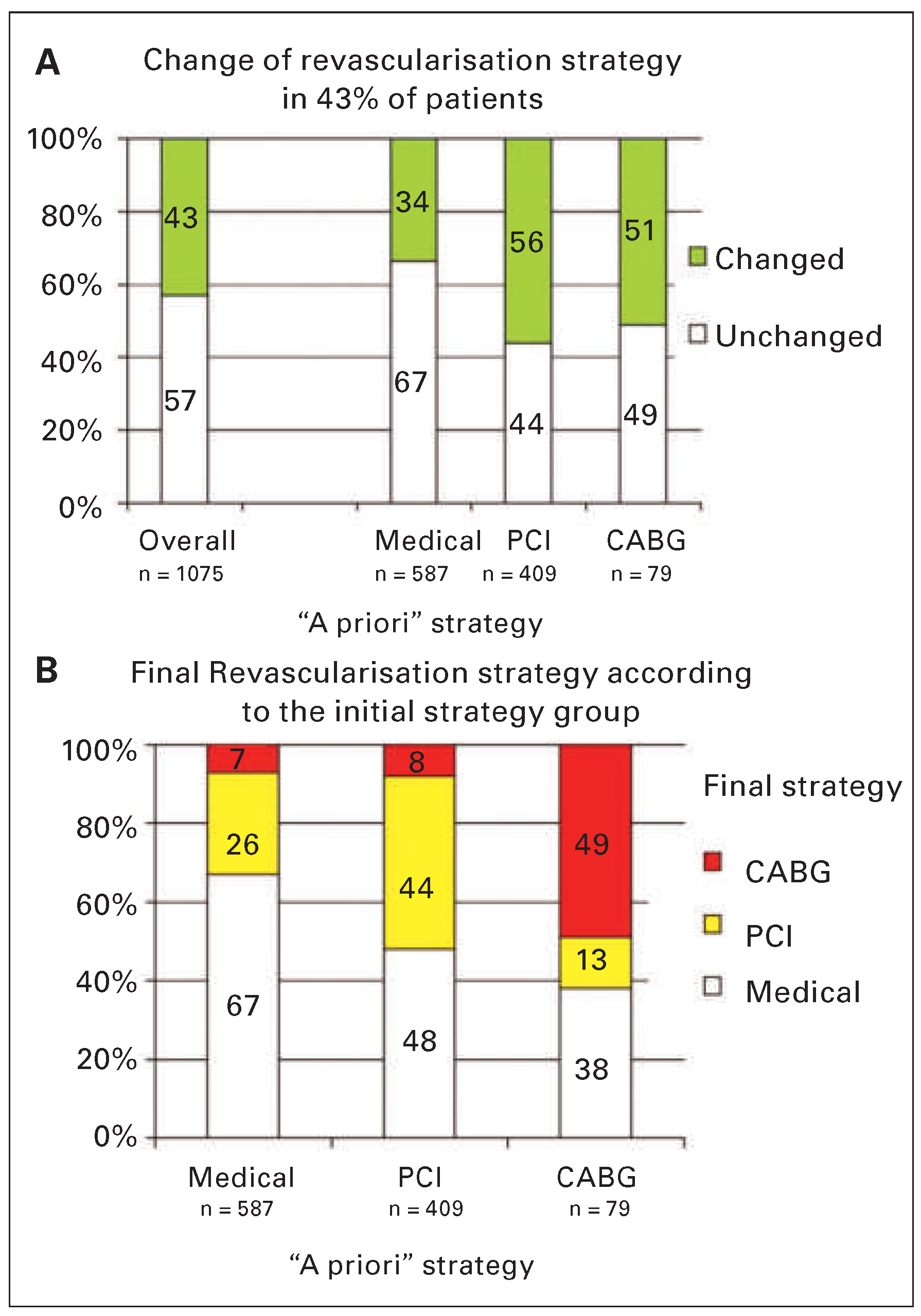
The recent study by Van Belle et al. was based on 1 075 patients from the French FFR registry. Interventional cardiologists had to prepare their initial therapy plan aher an angiogram and a subsequent plan aher the results of follow up FFR measurement. A highly significant 43% of patients had their treatment plan changed on the basis of the FFR results (Figure 4). Aher FFR, 51% of the treatment plans initially set for a coronary artery bypass grah were changed; plans were changed for 56% of patients set to undergo PCI and 33% of patients meant to have OMT. This study showed a dramatic reclassification of patients following FFR measurement and confirmed the poor diagnostic accuracy of the coronary angiogram for detecting ischaemia.

iFR Versus FFR

Hyperaemia is a fundamental aspect of FFR measurement. In order to facilitate maximum blood flow and microcirculation (and minimal resistance) during that measurement, the patient is given a pharmacological vasodilator, usually adenosine. Blood pressure is then measured proximal and distal to the stenosis, and their ratio is the FFR.
In April 2012, Sen et al. published the results of an alternative method of measurement known as the instantaneous wave free ratio or iFR [11]. In order to avoid the administration of adenosine, the authors looked for a period in the cardiac cycle where resistance (at rest) is naturally minimised. They identified a “wave free pe riod” with resistances similar in both magnitude and variability to those observed during FFR measurement; this allowed them to calculate the ratio of pressures distal and proximal to the lesion without adenosine. Jeremias et al. recently compared iFR and FFR in a study of 1 768 patients from 15 clinical sites (Table 1) [12]. They observed that with an iFR of 0.90 (corresponding to an FFR ≤0.80), the overall accuracy of this method was only 80.4% and, therefore, one patient in five was misdiagnosed. In our opinion, adenosine is neither a confounder in FFR measurement nor is it a danger to the patient, especially with intra-coronary administration. Accordingly, iFR should not currently be recommended as an alternative to FFR in routine clinical practice.

Conclusion

FFR measurement has become a validated diagnostic tool used in routine daily practice; it is easily reproducible, quick to measure, correlates very well with noninvasive tests, and has been extensively tested in different clinical trials. For these reasons, FFR measurement has a class I recommendation (level of evidence A) in the ESC and ACC/AHA guidelines for identifying haemodynamically relevant coronary lesions [3,4]. The iFR, however, is a new diagnostic tool that has only been studied for the past 2 years; it is questionable conceptually and lacks clinical and experimental validation and cannot yet be recommended to be used in routine clinical practice.

Disclosure Statement

No financial support and no other potential conflict of interest relevant to this article was reported.

References

  1. Shaw, L.J.; Berman, D.S.; Maron, D.J.; Mancini, G.B.J.; Hayes, S.W.; Hartigan, P.M.; et al. Optimal medical therapy with or without percutaneous coronary intervention to reduce ischemic burden: results from the Clinical Outcomes Utilizing Revascularization and Aggressive Drug Evaluation (COURAGE) trial nuclear substudy. Circulation. 2008, 117, 1283–91. [Google Scholar] [CrossRef] [PubMed]
  2. Hachamovitch, R.; Rozanski, A.; Shaw, L.J.; Stone, G.W.; Thomson, L.E.; Friedman, J.D.; et al. Impact of ischaemia and scar on the therapeutic benefit derived from myocardial revascularization vs. medical therapy among patients undergoing stress rest myocardial perfusion scintigraphy. Eur Heart J. 2011, 32, 1012–24. [Google Scholar] [CrossRef] [PubMed]
  3. Levine, G.N.; Bates, E.R.; Blankenship, J.C.; Bailey, S.R.; Bittl, J.A.; Cercek, B.; et al. 2011 ACCF/AHA/SCAI Guideline for Percutaneous Coronary Intervention: a report of the American College of Cardiology Foundation/American Heart Association Task Force on Practice Guidelines and the Society for Cardiovascular Angiography and Interventions. Circulation. 2011, 124, e574–651. [Google Scholar] [PubMed]
  4. Authors/Task Force m Windecker, S.; Kolh, P.; Alfonso, F.; Collet, J.P.; Cremer, J.; et al. 2014 ESC/EACTS Guidelines on myocardial revascularization: The Task Force on Myocardial Revascularization of the European Society of Cardiology (ESC) and the European Association for Cardio Thoracic Surgery (EACTS)Developed with the special contribution of the European Association of Percutaneous Cardiovascular Interventions (EAPCI). Eur Heart J. 2014, 35, 2541–2619. [Google Scholar]
  5. Pijls, N.H.; Van Gelder, B.; Van der Voort, P.; Peels, K.; Bracke, F.A.; Bonnier, H.J.; et al. Fractional flow reserve. A useful index to evaluate the influence of an epicardial coronary stenosis on myocardial blood flow. Circulation. 1995, 92, 3183–93. [Google Scholar] [CrossRef] [PubMed]
  6. Boden, W.E.; O’Rourke, R.A.; Teo, K.K.; Hartigan, P.M.; Maron, D.J.; Kostuk, W.J.; et al. Optimal medical therapy with or without PCI for stable coronary disease. N Engl J Med. 2007, 356, 1503–16. [Google Scholar] [CrossRef] [PubMed]
  7. Toth, G.; Hamilos, M.; Pyxaras, S.; Mangiacapra, F.; Nelis, O.; De Vroey, F.; et al. Evolving concepts of angiogram: fractional flow reserve discordances in 4000 coronary stenoses. Eur Heart J. 2014, 35, 2831–8. [Google Scholar] [CrossRef] [PubMed]
  8. Pijls; et al. Fractional Flow Reserve Versus Angiography for Guiding Percutaneous Coronary Intervention in Patients With Multivessel Coronary Artery Disease: 2 Year Follow Up of the FAME (Fractional Flow Reserve Versus Angiography for Multivessel Evaluation) Study. J Am Coll Cardiol. 2010, 56, 177 84. [Google Scholar] [CrossRef]
  9. Van, Nunen; et al. Fractional flow reserve versus angiography for guidance of PCI in patients with multivessel coronary artery disease (FAME): 5 year follow up of a randomised controlled trial. Lancet. 2015. [Google Scholar]
  10. De Bruyne, B.; Fearon, W.F.; Pijls, N.H.; Barbato, E.; Tonino, P.; Piroth, Z.; et al. Fractional flow reserve guided PCI for stable coronary artery disease. N Engl J Med. 2014, 371, 1208–17. [Google Scholar] [CrossRef]
  11. Sen, S.; Escaned, J.; Malik, I.S.; Mikhail, G.W.; Foale, R.A.; Mila, R.; et al. Development and validation of a new adenosine independent index of stenosis severity from coronary wave intensity analysis: results of the ADVISE (ADenosine Vasodilator Independent Stenosis Evaluation) study. J Am Coll Cardiol. 2012, 59, 1392–1402. [Google Scholar] [CrossRef] [PubMed]
  12. Jeremias, A.; Maehara, A.; Genereux, P.; Asrress, K.N.; Berry, C.; De Bruyne, B.; et al. Multicenter core laboratory comparison of the instantaneous wave free ratio and resting Pd/Pa with fractional flow reserve: the RESOLVE study. J Am Coll Cardiol. 2014, 63, 1253–61. [Google Scholar] [CrossRef] [PubMed]
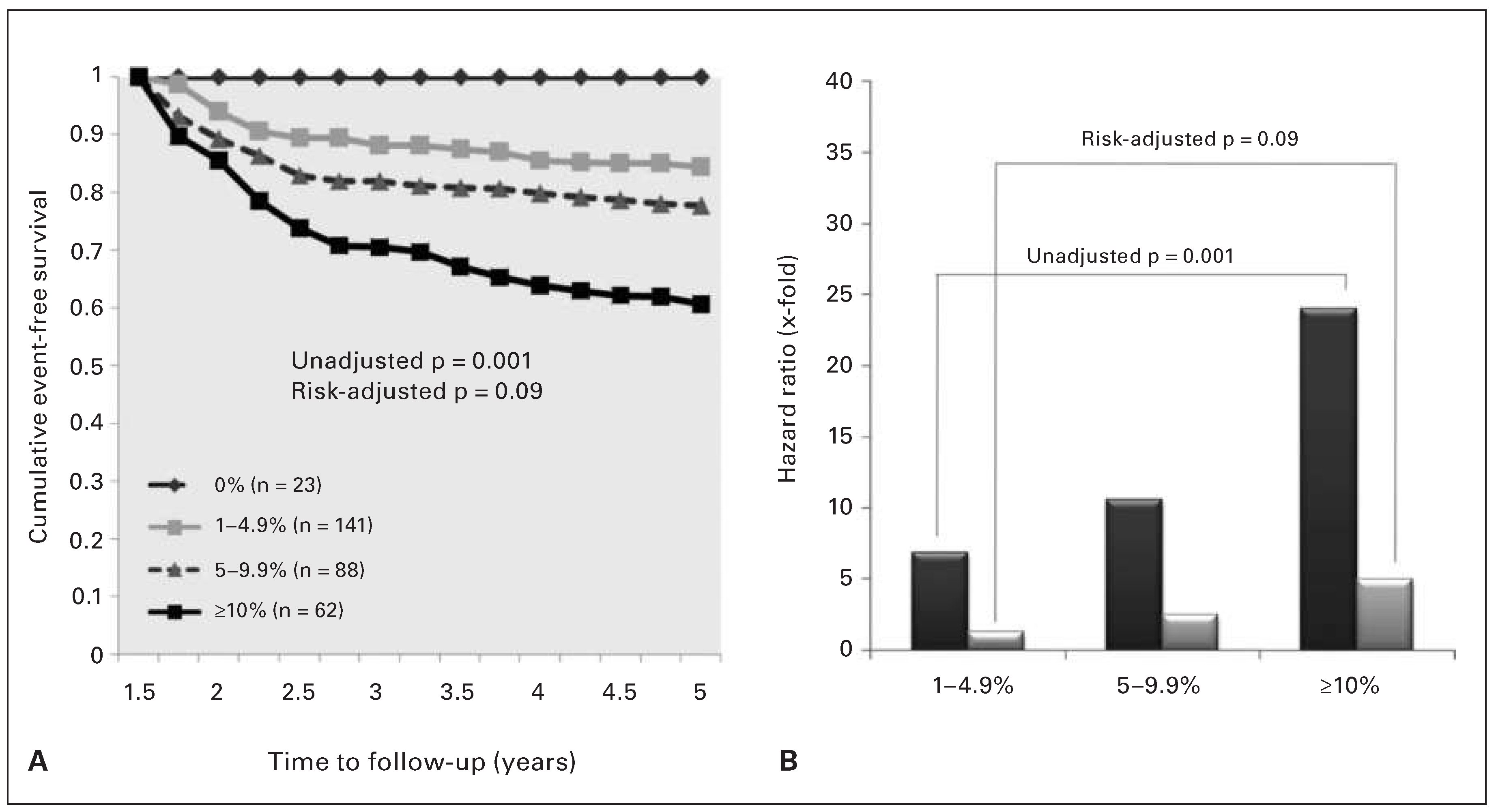
Figure 1. A. Kaplan-Meier survival plot for patients by residual ischaemia, including 0%, 1–4.9%, 5–9.9%, and ≥10% ischaemic myocardium, 6–18 months after PCI + OMT or OMT. Overall event-free survival was 100%, 84.4%, 77.7%, and 60.7%, respectively, for the groups with 0%, 1–4.9%, 5–9.9%, and ≥10% ischaemic myocardium (p = 0.001). In a risk-adjusted Cox model (controlling for randomised treatment), this difference was not significant (p = 0.09). B. Unadjusted (dark gray bars) and risk-adjusted (light gray bars) hazard ratios for the extent and severity of residual ischaemia at 6–18 months of follow-up. OMT = optimal medical treatment; PCI = percutaneous coronary intervention. Reprinted with permission from: Shaw LJ, Berman DS, Maron DJ, Mancini GBJ, Hayes SW, Hartigan PM, et al. Optimal medical therapy with or without percutaneous coronary intervention to reduce ischemic burden: results from the Clinical Outcomes Utilizing Revascularization and Aggressive Drug Evaluation (COURAGE) trial nuclear substudy. Circulation. 2008, 117, 1283–91. Promotional and commercial use of the material in print, digital or mobile device format is prohibited without the permission from the Publisher Wolters Kluwer Health. Please contact healthpermissions@wolterskluwer.com for further information.
Cardiovascmed 19 00067 g001
Figure 3. Correlation between diameter stenosis (DS) vs fractional flow reserve (FFR) in the overall population (A), and specifically in the left main stem. (B) and the three major branches (C–F). The x-axes indicate the functional metric (FFR), and the y-axes indicate the angiographic metrics (DS). Reprinted with permission of Oxford University Press from: Toth G, Hamilos M, Pyxaras S, Mangiacapra F, Nelis O, De Vroey F, et al. Evolving concepts of angiogram: fractional flow reserve discordances in 4000 coronary stenoses. Eur Heart J. 2014, 35, 2831–8.
Cardiovascmed 19 00067 g003
Figure 4. A. Reclasification of the revascularisation strategy according to the “a priori” strategy group. Despite minor overall changes, a change in strategy occurred for 43% of all patients (κ, 0.22; 95% confidence interval 0.17–0.27). B. Details of the final revascularisation strategy applied in each a priori strategy group. CABG = coronary artery bypass graft surgery. PCI = percutaneous coronary intervention. Reprinted with permission from: Van Belle E, Rioufol G, Pouillot C, Cuisset T, et al. Outcome impact of coronary revascularization strategy reclassification with fractional flow reserve at time of diagnostic angiography: insights from a large French multicenter fractional flow reserve registry. Circulation. 2014, 129(2):173–85. Promotional and commercial use of the material in print, digital or mobile device format is prohibited without the permission from the Publisher Wolters Kluwer Health. Please contact healthpermissions@ wolterskluwer.com for further information.
Cardiovascmed 19 00067 g004
Table 1. Individual Data From Included Studies and Individual Study Sites.
Study / participating SiteNo. of lesionsCut-off pointAUC from ROCOverall accuracy in %
Total15230.90.8180.4
ADVISE4320.910.8281.9
VERIFY6540.890.879.4
Seoul National University1790.920.8382.7
Stony Brook University1490.930.8179.2
Columbia University950.910.8482.1
AMC/VUMC/KCL840.90.7878.6