Next Article in Journal
What Are the Current Blood Pressure Targets?
Previous Article in Journal
Current Aspects of Atrial Fibrillation Surgery
 
 
Cardiovascular Medicine is published by MDPI from Volume 28 Issue 1 (2025). Previous articles were published by another publisher in Open Access under a CC-BY (or CC-BY-NC-ND) licence, and they are hosted by MDPI on mdpi.com as a courtesy and upon agreement with Editores Medicorum Helveticorum (EMH).
Case Report

Pulmonary Valve Stenosis Due to Undifferentiated Pleomorphic Sarcoma

by
Panagiotis Antiochos
1,*,
Pierre Monney
1,
Salah Dine Qanadli
3,
Leopold Schlueter
5,
Samuel Rotman
2,
Emilienne Descloux
2,
Lars Niclauss
4,
Michel Hurni
4 and
Olivier Muller
1
1
Division of Cardiology, Department of Internal Medicine, University Hospital of Lausanne (CHUV), Lausanne, Switzerland
2
Institute of Pathology, University Hospital of Lausanne (CHUV), Lausanne, Switzerland
3
Department of Radiology, University Hospital of Lausanne (CHUV), Lausanne, Switzerland
4
Division of Cardiovascular Surgery, Department of Surgery, University Hospital of Lausanne (CHUV), Lausanne, Switzerland
5
The Cecil Clinic, Hirslanden, Lausanne, Switzerland
*
Author to whom correspondence should be addressed.
Cardiovasc. Med. 2015, 18(6), 186; https://doi.org/10.4414/cvm.2015.00333
Submission received: 24 March 2015 / Revised: 24 April 2015 / Accepted: 24 May 2015 / Published: 24 June 2015

Abstract

Pulmonary artery sarcomas are uncommon intracardiac tumours with poor prognosis. We report the case of a 69-year-old woman in good health presenting with rapidly progressive dyspnoea and an unfamiliar systolic murmur. Echocardiography revealed pulmonary valve stenosis due to an obstructing mobile mass. Imaging studies confirmed the presence of a contrast-enhancing lesion adherent to the valve, extending into the pulmonary trunk and right ventricular outflow tract, and suggestive of malignancy. Endovascular biopsy was attempted with no success. Surgical resection with autologous graft valve replacement and pulmonary artery reconstruction was performed. Postoperative histological examination confirmed the diagnosis of an undifferentiated pleomorphic sarcoma. Pulmonary artery sarcoma should be considered as a rare differential diagnosis in patients presenting with dyspnoea and a crescendo–decrescendo systolic murmur increasing with inspiration. Echocardiography is a useful first diagnostic approach but multi-imaging assessment is almost always necessary for definite diagnosis. Our case provides insights into the challenges met by cardiologists, radiologists and cardiac surgeons in the management of such cases.

Case description

A 69-year-old Caucasian woman presented to her general practitioner with new onset fatigue and rapidly progressive dyspnoea on exertion. Upon physical examination the patient was afebrile, blood pressure was 116/72 mm Hg, pulse rate 76/min and respiration rate 12/min. Heart sounds were normal, but a new, 3/6, crescendo–decrescendo systolic murmur, which increased during deep inspiration and was maximal along the left sternal border, was noted. Laboratory studies of electrolytes, renal function, liver and cardiac enzymes, acute phase reactants and complete blood count were within normal limits, as were a 12-lead ECG and a chest radiograph.
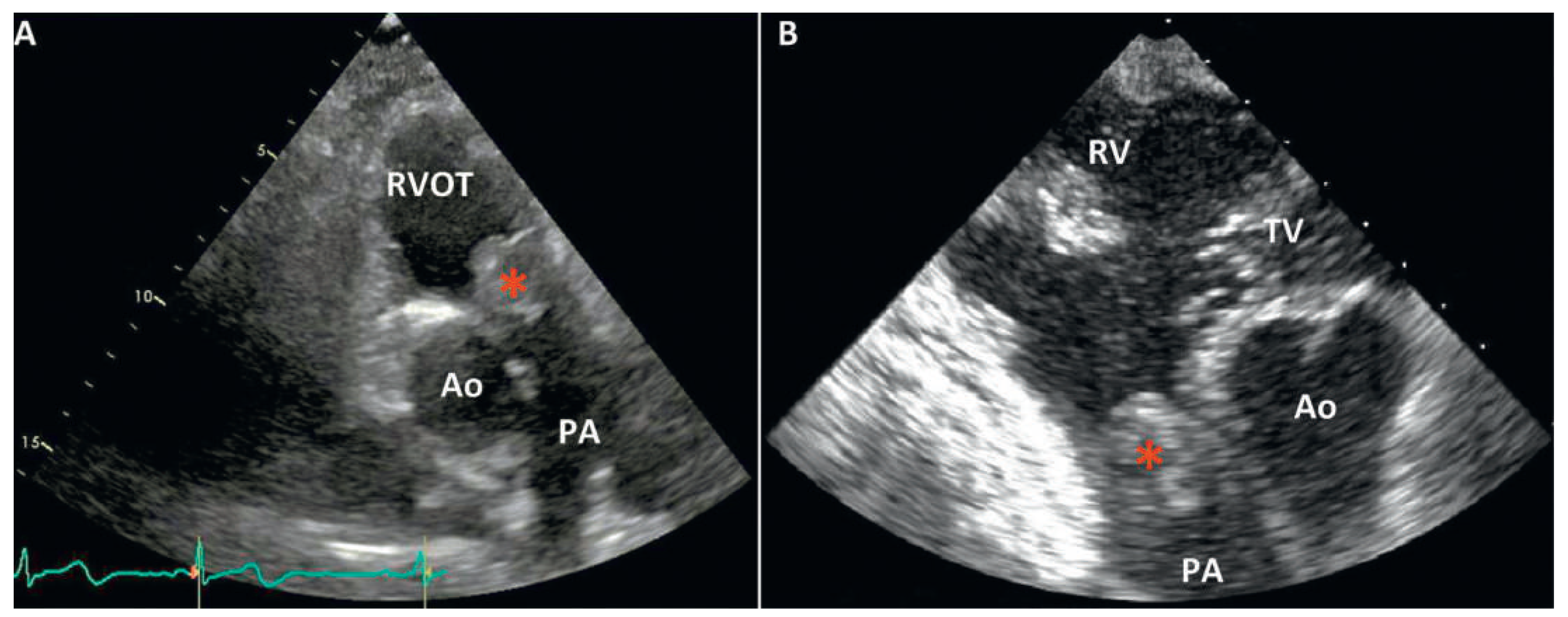
Transthoracic echocardiography (TTE) revealed a moderate pulmonary valve stenosis caused by a globular mass protruding into the right ventricular outflow tract (RVOT) during diastole (Figure 1). The maximum pulmonary pressure gradient was measured at 42 mm Hg (mean 30 mm Hg). Left as well as right ventricle size and function were normal.
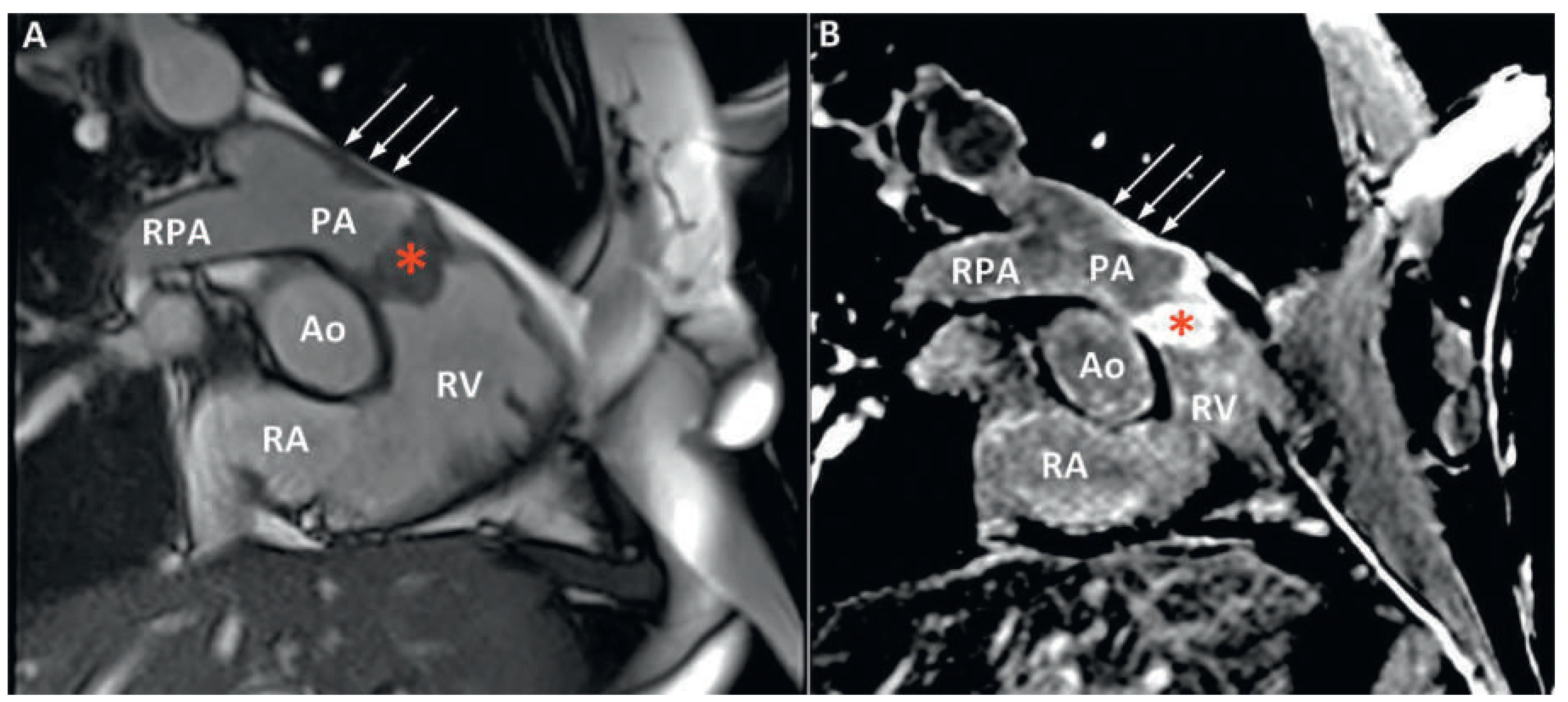
The patient was promptly referred to our institution where cardiac computed tomography (CT) revealed a 25 × 35 × 24 mm mass attached to the RVOT and extending into the pulmonary trunk through the pulmonary valve (Figure 2). After intravenous injection of 60–80 ml iodinated contrast material, late image acquisition showed inhomogeneous enhancement of the mass and mural enhancement of the pulmonary trunk suggestive of infiltration. This technique in the context of cardiac CT consists of acquiring images 60 seconds after CT angiography in order to optimise tissue enhancement. Fludeoxyglucose (18F) positron emission tomography-CT (18F-FDG PET-CT) revealed mild metabolic activity of the mass, with no signs of metastasis (Figure 3). Finally, cardiac magnetic resonance imaging (MRI) confirmed proximal extension of the valve mass, 4 cm towards the pulmonary trunk, without apparent invasion of the bifurcation (Figure 4). A hyperintense signal in T2-weighted images indicated oedema, whereas moderate signal-enhancement during resting perfusion suggested rich vascularisation. Intense signals on gadolinium enhancement sequences suggested rich extracellular content (fibrosis or extracellular matrix), but no significant necrosis (Figure 5).
A provisory diagnosis of intracardiac malignancy was made on clinical (rapid installation of symptoms) and imaging (vascular infiltrative mass) grounds, and endovascular biopsy was attempted. The procedure was uneventful, but yielded limited material for histology. The case was discussed by a multidisciplinary panel involving cardiologists, radiologists and a cardiothoracic surgeon, and surgery was decided on. The time delay from first diagnosis on TTE to the operation was 22 days. During the procedure, a bulky, white mass, densely adherent and infiltrating the pulmonary valve and all of the pulmonary infundibulum was identified and removed, along with the deformed pulmonary valve, part of the RVOT, as well as most of the pulmonary trunk as proximally as possible to the bifurcation. An autologous graft was used to replace the valve; the right ventricle was then anastomosed to the distal part of the pulmonary artery. Postoperative histology revealed a mesenchymal tumour consisting of highly atypical spindle cells without specific differentiation (Figure 6) [1]. Given the retrograde extension of the mass and the possibility of residual fragments very low inside the RVOT, the patient was referred for adjuvant chemotherapy and radiotherapy after six weeks of convalescence in a cardiac rehabilitation centre.

Discussion

Our case highlights the major impediments encountered during management of these rare and often fatal intracardiac tumours; from timely and accurate diagnosis to surgical resection and adjuvant care, pulmonary artery sarcomas (PAS) represent a diagnostic and therapeutic challenge for cardiologists, radiologists, cardiac surgeons and oncologists.
Typical onset age is 45–55 years with a female to male ratio of 2:1. The tumour typically arises within the pulmonary trunk and spreads into the pulmonary arteries. Embryologically, it originates from the bulbus cordis, a structure that later gives birth to both the RVOT and pulmonary trunk, which could, in our case, explain valve involvement and RVOT extension. As with the majority of intracardiac tumours, presentation is non-specific, with dyspnoea on exertion, chest or back pain, cough and haemoptysis being common symptoms. In many cases, the clinical presentation mimics chronic thromboembolic pulmonary hypertension, and in advanced stages constitutional symptoms appear. Physical examination remains unremarkable until late. Our case is unique in the sense that it presented as a new unfamiliar murmur that alarmed the primary physician. Such a finding is extremely rare and highlights the paramount importance of good auscultation skills and a clinical acumen. A new harsh systolic murmur that increases with inspiration indicates pulmonary stenosis and warrants further investigation with echocardiography. TTE may show an increased gradient across a narrowed RVOT [2]. The presence of a pedunculated lesion arising from the RVOT, or pulmonary valve or trunk should always raise suspicion of PAS [3] and prompt a thoracic CT or a cardiac MRI.
CT findings can be misleading in over 50% of cases, as PAS is confused with the far more common condition of central pulmonary embolism. Both entities present with a filling defect in the pulmonary arteries. Suggestive of PAS are an inhomogeneous, soft tissue density, with the filling defect occupying the entire lumen of the pulmonary trunk, along with delayed contrast enhancement on angiography [4]. Once suspicion is raised, PET-CT and MRI are helpful to differentiate fully the mass. Preoperative histological diagnosis is generally not possible although it should be attempted through CT-guided transthoracic aspiration, transvenous catheter biopsy or transbronchial biopsy. In most cases definitive diagnosis is made only during surgery or at autopsy. Surgery remains the cornerstone of management, offering definite diagnosis, clinical improvement and the best prognosis [5]. Even if curative resection is not possible, palliative resection can provide symptom relief by restoring haemodynamics. Reported interventions range from bilateral pneumonectomy, pulmonary endarterectomy (PEA) with or without pneumonectomy and with or without reconstruction of the pulmonary artery to debulking and palliative stenting. Regarding adjuvant therapy, combined chemoand radiotherapy should be adopted in nearly every case to extend palliation. Overall, PAS represents a fatal disease, with a mean survival time without surgical intervention of 1.5 months. Survival of 36.5 ± 20.2 months has been reported for patients benefiting from a “curative” resection while 11 ± 3 months for those undergoing “incomplete” resection [5].
In conclusion, it is important that physicians recognise the clinical features associated with PAS and include it as a differential diagnosis in the appropriate setting. Early referral for multidisciplinary management in a tertiary centre offers the only chance for prolonged survival and improved quality of life.

Disclosures

No financial support and no other potential conflict of interest relevant to this article was reported.
References

A full list of references is available in the online version of this article.
    Figure 1. (A) TTE parasternal long axis view of the pulmonary artery. The tumour is recognised as a large round mass invading the pulmonary valve. (B) Transoesophageal echocardiography (TOE) in transgastric inflow–outflow view of the right ventricle. The tricuspid valve and the right ventricular apex are seen at the top of the picture. The pulmonary valve is replaced by a ball-like tumour (*) obstructing the RVOT. Ao = aorta; PA = pulmonary artery; RV = right ventricle; RVOT = right ventricle outflow tract; TV = tricuspid valve.
    Figure 1. (A) TTE parasternal long axis view of the pulmonary artery. The tumour is recognised as a large round mass invading the pulmonary valve. (B) Transoesophageal echocardiography (TOE) in transgastric inflow–outflow view of the right ventricle. The tricuspid valve and the right ventricular apex are seen at the top of the picture. The pulmonary valve is replaced by a ball-like tumour (*) obstructing the RVOT. Ao = aorta; PA = pulmonary artery; RV = right ventricle; RVOT = right ventricle outflow tract; TV = tricuspid valve.
    Cardiovascmed 18 00186 g001
    Figure 2. Cardiac computed tomography, 2d multi-planar reformation showing the voluminous mass (*) attached to the pulmonary valve (A) and protruding in the pulmonary trunk during systole (B). Ao = aorta; LV = left ventricle; PA = pulmonary artery; RVOT = right ventricle outflow tract.
    Figure 2. Cardiac computed tomography, 2d multi-planar reformation showing the voluminous mass (*) attached to the pulmonary valve (A) and protruding in the pulmonary trunk during systole (B). Ao = aorta; LV = left ventricle; PA = pulmonary artery; RVOT = right ventricle outflow tract.
    Cardiovascmed 18 00186 g002
    Figure 3. Fludeoxyglucose (18F) positron emission tomography-computed tomography (18F-FDG PET-CT) showing discrete metabolic activity of the mass (arrow), with no evidence of other lesion. Ao = aorta; PA = pulmonary artery; RV = right ventricle.
    Figure 3. Fludeoxyglucose (18F) positron emission tomography-computed tomography (18F-FDG PET-CT) showing discrete metabolic activity of the mass (arrow), with no evidence of other lesion. Ao = aorta; PA = pulmonary artery; RV = right ventricle.
    Cardiovascmed 18 00186 g003
    Figure 4. (A) Cardiac magnetic resonance imaging (MRI) steady-state free precession (SSFP) cine acquisition in inflow–outflow view of the right ventricle. The tumour is seen as a massive thickening of the pulmonary valve (*). Note also the thickening of the anterior wall of the pulmonary trunk (arrows). (B) Corresponding acquisition with a phase-sensitive inversion-recovery sequence after contrast injection. There is homogenous late gadolinium enhancement of the tumour (*) and of the anterior wall of the pulmonary trunk (arrows). Ao = aorta; PA = pulmonary artery; RPA = right pulmonary artery; RA = right atrium; RV = right ventricle.
    Figure 4. (A) Cardiac magnetic resonance imaging (MRI) steady-state free precession (SSFP) cine acquisition in inflow–outflow view of the right ventricle. The tumour is seen as a massive thickening of the pulmonary valve (*). Note also the thickening of the anterior wall of the pulmonary trunk (arrows). (B) Corresponding acquisition with a phase-sensitive inversion-recovery sequence after contrast injection. There is homogenous late gadolinium enhancement of the tumour (*) and of the anterior wall of the pulmonary trunk (arrows). Ao = aorta; PA = pulmonary artery; RPA = right pulmonary artery; RA = right atrium; RV = right ventricle.
    Cardiovascmed 18 00186 g004
    Figure 5. (A) T1-weighted image; the mass (arrow) has a signal iso-intense to the myocardium. (B) T2-weighted image; the mass has a signal hyper-intense to the myocardium indicating presence of tissue oedema. (C) Late gadolinium enhancement sequence; the mass shows bright enhancement indicating rich vascularisation. (D to F) Perfusion imaging; (D) contrast in the right cardiac chambers; (E) contrast in the left cardiac chambers; (F) contrast enhancing the myocardium: the mass shows moderate first-pass enhancement with peak signal intensity lower than that of the myocardium.
    Figure 5. (A) T1-weighted image; the mass (arrow) has a signal iso-intense to the myocardium. (B) T2-weighted image; the mass has a signal hyper-intense to the myocardium indicating presence of tissue oedema. (C) Late gadolinium enhancement sequence; the mass shows bright enhancement indicating rich vascularisation. (D to F) Perfusion imaging; (D) contrast in the right cardiac chambers; (E) contrast in the left cardiac chambers; (F) contrast enhancing the myocardium: the mass shows moderate first-pass enhancement with peak signal intensity lower than that of the myocardium.
    Cardiovascmed 18 00186 g005
    Figure 6. (A) (Haematoxylin and eosin; × 40) Postoperative histology revealed a poorly differentiated tumour composed of a proliferation of spindle cells with fibroblastic differentiation (black arrows). Some areas have a fibromyxoid pattern (*). (B) (haematoxylin and eosin; × 200); Highly atypical giant spindle cell (white arrows). (C) Macroscopic aspect of the mass.
    Figure 6. (A) (Haematoxylin and eosin; × 40) Postoperative histology revealed a poorly differentiated tumour composed of a proliferation of spindle cells with fibroblastic differentiation (black arrows). Some areas have a fibromyxoid pattern (*). (B) (haematoxylin and eosin; × 200); Highly atypical giant spindle cell (white arrows). (C) Macroscopic aspect of the mass.
    Cardiovascmed 18 00186 g006

    Share and Cite

    MDPI and ACS Style

    Antiochos, P.; Monney, P.; Qanadli, S.D.; Schlueter, L.; Rotman, S.; Descloux, E.; Niclauss, L.; Hurni, M.; Muller, O. Pulmonary Valve Stenosis Due to Undifferentiated Pleomorphic Sarcoma. Cardiovasc. Med. 2015, 18, 186. https://doi.org/10.4414/cvm.2015.00333

    AMA Style

    Antiochos P, Monney P, Qanadli SD, Schlueter L, Rotman S, Descloux E, Niclauss L, Hurni M, Muller O. Pulmonary Valve Stenosis Due to Undifferentiated Pleomorphic Sarcoma. Cardiovascular Medicine. 2015; 18(6):186. https://doi.org/10.4414/cvm.2015.00333

    Chicago/Turabian Style

    Antiochos, Panagiotis, Pierre Monney, Salah Dine Qanadli, Leopold Schlueter, Samuel Rotman, Emilienne Descloux, Lars Niclauss, Michel Hurni, and Olivier Muller. 2015. "Pulmonary Valve Stenosis Due to Undifferentiated Pleomorphic Sarcoma" Cardiovascular Medicine 18, no. 6: 186. https://doi.org/10.4414/cvm.2015.00333

    APA Style

    Antiochos, P., Monney, P., Qanadli, S. D., Schlueter, L., Rotman, S., Descloux, E., Niclauss, L., Hurni, M., & Muller, O. (2015). Pulmonary Valve Stenosis Due to Undifferentiated Pleomorphic Sarcoma. Cardiovascular Medicine, 18(6), 186. https://doi.org/10.4414/cvm.2015.00333

    Article Metrics

    Back to TopTop