Next Article in Journal
Score Calcique et PET/CT
Previous Article in Journal
Re: “Chronic Total Occlusion: Current Methods of Revascularisation” by Patrick T. Siegrist and Satoru Sumitsuji (Cardiovascular Medicine. 2014;17(12):347–356)
 
 
Cardiovascular Medicine is published by MDPI from Volume 28 Issue 1 (2025). Previous articles were published by another publisher in Open Access under a CC-BY (or CC-BY-NC-ND) licence, and they are hosted by MDPI on mdpi.com as a courtesy and upon agreement with Editores Medicorum Helveticorum (EMH).
Case Report

Radiofrequency Ablation of Atrial Tachycardia from ''No-Man's Land"

by
Nikola Pavlović
1,2,
Sven Knecht
1,
Aline Mühl
1,
Tobias Reichlin
1,
Beat Schaer
1,
Stefan Osswald
1,
Christian Sticherling
1 and
Michael Kühne
1,*
1
Cardiology/Electrophysiology, University Hospital Basel, Basel, Switzerland
2
Cardiology/Electrophysiology, University Hospital “Sestre milosrdnice”, Zagreb, Croatia
*
Author to whom correspondence should be addressed.
Cardiovasc. Med. 2015, 18(3), 103; https://doi.org/10.4414/cvm.2015.00322 (registering DOI)
Submission received: 18 December 2014 / Revised: 18 January 2015 / Accepted: 18 February 2015 / Published: 18 March 2015

Abstract

Atrial tachycardias can originate from almost any part of the atria, with a predilection for certain anatomical structures. Tachycardias originating from the peri-atrioventricular nodal region are rare, and can be challenging to ablate owing to the potential risk of atrioventricular block. We describe a patient with peri-nodal atrial tachycardia that was successfully ablated from the non-coronary aortic cusp.

Case report

A 78-year-old male with a history of hypertension and coronary artery disease was evaluated for frequent, daily palpitations lasting up to several hours. His basic laboratory results and 12-lead electrocardiogram (ECG) were normal and echocardiography showed normal left ventricular function. Twentyfour-hour Holter monitoring showed a narrow QRS and long RP tachycardia occurring throughout the day, more than 50% of time. Until admission, tachycardia was not verified in a 12-lead ECG.
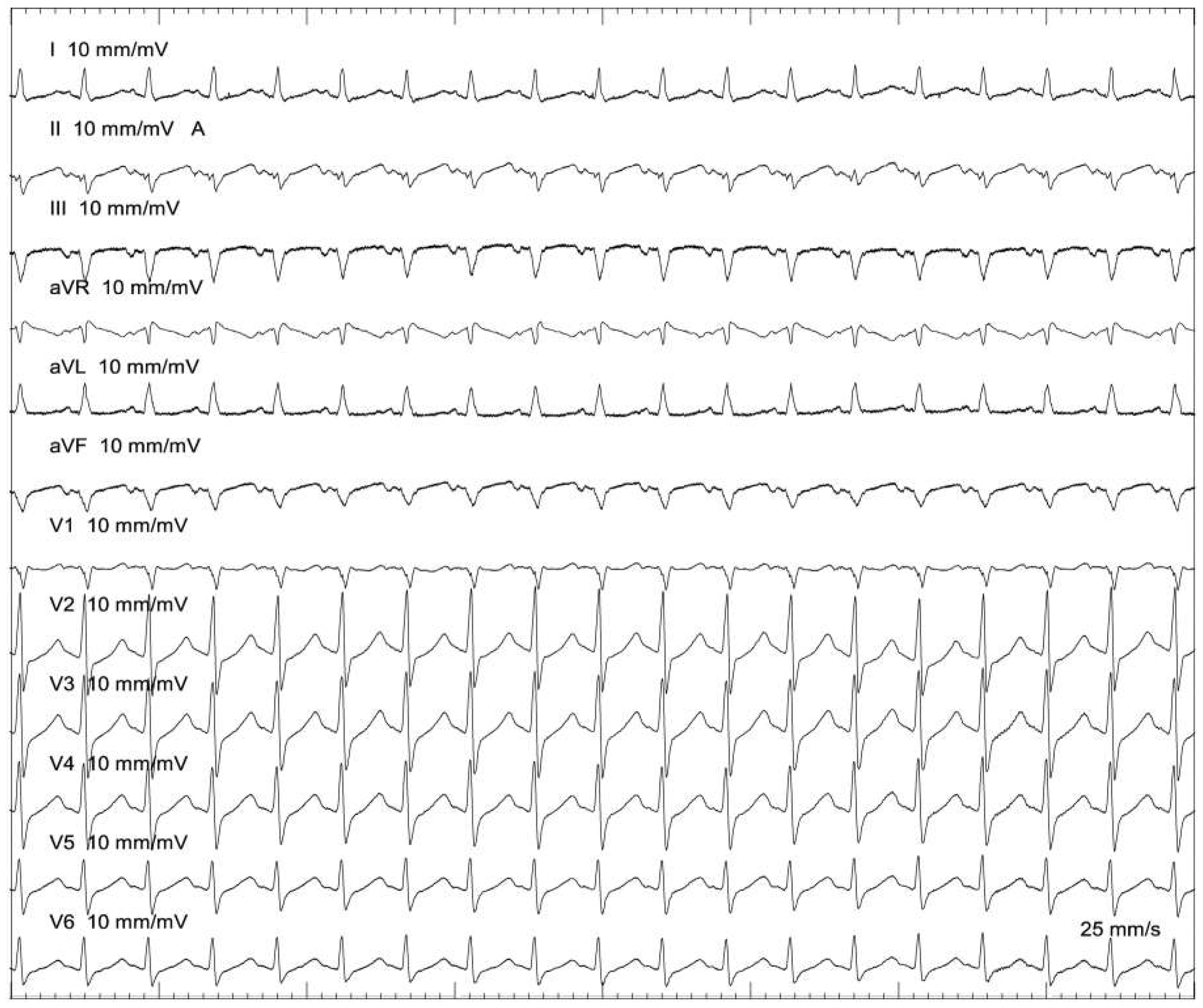
The patient was scheduled for an electrophysiological study. Catheters were placed in the high right atrium (HRA), the His bundle (His), the right ventricular apex (RVA) and coronary sinus (CS). At the beginning of the procedure, sustained tachycardia was verified with 1:1 atrioventricular (AV) conduction, a long RP interval and mostly negative P waves in the inferior leads (Figure 1). With entrainment from the RVA, tachycardia was either dissociated from the ventricle or a VAAV response was evoked as a criterion for atrial tachycardia. The tachycardia was reproducibly inducible and the earliest activation was found on the His catheter.
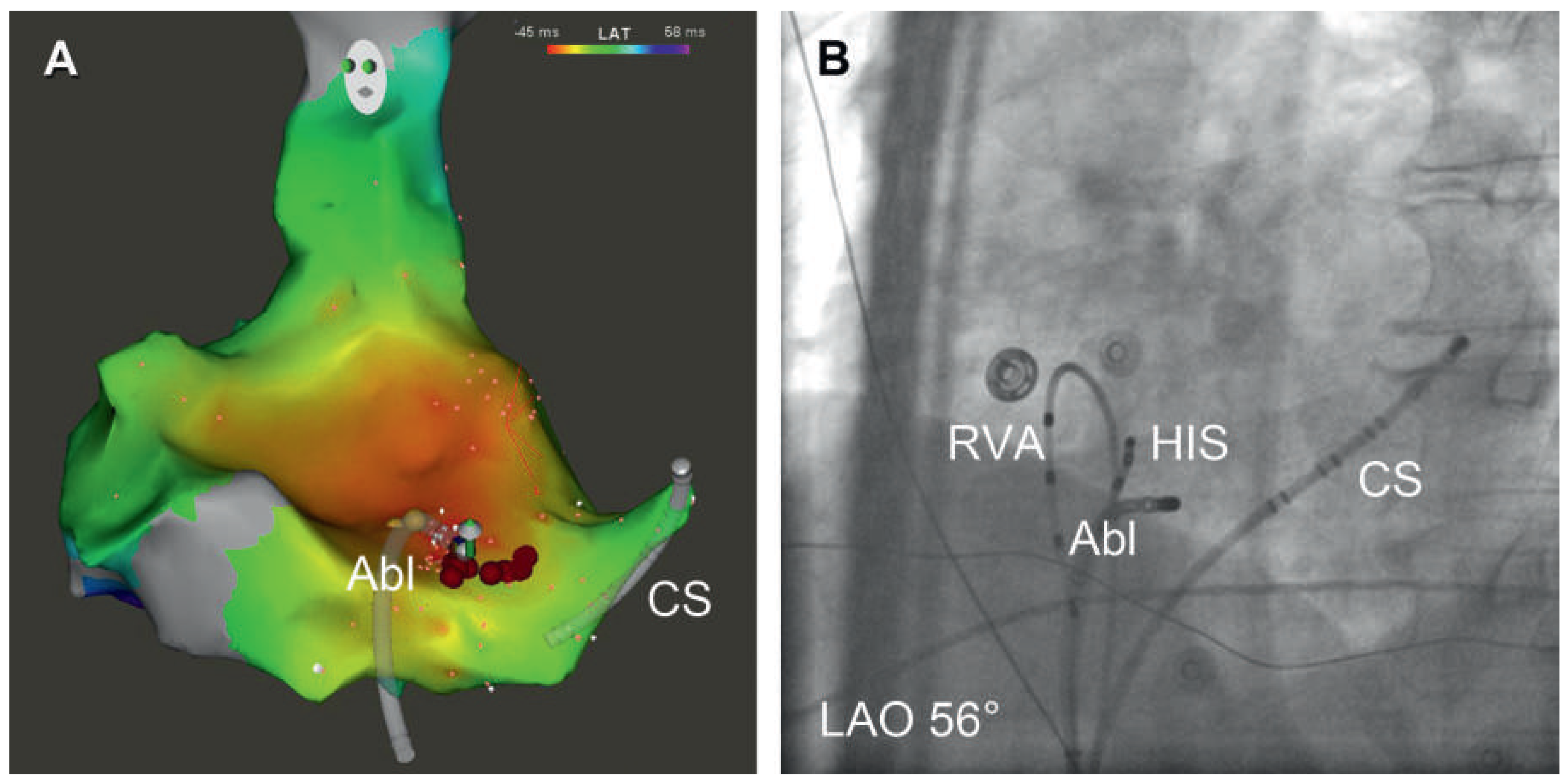
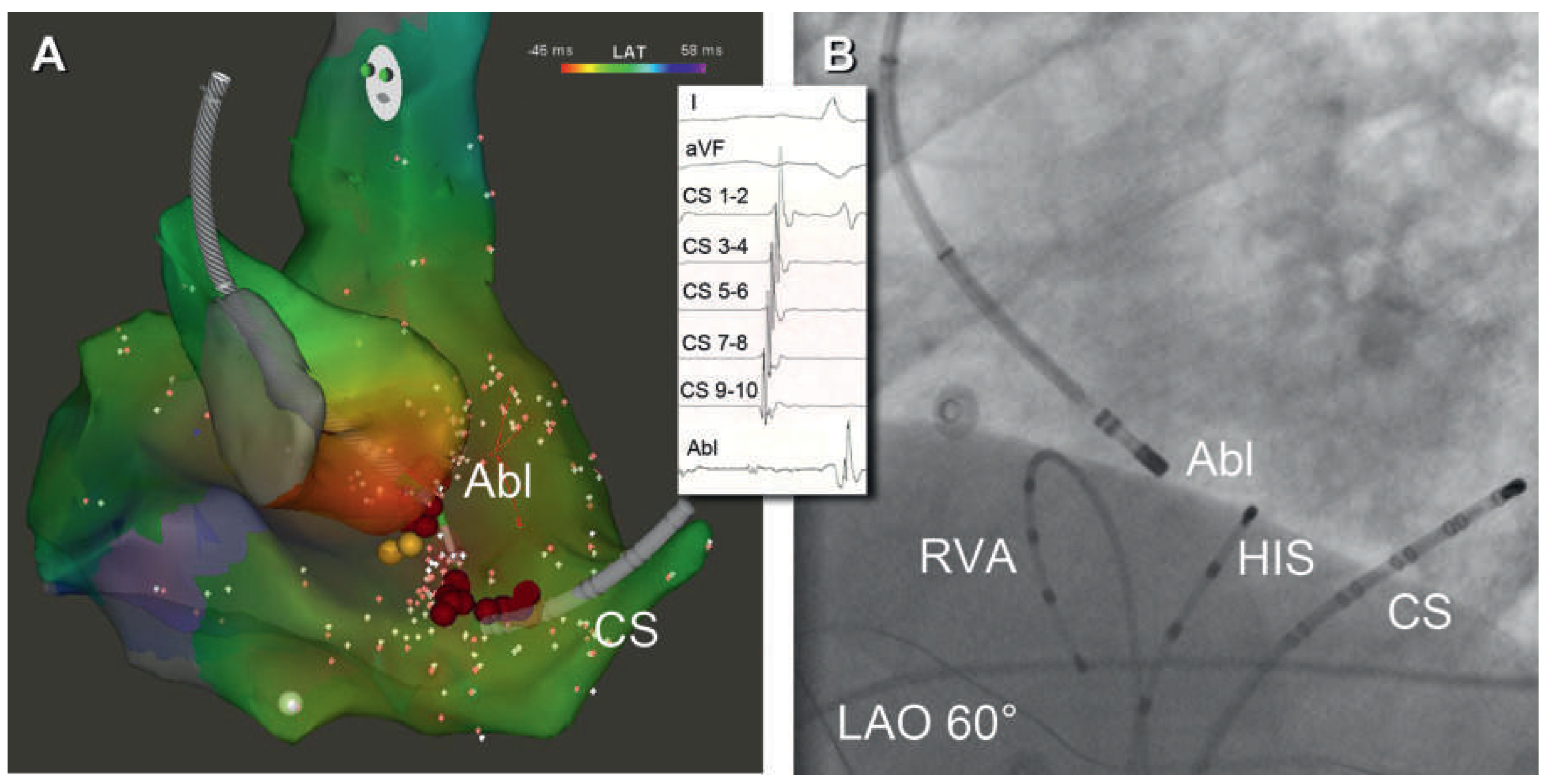
Mapping was performed with a 3.5 mm irrigated-tip catheter (7.5F, Thermocool Smart Touch, Biosense Webster, Diamond Bar, CA) in conjunction with a 3-dimensional mapping system (CARTO 3, Biosense Webster). After mapping the right atrium, earliest activation was found in the peri-AV nodal region. Since ablation in this area carries a risk of AV block, only limited ablation was performed in close proximity to the His, but without effect (Figure 2). As a next step, the aortic root was mapped using a retrograde aortic approach, and early activation was found in the non-coronary cusp (Figure 3). A low amplitude, slightly fragmented atrial electrogram was found and the activation time in the non-coronary cusp was identical to the time measured at the His region when mapping the right atrium. Ablation in the noncoronary cusp (contact force 23 g) in a power-controlled mode (15 W with increase to 25 W) terminated the tachycardia after 3 seconds of the first radiofrequency energy application (total of 183 s) and the tachycardia remained non-inducible after the ablation. The local electrogram at the successful site is shown in Figure 3. The patient was discharged the day after the procedure, and an overnight Holter ECG showed no atrial arrhythmia. The patient remained free of arrhythmia during a follow-up of three months.

Discussion

Atrial tachycardia can originate from almost any part of the atria with a predilection for certain anatomical structures such as the crista terminalis, tricuspid and mitral annulus, the coronary sinus musculature, superior vena cava and the pulmonary veins. Although tachycardias originating from the peri-AV nodal region are rare, they have been described in up to 18% of selected patients [1]. Whereas most atrial tachycardias are amenable for ablation with relatively high success rates, tachycardias from the peri-AV nodal region can be challenging owing to the potential risk of AV block [2].
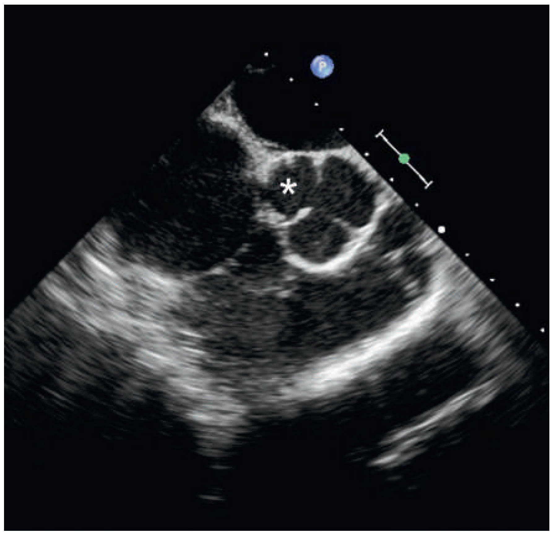
The peri-AV nodal region is the anterior part of the interatrial septum, whereas the aortic root has a central location in the heart, located antero-superior to the mitral and tricuspid annulus. Its non-coronary cusp is in contact with both the left and right atria and with the anterior part of interatrial septum [1]. If ablation of a tachycardia thought to originate from the peri-nodal region is unsuccessful when ablating from the right atrium and/or the risk of AV block is high (when a His spike is recorded at the site of earliest activation in the right atrium, as in our case), mapping the non-coronary cusp should be considered. The proximity of these structures can be appreciated in Figure 4.
Although rare, atrial tachycardias that are amenable for ablation from the non-coronary cusp have been described. There are significant differences in reported P wave morphologies. Ouyang et al. described positive P waves in lead I and aVL and biphasic P waves in V1 [3] whereas Chen et al. described biphasic P waves in V1 and positive P waves in the inferior leads [4]. In the studies performed by Das et al. [1] and Marrouche et al. [5], characteristic P wave morphologies for left septal and non-coronary cusp tachycardias could not be identified. Typical findings of these tachycardias are early activation in the His region found during mapping, and local activation in the non-coronary cusp preceding the onset of the P wave by up to 40 ms [1]. Also, these tachycardias are reported to be incessant, easily inducible by atrial stimulation and may be terminated by adenosine [6]. In our case, the local activation time was identical on the His catheter and the non-coronary cusp, suggesting an origin of the tachycardia in the non-accessible “no-man’s land” between the His catheter and the non-coronary cusp. Owing to the high risk of AV nodal injury with ablation from the right atrium and no success with limited ablation in close proximity to the His, application of radiofrequency energy from within the non-coronary cusp (where local activation preceded the P wave by 36 ms) eliminated the tachycardia. Contrast injection into the aortic root and/or diagnostic coronary angiography may enhance the safety and accuracy of the mapping and ablation procedure in this area but is not considered mandatory.

Conclusion

Atrial tachycardias arising from the peri-nodal region may not only be targeted from the right and left atria but also from the non-coronary cusp. Early activation in the peri-AV nodal or para-Hisian region should prompt the operator to map the non-coronary cusp where effective and safe ablation of the tachycardia may be feasible.

Disclosures

Nikola Pavlović was supported by an educational grant of the European Heart Rhythm Association. Sven Knecht: none. Aline Mühl is a former employee of Biosense Webster. Tobias Reichlin: none. Christian Sticherling has served on the speakers’ bureau for Medtronic, Biotronik, Boston Scientific, Sorin and Sanofi Aventis, has received study grants from Boston Scientific and Biotronik and has received consulting fees from Sanofi Aventis and Medtronic. Michael Kühne has served on the speakers’ bureau for Boston Scientific, St. Jude Medical and Biotronik and serves as a proctor for Medtronic. He has received lecture/consulting fees from Sorin, Boehringer Ingelheim, Bayer, Sanofi Aventis, Novartis and MSD. He has received unrestricted grants from Sanofi Aventis, Bayer and Boehringer Ingelheim.

References

The reference list can be found in the on-line version of this article.
    Figure 1. 12-lead ECG of the tachycardia in the electrophysiology lab. P waves are biphasic in V1, V2 and aVR, biphasic but more negative in the inferior leads, and positive in aVL and lead I.
    Figure 1. 12-lead ECG of the tachycardia in the electrophysiology lab. P waves are biphasic in V1, V2 and aVR, biphasic but more negative in the inferior leads, and positive in aVL and lead I.
    Cardiovascmed 18 00103 g001
    Figure 2. (A) Left anterior oblique (LAO) view of a 3-dimensional electroanatomic activation map of the right atrium showing the His position (yellow dots) and the unsuccessful ablation site close to the His (red dots). The ablation catheter (Abl) and the coronary sinus catheter (CS) are visualised on the map. (B) Fluoroscopic LAO view showing the ablation catheter (Abl) positioned close to the His catheter (HIS). Diagnostic catheters at the right ventricular apex (RVA) and a decapolar coronary sinus catheter (CS) can also be seen.
    Figure 2. (A) Left anterior oblique (LAO) view of a 3-dimensional electroanatomic activation map of the right atrium showing the His position (yellow dots) and the unsuccessful ablation site close to the His (red dots). The ablation catheter (Abl) and the coronary sinus catheter (CS) are visualised on the map. (B) Fluoroscopic LAO view showing the ablation catheter (Abl) positioned close to the His catheter (HIS). Diagnostic catheters at the right ventricular apex (RVA) and a decapolar coronary sinus catheter (CS) can also be seen.
    Cardiovascmed 18 00103 g002
    Figure 3. (A) LAO view of a partial 3-dimensional electroanatomic activation map of the aortic root with the ablation catheter (Abl) positioned at the successful site in the non-coronary cusp (slightly above the yellow His tags). (B) Fluoroscopic LAO view with the ablation catheter (Abl) positioned at the successful ablation site in the non-coronary cusp with a retrograde aortic approach. Inset: Surface ECG leads I, aVF, recordings from the decapolar coronary sinus catheter (CS) as well as the slightly fragmented electrogram recorded at the successful ablation site (Abl) in the non-coronary cusp are shown. After 3 seconds of radiofrequency energy application, tachycardia terminated.
    Figure 3. (A) LAO view of a partial 3-dimensional electroanatomic activation map of the aortic root with the ablation catheter (Abl) positioned at the successful site in the non-coronary cusp (slightly above the yellow His tags). (B) Fluoroscopic LAO view with the ablation catheter (Abl) positioned at the successful ablation site in the non-coronary cusp with a retrograde aortic approach. Inset: Surface ECG leads I, aVF, recordings from the decapolar coronary sinus catheter (CS) as well as the slightly fragmented electrogram recorded at the successful ablation site (Abl) in the non-coronary cusp are shown. After 3 seconds of radiofrequency energy application, tachycardia terminated.
    Cardiovascmed 18 00103 g003
    Figure 4. Transoesophageal echocardiography image showing the right and left atrium, interatrial septum and the aortic cusps. The relationship between the interatrial septum, the left and right atrium and the non-coronary cusp (*) can be appreciated. Echocardiographic guidance during ablation was not used in our case.
    Figure 4. Transoesophageal echocardiography image showing the right and left atrium, interatrial septum and the aortic cusps. The relationship between the interatrial septum, the left and right atrium and the non-coronary cusp (*) can be appreciated. Echocardiographic guidance during ablation was not used in our case.
    Cardiovascmed 18 00103 g004

    Share and Cite

    MDPI and ACS Style

    Pavlović, N.; Knecht, S.; Mühl, A.; Reichlin, T.; Schaer, B.; Osswald, S.; Sticherling, C.; Kühne, M. Radiofrequency Ablation of Atrial Tachycardia from ''No-Man's Land". Cardiovasc. Med. 2015, 18, 103. https://doi.org/10.4414/cvm.2015.00322

    AMA Style

    Pavlović N, Knecht S, Mühl A, Reichlin T, Schaer B, Osswald S, Sticherling C, Kühne M. Radiofrequency Ablation of Atrial Tachycardia from ''No-Man's Land". Cardiovascular Medicine. 2015; 18(3):103. https://doi.org/10.4414/cvm.2015.00322

    Chicago/Turabian Style

    Pavlović, Nikola, Sven Knecht, Aline Mühl, Tobias Reichlin, Beat Schaer, Stefan Osswald, Christian Sticherling, and Michael Kühne. 2015. "Radiofrequency Ablation of Atrial Tachycardia from ''No-Man's Land"" Cardiovascular Medicine 18, no. 3: 103. https://doi.org/10.4414/cvm.2015.00322

    APA Style

    Pavlović, N., Knecht, S., Mühl, A., Reichlin, T., Schaer, B., Osswald, S., Sticherling, C., & Kühne, M. (2015). Radiofrequency Ablation of Atrial Tachycardia from ''No-Man's Land". Cardiovascular Medicine, 18(3), 103. https://doi.org/10.4414/cvm.2015.00322

    Article Metrics

    Back to TopTop