Next Article in Journal
Heart Failure in a Young Lady from Tibet
Previous Article in Journal
Live Coronary Occlusion During Coronary Angiography
 
 
Cardiovascular Medicine is published by MDPI from Volume 28 Issue 1 (2025). Previous articles were published by another publisher in Open Access under a CC-BY (or CC-BY-NC-ND) licence, and they are hosted by MDPI on mdpi.com as a courtesy and upon agreement with Editores Medicorum Helveticorum (EMH).
Case Report

Fluoroscopy-Free Ablation of Premature Ventricular Contractions

by
Michael Kühne
*,
Sven Knecht
,
Aline Mühl
,
Tobias Reichlin
,
Christian Sticherling
and
Stefan Osswald
Cardiology/Electrophysiology, University Hospital Basel, Basel, Switzerland
*
Author to whom correspondence should be addressed.
Cardiovasc. Med. 2015, 18(12), 357; https://doi.org/10.4414/cvm.2015.00377
Submission received: 23 September 2015 / Revised: 23 October 2015 / Accepted: 23 November 2015 / Published: 23 December 2015

Abstract

Catheter ablations of complex arrhythmias are commonly performed using an electroanatomic mapping system in combination with fluoroscopic guidance, resulting in exposure to ionising radiation for patients, physicians and staff in the electrophysiology laboratory. We present a case demonstrating that completely fluoroscopy-free catheter ablation is feasible without using additional technology such as periprocedural intracardiac or transoesophageal echocardiography.

Case report

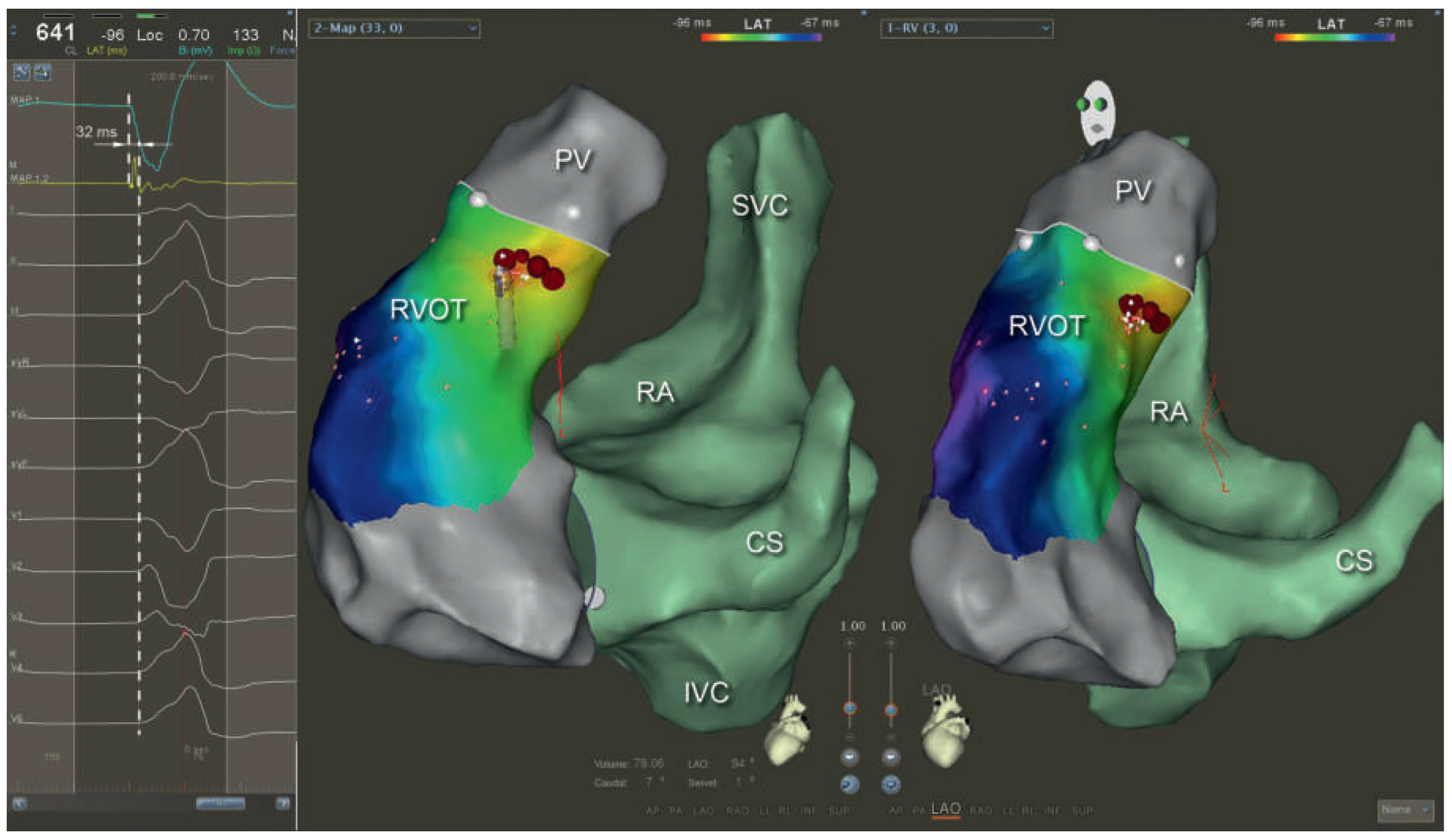
A 60-year-old woman was referred for ablation of symptomatic premature ventricular contractions (PVCs) originating from the right ventricular outflow tract (RVOT) (Figure 1). The PVC burden on 24-hour Holter monitoring was 19%, and left ventricular ejection fraction was 58%. To perform fluoroscopy-free ablation, a 3D electroanatomical mapping system (Carto3, Biosense Webster, Diamond Bar, USA) was used. After obtaining vascular access, the ablation catheter (Navistar Thermocool, Biosense Webster) was slowly advanced to the right atrium (RA), avoiding any unusual resistance. As soon as the catheter was visualised on the electroanatomic mapping (EAM) system, catheter orientation was monitored and fast anatomical mapping (FAM) was started to map the course of the inferior vena cava (IVC). As with conventional fluoroscopy-guided mapping, the intracardiac electrogram on the catheter tip was monitored until atrial signals were recorded. The earliest point with near-field atrial electrograms was tagged as the entrance of the IVC into the RA. The catheter was advanced into the superior vena cava (SVC) and the SVC–RA junction was marked where atrial signals on the catheter tip disappeared. The RA was then anatomically mapped by advancing the catheter into the SVC and pulling it into the IVC in different axial rotations with a slight deflection. The proximal coronary sinus was mapped by deflection and clockwise rotation of the catheter, and the tricuspid valve was annotated on the map. Duration from vascular access to the end of the RA mapping was 14 minutes. The reconstruction of the RA and the tricuspid annulus on the electroanatomical mapping system served as a roadmap for accessing the right ventricle and for navigation during subsequent mapping of the RVOT, which lasted 28 minutes. The earliest activation of the PVCs was identified below the pulmonary valve (white line) with a local endocardial activation time of −32 ms before onset of the QRS complex on the surface electrocardiogram (Figure 2). Radio-frequency energy delivery (133 s, 25 W) at that site eliminated the PVC. The overall procedure duration, including 30 minutes of waiting period, was 75 minutes. Twenty-four-hour Holter monitoring 3 months after the ablation showed no recurrence of the PVCs.

Discussion

As shown recently, left-sided procedures can also be performed without the use of fluoroscopy by relying solely on a 3D electroanatomical mapping system without any additional technical equipment such as intracardiac or transoesophageal echocardiography [1,2]. In the presented case, mapping the IVC–SVC axis, the RA, the proximal coronary sinus and the tricuspid annulus enabled the operator to access the right ventricle and the RVOT in order to map and ablate RVOT PVC without the use of fluoroscopy. Usage of a contact-force sensing catheter may be a useful tool in addition to tactile feedback during navigation and ablation. Although especially attractive for younger patients or even pregnant women, a fluoroscopy-free approach may be considered for any patient with normal cardiac anatomy and without implanted leads in the chamber of interest, in order to reduce the radiation burden to patients, physicians and staff in the electrophysiology laboratory. Knowing the cardiac anatomy before the procedure by performing magnetic resonance imaging may be advantageous but is not a sine qua non. Finally, the fluoroscopy-free approach costs more than a fluoroscopy-guided approach. Whether these additional costs are justified is difficult to determine because of the long-term effects of ionising radiation.

Disclosure statement

Stefan Osswald received unrestricted grants from Medtronic, Boston Scientific, Biotronik, and St. Jude Medical. Christian Sticherling received educational grants from Biosense Webster and Biotronik. Michael Kühne received educational grants from Biosense Webster. The other authors reported no potential conflicts of interest related to this article.

References

  1. Mühl, A.; Kühne, M.; Sticherling, C.; Knecht, S. Fluoroscopy-Free PVI With nMARQ in a Patient With a PFO. J Cardiovasc Electrophysiol. 2015, 26, 906. [Google Scholar] [CrossRef] [PubMed]
  2. Knecht, S.; Sticherling, C.; Reichlin, T.; Pavlovic, N.; Mühl, A.; Schaer, B.; et al. Effective reduction of fluoroscopy duration by using an advanced electroanatomic-mapping system and a standardized procedural protocol for ablation of atrial fibrillation: ‘the unleaded study’. Europace. 2015. Epub ahead of print.
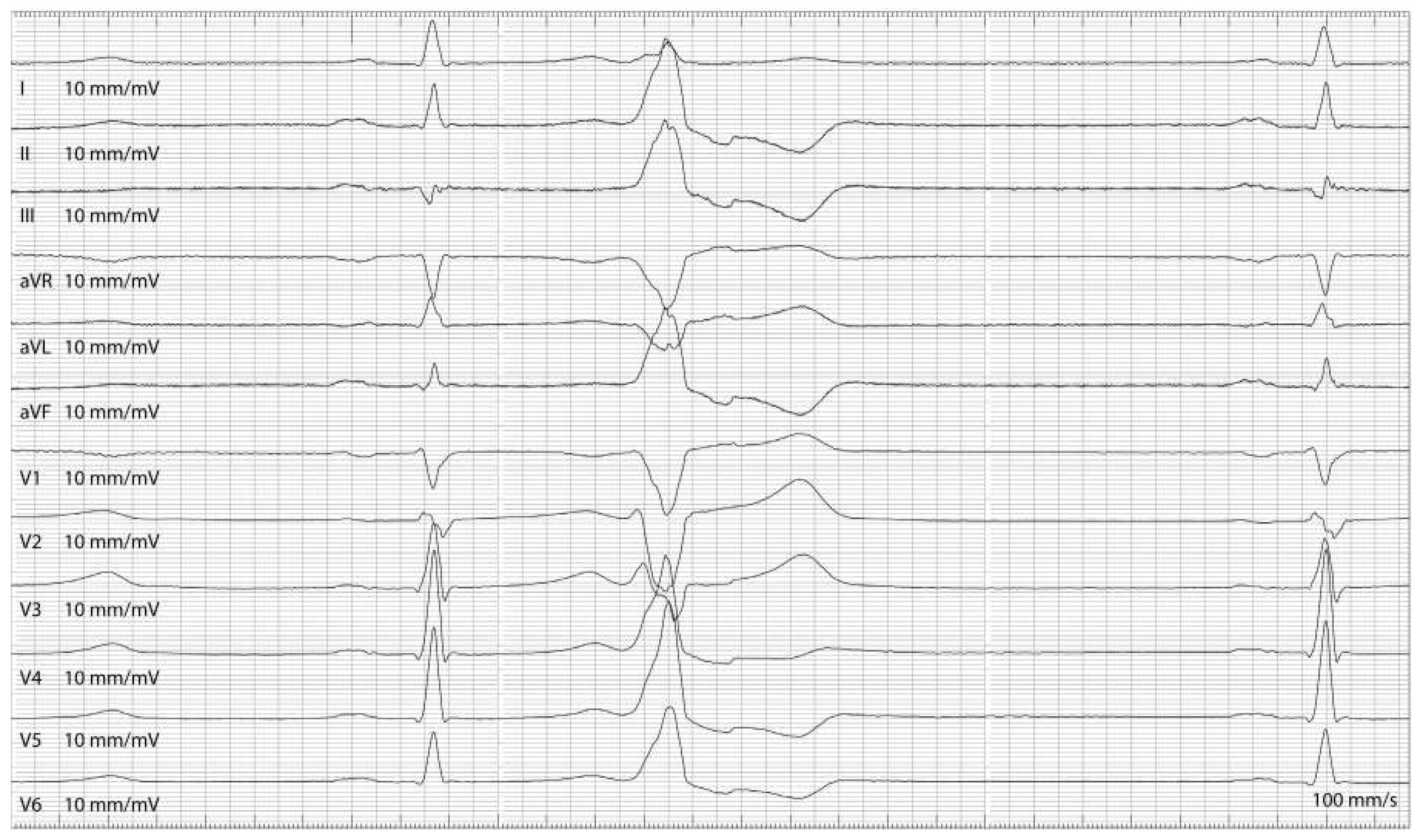
Figure 1. Twelve-lead ECG of the premature ventricular contraction.
Cardiovascmed 18 00357 g001
Figure 2. Screenshot of the Carto3 system with the earliest endocardial activation of –32 ms before the onset of the QRS complex (left column) and a left lateral and left anterior oblique view of the color-coded activation map of the right ventricular outflow tract and the right atrium (green). Red dots represent ablation sites. The ablation catheter is shown at the earliest site prior to ablation. CS = coronary sinus; IVC = inferior vena cava; PV = pulmonary valve; RA = right atrium; RVOT = right ventricular outflow tract; SVC = superior vena cava.
Cardiovascmed 18 00357 g002