Next Article in Journal
Prolonged Complete AV Block After Balloon Aortic Valvuloplasty in a Child
Previous Article in Journal
Timely Diagnosis of Congenital Heart Disease–Did We Improve?
 
 
Cardiovascular Medicine is published by MDPI from Volume 28 Issue 1 (2025). Previous articles were published by another publisher in Open Access under a CC-BY (or CC-BY-NC-ND) licence, and they are hosted by MDPI on mdpi.com as a courtesy and upon agreement with Editores Medicorum Helveticorum (EMH).
Review

The Weak Heart: Perioperative Management

by
Dominique Anne Bettex
1,*,
Pierre-Guy Chassot
2 and
Alain Rudiger
1
1
Institute for Anaesthesiology, University Hospital Zurich, Rämistrasse 100, CH-8091 Zurich, Switzerland
2
Department of Anaesthesiology, University Hospital of Lausanne, Lausanne, Switzerland
*
Author to whom correspondence should be addressed.
Cardiovasc. Med. 2015, 18(10), 273; https://doi.org/10.4414/cvm.2015.00362
Submission received: 21 July 2015 / Revised: 21 August 2015 / Accepted: 21 September 2015 / Published: 21 October 2015

Abstract

Heart failure (HF) is known to be a major risk factor in perioperative care. It should be subdivided into systolic or diastolic dysfunction as well as left or right ventricular failure. The perioperative management of HF patients is complex, consisting of prevention, diagnosis and therapy. The adequacy of the perioperative management determines the late postoperative outcome and will be presented in this review.

Introduction

Epidemiology 

Heart failure (HF) has an incidence of 1 to 2% in the general population [1]. Its prevalence increases further with age, rising from approximately 2% in individuals 65–69 years old to >8% in those ≤85 years [2]. The mortality rate of systolic HF is 14% at 1 year and up to 50% at 5 years, when symptomatic [2–4]. The average duration of survival after one hospitalisation is 2.4 years and drops to 0.6 year after the fourth hospitalisation [5]. About 25–40% of patients with congestive HF have a preserved ejection fraction and suffer from diastolic failure; their mortality is 50% lower than in the case of reduced ejection fraction [4].

Heart failure as a perioperative risk factor 

HF is known to be a major risk factor in perioperative care and is found in 2.5 to 10% of noncardiac surgical patients [6–8]. The incidence of severe cardiac events after major noncardiac surgery is between 2 and 8% [6,9,10]. In cardiac surgery, the incidence rises to over 20% [11]. In vascular surgery, it is 18% in the case of isolated diastolic failure, 23% in the case of asymptomatic systolic insufficiency and 49% in the case of congestive systolic failure [12]. In a retrospective study including 1 532 HF patients and 1757 coronary artery disease (CAD) patients undergoing major noncardiac surgery, the risk-adjusted mortality within 30 days was 11.7% in HF, 6.6% in CAD and 6.2% in control patients. The risk-adjusted 30-day readmission rate was 20% for HF, 14.2% for CAD and 11% for control patients [13]. HF patients undergoing major noncardiac surgery suffered substantial morbidity and mortality despite advances in perioperative care, whereas patients with CAD without HF had similar mortality to a more general population [10,13,14]. It is not only the perioperative cardiac risk which is high in HF patients but also the noncardiac complication incidence is higher than in patients without CAD or HF, as shown recently [14]. In this retrospective series, a near doubling of postoperative death was reported in HF patients, as well as a 40 to 69% increased risk of sepsis, and pulmonary and renal complications, but not of myocardial infarction. Perioperative management may have been focused on preventing myocardial ischaemia at the expense of other organ systems.
Interestingly, the newly published European Society of Cardiology (ESC) / European Surgical Association (ESA) guidelines on noncardiac surgery define the perioperative cardiac risk as follows: “cardiac complications can arise in patients with documented or asymptomatic ischaemic heart disease, left ventricular dysfunction, valvular heart disease and arrhythmias, who undergo surgical procedures that are associated with prolonged haemodynamic and cardiac stress” [15]. We may wonder why the role of right ventricular failure (RVF) in perioperative cardiac risk is so neglected. There is a plethora of literature and guidelines on the perioperative management of patients with coronary artery disease and left ventricular failure (LVF), but until now, there is a lack of guidelines on the perioperative management of the patient with RVF. However, RVF has been clearly associated with increased mortality among cardiac surgical patients as well as in the non-cardiac setting and in the intensive care unit (ICU) [16]; it is present in approximately 40% of postcardiotomy cardiogenic shock [11,17,18].

Risk assessment 

Preoperative risk assessment is essential, considering the risks of surgery (emergency, type, invasiveness, duration and potential blood loss), and the risks of the patient, considering their personal history (history of ischaemic cardiac disease, HF, cerebrovascular disease, insulin-dependent diabetes mellitus and impaired renal function) and their functional capacity (in metabolic equivalents, METs) [19]. Many di erent indices have been designed since the Revised Cardiac Risk Index of Lee [20], but their accuracy and concordance are limited [21]. According to the 2014 ESC/ESA guidelines, patients with established or suspected HF scheduled for noncardiac intermediate or high-risk surgery should undergo transthoracic echocardiography and/ or assessment of natriuretic peptides, and should be therapeutically optimised as necessary, using betablockers, angiotensin-converting enzyme inhibitors, angiotensin receptor blockers, mineralocorticoid antagonists and diuretics [15]. In cardiac surgery, indicators of major clinical risk in the perioperative period are: unstable coronary syndromes, decompensated HF, significant arrhythmias and severe valvular disease [19]. The EuroSCORE II is currently used for perioperative risk assessment in cardiac surgery [22].

Definition and diagnosis of heart failure

Definition 

HF is a functional and structural impairment of ventricular filling and/or ejection, leading to a failure of oxygen and nutrient delivery at a rate in accordance with the requirements of the tissues. It should be subdivided into left and right ventricular failure as well as systolic or diastolic dysfunction. Diastolic dysfunction resulting in depressed filling of the ventricle is mostly a disease of the left ventricle. Systolic dysfunction results in poor ventricular ejection and a ects both ventricles. Whereas diastolic failure may occur without overt systolic failure, the latter is always accompanied by diastolic failure. Basically, five myocardial mechanisms lead to HF: arrhythmias, pressure overload, volume overload, coronary disease and primary myocardial disease (cardiomyopathy). Clinically, acute HF presents frequently as pulmonary oedema, left/right congestive HF or cardiogenic shock with progressive end-organ malperfusion and possible failure [23]. Both the American College of Cardiology Foundation (ACCF) / American Heart Association (AHA) stages of HF and the New York Heart Association (NYHA) functional classification are useful complementary information about the presence and the severity of HF (Table 1) [2]. HF patients commonly suffer from comorbidities among which the 10 most frequent are: systemic hypertension (84%), ischaemic heart disease (72%), hyperlipidaemia (60%), anaemia (50%), diabetes (46%), arthritis (43.5%), chronic kidney disease (42%), chronic obstructive pulomonary disease (COPD) (30%), atrial fibrillation (28.5%) and Alzheimer’s disease / dementia (28%) [19].

Diagnosis 

The diagnosis of acute HF is based on clinical signs, echocardiography and cardiac biomarkers. Among the clinical signs are: orthopnoea, rales, abdominal discomfort, peripheral oedema, hypotension, tachycardia, oliguria, cyanosis, mottling and disorder of consciousness. Perioperative echocardiography should be performed as early as possible and will quickly provide information on regional and global ventricular function, right or left ventricular dysfunction, valvular dysfunction, tamponade and volume status. Whereas an increase in troponin level is highly correlated with postoperative major adverse cardiac events (MACE), the diagnostic role of natriuretic peptides (BNP, NT-pro-BNP) in the perioperative period remains to be demonstrated [24].

Perioperative triggers of acute cardiac decompensation

In the perioperative period, HF patients face numerous triggers of acute cardiac decompensation including hypertension, tachyarrhythmias, anaemia, hypercoagulability, inappropriate fluid management, pain, surgical stress, pulmonary hypertension (PHT), pulmonary or fat emboli and myocardial ischaemia [19]. Cardiac surgery may be complicated by coronary bypass occlusion, intracoronary air embolism, paravalvular or residual valvular regurgitation, PHT, tamponade, haemo- or pneumothorax. Patients with endstage or advanced HF who are symptomatic at rest despite maximal medical therapy (stage IV) present a unique challenge for perioperative management, and are at high risk for perioperative mortality (10–30%); however, their mortality drops to 6% when they are in a compensated stage (I–II) [10]. If not an emergency, surgery should be postponed in patients with decompensated, new onset or untreated HF [13]. Anaesthetising patients with ongoing therapy for acute HF requires a good knowledge of the disease and its treatments, as well as possible haemodynamic consequences of anaesthesia [25]. An assessment of a patient’s prognosis linked to his or her cardiac disease can help to determine the role of interventions for noncardiac diseases, meaning that patients with a very poor cardiac prognosis may not survive long enough to benefit from some noncardiac procedures [26]. The optimal perioperative course of high-risk cardiovascular patients should be based on close cooperation between cardiologists, surgeons, anaesthesiologists and intensivists.
Any patients with RVF or LVF poorly tolerate circulatory overload; overzealous fluid infusion could explain some forms of iatrogenic perioperative HF. If LVF patients nicely tolerate the vasodilating effects of anaesthetics agents and LV unloading with mechanical ventilation, the most vulnerable time is the period of weaning from the ventilator and extubation. In contrast, RVF patients poorly tolerate the negative inotropic effect of anaesthetic agents and the increase in right ventricular afterload resulting from mechanical ventilation; any significant drop in systemic pressure (right ventricular perfusion) or hypoxic event (intraoperative mishaps or postoperative pulmonary complications) could precipitate or aggravate ongoing right ventricular decompensation.
Since right and left ventricular failure represent di erent entities with di erent management, they will be discussed separately.

Specific perioperative considerations of right ventricular failure

Premedication 

In the absence of evidence-based studies, similar perioperative management can be recommended in patients with LVF with preserved ejection fraction as in patients with LVF and reduced ejection fraction. The perioperative management of LVF starts with the preoperative visit. The first step will be to identify preexisting systolic or diastolic abnormalities of the left ventricular function and establish the diagnosis of the underlying cardiac disease (Table 2). Clinical signs of left ventricular dysfunction, although nonspecific, (tachypnoea, orthopnoea, rales, legs oedema, hypotension, tachycardia, oliguria, cyanosis, mottling and somnolence), and the electrocardiogram (ECG; arrhythmias, tachycardia, signs of old or recent myocardial infarction, strain pattern, left bundle-branch block), chest X ray (cardiomegaly, pulmonary oedema), transthoracic echocardiography (TTE), coronary angiogram if relevant, as well as laboratory data (troponin, BNP or proNT-BNP, kidney and liver parameters, haematology values) will all be studied carefully. TTE is the screening investigation of choice. Individualised premedication and anxiolytic therapy is then administered, avoiding stress, but also respiratory depression and hypotension. Preoperative HF therapy other than diuretics (beta-blockers, angiotensin converting-enzyme inhibitors, angiotensin receptor blockers) should be further administered over the perioperative phase. When angiotensin converting-enzyme inhibitors and angiotensin receptor blockers are prescribed for systolic ventricular failure, the risk of hypotension during anaesthesia is much less than when they are prescribed for systemic hypertension; in this latter case, their transient discontinuation 24 hours before surgery should be considered [15].

Intraoperative prevention 

Intraoperatively, the next step will be to prevent any worsening of left ventricular function. The haemodynamic goals in the case of left ventricular failure are to optimise left ventricular preload and contractility, to reduce afterload, and to avoid tachycardia, bradycardia or arrhythmias. Simultaneously, hypoperfusion of major organs and reduction of coronary blood flow must be prevented. Particular attention on avoiding overdose of drugs should be kept in mind, particularly during induction, as the patient’s sensitivity is high and circulation time is slow. Frail patients maintain an acceptable cardiac output only within very restricted limits at rest, but have lost all physiological reserve in case of increased demand such as during surgery. Therefore, it is of the utmost importance to maintain rigorously a stable haemodynamic status, to immediately correct any significant deviation, and to equilibrate cardiac output with metabolic requirements. Major surgery can be successfully undertaken in patients with a depressed haemodynamic condition as long as no intercurrent complication supervenes. Oxygen debt, acidosis and hypoperfusion intraoperatively will lead to multiple organ failure appearing later in the postoperative course. Fatal issue usually happens after a few days of intensive care, which may leave the wrong impression that anaesthesia management is not involved in this dismal outlook.
A recent meta-analysis showed that goal-directed therapy (GDT) in high-risk surgery is beneficial in reducing cardiovascular events (odds ratio 0.54), irrespective of the choice of monitored physiological parameter or haemodynamic monitor in use [27]. The benefit was most pronounced in patients receiving fluid and inotrope therapy to achieve a supranormal oxygen delivery target, with the use of minimally invasive cardiac output monitoring. However, a Cochrane review found no di erences in the rate of arrhythmias, myocardial infarction, congestive HF or pulmonary oedema between patients treated with perioperative GDT and control patients [28]. These inconsistent results regarding clinical outcomes may be explained by poor adherence by clinicians to the protocol or the inappropriateness of the proposed algorithm in selected high-risk patients. The current evidence does not support widespread implementation of GDT to reduce mortality but does suggest that complications and duration of hospital stay are reduced [28]. Large randomised studies are needed to solve this question definitely. Until then, individualised haemodynamic goals should be defined, depending upon the patient’s characteristics and institutional preferences, considering optimisation of venous saturation (central or mixed), blood lactate, stroke volume index and/or cardiac index.

Monitoring 

The choice of monitoring for these patients should be discussed. In addition to the standard ASA surveillance, extended haemodynamic monitoring must be adapted to the LVF patient and to the type of surgery. An arterial catheter and a multilumen central catheter will be necessary in most cases. The current transoesophageal echocardiography (TEE) guidelines recommend the use of TEE in noncardiac surgery if severe haemodynamic, pulmonary and neurological compromises are anticipated, or in the case of life-threatening circulatory instability unresponsive to conventional interventions [9,29]. It is recommended in any openheart surgery and may be considered for coronary artery bypass surgery. The authors believe that TEE is mandatory in any perioperative case of LVF, except in the case of absolute contraindications. Details on the TEE assessment of left ventricular function can be found elsewhere [30,31].
There is no convincing evidence for the use of a pulmonary artery catheter (PAC) in perioperative patients during noncardiac surgery. In a case-control analysis, the perioperative use of a PAC was associated with a higher incidence of postoperative HF and noncardiac events than in a matched control group [32], and a meta-analysis of 13 randomised studies showed a null effect of PAC on outcome, when used in a random fashion during major surgery among critically ill patients [33]. However, these negative results do not preclude the use of PAC in selected cases. The use of less invasive perioperative cardiac output monitoring techniques (PiCCO™, Oesophageal Doppler, Flow-Track™ Vigileo) could be associated with a reduction in length of stay and complications [34], but large randomised studies are still lacking [15]. It should be reminded that the impact of a monitoring device on clinical outcome relies entirely on the interpretation of the data made by the physician and on the therapeutic algorithm implanted in the institution, but not on the technique itself.

Choice of anaesthesia 

In the general population, the choice of the anaesthetic agent has been considered to be of little importance in terms of patient outcome, provided that vital functions are adequately supported. In compromised patients, however, induction agents are best chosen among the substances with the least haemodynamic effects. Most volatile and intravenous anaesthetic agents reduce preload, afterload and contractility, and require proper management to ensure maintenance of organ flow and perfusion pressure. Induction agents may be classified by increasing order of negative inotropic action as follows: etomidate, midazolam, propofol, ketamine, and thiopental. The key conditions for a stable induction are: a reduction of the dose according to the degree of ventricular dysfunction, and a slowdown in the rate of drug administration, completing very progressively the dose required for intubation. Opioids have no known adverse effects on the left ventricular function. In patients suffering from ischaemia-induced ventricular failure, use of halogenated gases may be advised because of their preconditioning effect [35]. In coronary artery bypass grah (CABG) surgery, they tend to improve myocardial performance recovery [36]; whether this can be extrapolated to noncardiac surgery is still debated. Intermittent positive pressure ventilation (IPPV) is well tolerated in left ventricular failure because the increase in intrathoracic pressure corresponds to a decrease in left ventricular afterload. If a neuraxial technique is chosen, the local anaesthetic agent should be introduced slowly to avoid systemic vasodilation. When the blockade reaches the fourth thoracic dermatome, a reduction in cardiac sympathetic tone may occur with a decrease in myocardial contractility, heart rate, and change in loading conditions. The ESC/ESA guidelines have estimated, however, that neuraxial anaesthesia and analgesia may be considered for the management of patients with cardiovascular risk factors or diseases [15].

Perioperative LVF management 

Early identification of LVF and the underlying cardiac disease, as well as prompt and aggressive management, decrease postoperative morbidity and mortality. The search for reversible conditions is essential: myocardial ischaemia or infarction, acute valvular dysfunction, left ventricular tract obstruction with systolic anterior motion of the mitral valve, septic shock, aortic dissection type I or postcardiotomy in the case of cardiac surgery must be treated appropriately. Resuscitation measures must be undertaken immediately. Similarly to sepsis therapy, the concept of the “golden hours” for acute HF management is essential [19]. Oxygenation and ventilation should be immediately maximised and acid-base as well as electrolyte abnormalities should be corrected. Optimisation of preload, afterload and contractility (Table 3), ideally under echocardiographic monitoring, and control of the heart rate and rhythm should stabilise the left ventricle. Positive inotropic agents must be used with caution, with carefull consideration of their risk–benefit ratio; several studies indicate an association between prescription of inotropes and poor clinical outcome [37]. It remains of great use in patients with acute systolic dysfunction and low cardiac output, and evidence of systemic hypoperfusion or congestion. The dosage should be kept as low as possible and the possibility of weaning should be regularly assessed (Table 4). Until now, no catecholamines have been shown to improve outcome of patients, except levosimendan [38,39]. In a recent large meta-analysis considering 45 randomised controlled trials and analysing 5480 patients, levosimendan was shown to reduce mortality of adult patients in cardiology and cardiac surgery settings [40]. In a consensus of experts, levosimendan was included among eight nonsurgical ancillary drugs, techniques or strategies that might decrease mortality in cardiac surgery [41]. It has the great advantage of acting as both a positive inotrope and an afterload reduction agent, without increasing myocardial oxygen consumption. If LVF persists, mechanical assistance should be started as soon as possible and preferentially before organ dysfunction. Intra-aortic balloon counterpulsation (IABP) improves coronary perfusion through augmentation of diastolic pressure, decreases afterload and, consequently, reduces myocardial oxygen consumption and increases cardiac output. It has been the most widely used mechanical circulatory support device for nearly five decades, particularly during and after cardiac surgery. Following the results of the first large randomised, open-label trial on the use of IABP in acute myocardial infarction complicated by cardiogenic shock (IABP-SHOCK II), the indications for IABP have been greatly restricted [42,43]. In the current guidelines, IABP is indicated only in cardiogenic shock complicating myocardial infarction [15]. The use of IABP in noncardiac surgery is founded on case reports and small case series [44,45], with good results in the acute perioperative time. In cases of refractory low cardiac output syndrome, extracorporeal life support (ECLS) might become an option. It should be inserted before irreversible organ dysfunction develops, as a bridge to decision, to recovery, to ventricular assist device (LVAD) or to transplantation.
The early postoperative care of LVF patients should be conducted in a high-acuity nursing environment, with invasive monitoring and requisite assessment of cardiac biomarkers (e.g., troponin and brain natriuretic peptides) [7]. Recent meta-analyses demonstrated that increased postoperative troponin and BNP concentrations after noncardiac surgery were associated with a significantly increased risk of mortality [15,46,47]. Preoperatively and postoperatively, patients who could most benefit from BNP or troponin measurements are those with METs ≤4 or with a revised cardiac risk index value >1 for vascular surgery and >2 for nonvascular surgery [15].
Diastolic dysfunction and failure are initially characterised by a maintained cardiac output, but by restrictive filling conditions (lack of relaxation, sti ventricle). Elevated filling pressures, intolerance to tachycardia or bradycardia, and extreme dependence of stroke volume from preload (intolerance to hypovolaemia, large variations of arterial pressure with positive pressure ventilation) define their haemodynamic behaviour. Diastolic function is best evaluated with echocardiography. Unfortunately, there are no efficient means to improve diastolic function [48].

Specific perioperative considerations of right ventricular failure

The anaesthesiologist will be confronted with patients with right venticular failure in various circumstances (Table 2 and Table 5):
In noncardiac surgery, perioperative right ventricular failure is most often, although not exclusively, secondary to acute PHT; a normal right ventricle can cope for only 1–2 hours with a mean positive airway pressure of ≤40 mm Hg [49].
In cardiac surgery, right ventricular failure may be secondary to acute PHT, but also frequently to volume overload, myocardial ischaemia, preexisting right ventricular dysfunction or arrhythmias. Among cardiac surgery patients, those undergoing cardiac transplantation or LVAD implantation are at higher risk.
Grown-up congenital heart disease (GUCH) patients, for cardiac or noncardiac surgery.

Premedication 

The perioperative management of right ventricular failure, like the management of left ventricular failure, consists of several successive steps, starting with a preoperative visit. The first step is to identify preexisting abnormalities of the right ventricular function and of the pulmonary vasculature, knowing that perioperative risk factors for right ventricular decompensation include a preexisting right ventricular dysfunction, or severe PHT without right ventricular dysfunction. A thorough history and clinical examination is required. Clinical signs of right ventricular dysfunction, although nonspecific (dyspnoea, hypotension, right upper quadrant discomfort, ascites, jugular vein distension), and ECG (sinus tachycardia, T-wave inversion in III and aVF or V1 to V4, right bundle-branch block, rightward axis), chest X ray (pulmonary artery, right atrium and right ventricular dilation), transthoracic echocardiography (TTE), right heart catheterisation in cases of moderate to severe right ventricular dysfunction combined with severe PHT [50] and laboratory values, must all be studied carefully. TTE is the screening investigation of choice. Individualised premedication and anxiolytic therapy is then administered, avoiding stress, but also respiratory depression with the risk of secondary PHT. Preoperative chronic PHT therapy should be further administered over the perioperative period.

Intraoperative prevention 

Intraoperatively, the next step will be to prevent any aggravation of possible pre-existing RV dysfunction. In this view, any increase in pulmonary vascular resistance (PVR) and right ventricular myocardial ischaemia, which exacerbate each other, should be avoided. The haemodynamic goals are to maintain right ventricular preload and contractility, minimising the PVR and avoiding right ventricular coronary hypoperfusion.

Monitoring 

As for the patients in left ventricular failure, an arterial catheter and a multilumen central venous catheter will be useful, particularly in the case of IPPV. Intraoperative TEE is mandatory in all patients with RVF, except in the case of absolute contraindication [51]. Detail on the TEE assessment of right ventricular function can be found in previous publications [52–54] as well as on the ASE website (www.asecho.org).
A PAC may be indicated in case of severe PHT and RVF, but should be used with caution considering the risk for arrhythmias and catheter-induced pulmonary artery rupture [55]. The measurement of cardiac output may be altered in the presence of tricuspid regurgitation. The use of continuous right ventricular pressure waveform monitoring might be helpful, as described by Denault et al. [16] (Figure 1). In cases of right ventricular dysfunction, a progressive change in the diastolic pressure slope from horizontal to obliquely ascending will be observed. As right ventricular function deteriorates, the slope will change to a square root shape, and finally right ventricular and pulmonary diastolic pressures will equalise [16]. Combining right ventricular pressure waveform and TEE monitoring allows rapid determination of the cause of right ventricular systolic and diastolic dysfunction.

Choice of anaesthesia 

The choice of anaesthetic is equally relevant in the prevention of RVF. The depth of anaesthesia and postoperative analgesia should be sufficient to avoid large sympathetic haemodynamic responses to pain and surgery. Volatile anaesthetics may all worsen right ventricular dysfunction by reducing preload, afterload and contractility. The PVR is increased by both desflurane and nitrous oxide [56,57]. Ketamine seems to increase PVR in adults but not in children [58,59]. Although the potential deleterious effect on PVR remains a concern, it must be balanced against the potential benefits of combined analgesia and hypnosis and its absence of significant myocardial depression and vasodilation [60]. Etomidate has been advocated as the induction agent of choice although there is no comparative data [61,62]. Opioids have no known adverse effects on the right ventricular function. If a neuraxial technique is chosen, a local anaesthetic agent should be introduced slowly to avoid inconsiderate systemic vasodilation.

Perioperative RVF management 

The third step consists of early identification of RVF. The functional state of the right ventricle cannot be determined from the severity of PHT alone. In a patient with known PHT developing RVF, the PAP will pseudonormalise as right ventricular function fails. In other words, while a falling PAP may be due to a reduction in PVR or left atrial pressure (LAP), it might also be a sign of a failing right ventricle that cannot build up the pressure any more. Conversely, an increase in cardiac output in the face of a high, relatively fixed PVR will increase the PAP. In cases of progressive RVF, PAP may appear normal but right atrial pressure (RAP) will be elevated. Both systemic and pulmonary artery pressures will be reduced to a similar degree; the ratio of systemic to pulmonary mean arterial pressures may better reflect the severity of PHT. In cardiac surgery, this ratio has been shown to be a better predictor of postoperative complications than the absolute values [63]. A search for possible underlying reversible conditions is essential: pulmonary embolism, lung infection, bronchial asthma, COPD, ventilation/perfusion mismatch, valvulopathy, myocardial ischaemia, intracardiac shunt and left ventricular diastolic or systolic dysfunction can all contribute to RVF.
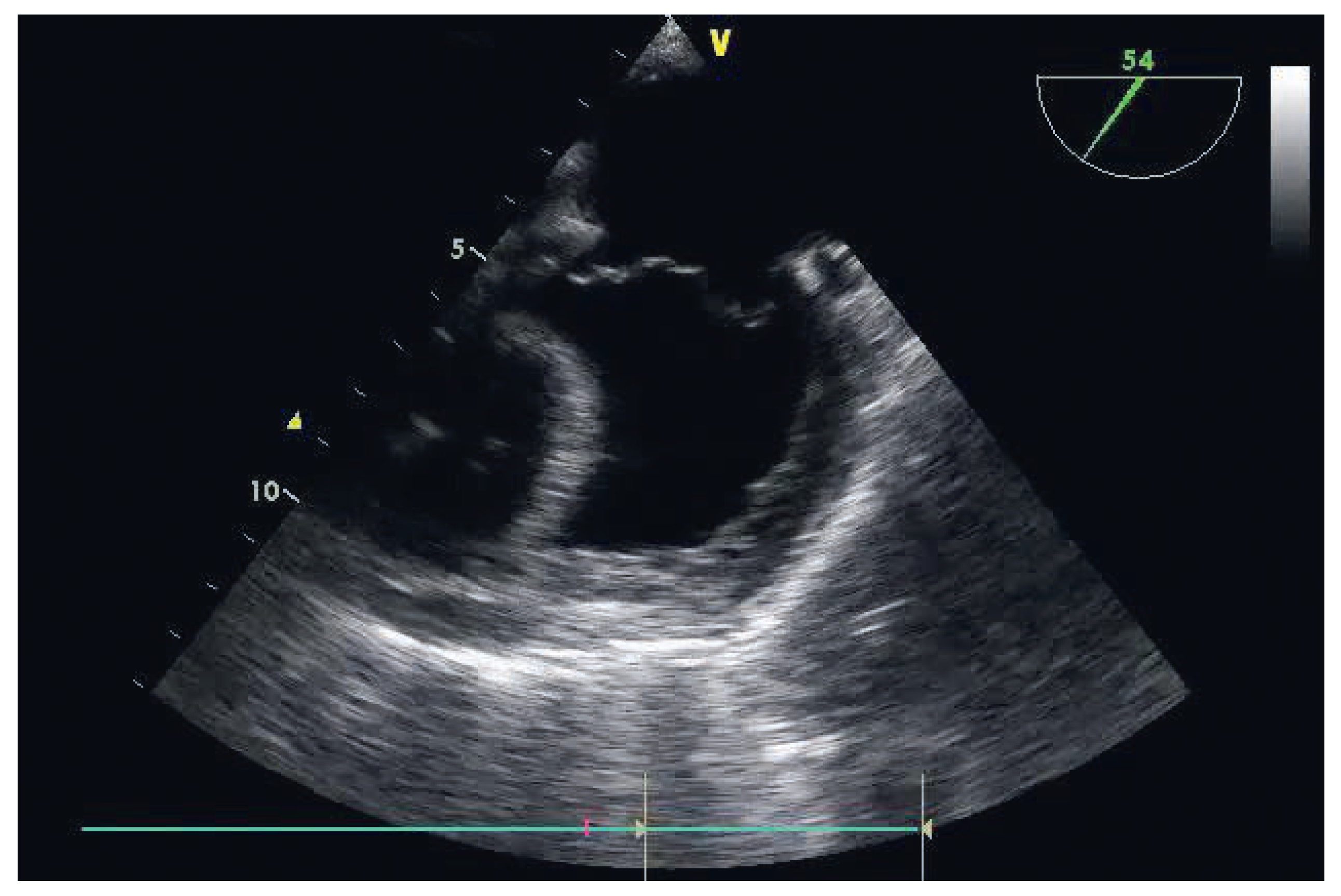
Finally, in presence of progressive RVF, standard therapy is initiated (Table 3), keeping in mind that one of the main goals of management is the maintenance of the ventricular interaction. Right ventricular function is significantly reduced if the septum is dysfunctional; the maximum right ventricular developed pressure is reduced by 30% when the septum is inactivated [64]. Conditions that reduce the left ventricular systolic pressure or increase the right ventricular systolic pressure reverse the trans-septal gradient and lead to further RVF [65,66]. Although the right ventricle is highly preload-dependent, overfilling can cause right ventricular dilation and secondary tricuspid regurgitation with a resulting increase in right ventricular wall stress, decrease in left ventricular compliance and progressive reduction of cardiac output, leading to further right ventricular dilation (Figure 2). Preload is optimised, observing the effect of a volume tolerance test on central venous pressure, PAP and right ventricular filling (TEE/TTE). However, pulse pressure variation cannot be used for fluid assessment in patients with RVF, as pulsus paradoxus might result from RVF and ventricular interdependence [67,68]. Sinus rhythm is maintained as well as a heart rate of at least 90/min. The right ventricular afterload is decreased, initially using 10O% FiO2, mild hyperventilation, alkalinisation, and adaptation of ventilation settings: the relationship between tidal volume and PVR has a unique “U” shape, PVR being minimal at functional residual capacity and maximal in the event of hypoventilation or hyperinflation [52,53]. Any sympathetic stimulation such as stress, pain, anxiety, hypothermia and shivering is suppressed, and anaesthetics potentially associated with augmentation of PVR are avoided. If these diverse measures do not allow a stabilisation of the right ventricular function, pulmonary vasodilatation is then started, preferentially using inhalational substances such as nitric oxide (NO), iloprost or milrinone, to avoid systemic vasodilation. Because of their di erent modes of action, combination of inhalational vasodilators might be synergistic [50,62]. Keep in mind that the key haemodynamic sign of a therapeutic response to inhaled pulmonary vasodilators is not a reduction in pulmonary artery pressure but a decrease in CVP and an increase in cardiac output [62]. Considering further ventricular interdependence [69,70] as well as the coronary perfusion of the right ventricular myocardium, the systemic arterial pressure is maintained elevated to avoid right ventricular myocardial ischaemia, using vasopressors as needed (noradrenaline, vasopressin). The benefit of systemic vasoconstriction has to be balanced with the risk of pulmonary vasoconstriction; fortunately, the pulmonary arterial tree has fewer alpha-receptors than the systemic arteries and is devoid of receptors for vasopressin [50]. Finally, inotropic agents are deployed as needed: dobutamine, adrenaline, phosphodiesterases III inhibitors (milrinone) or levosimendan have all been shown to be effective in case of acute RVF. However, as levosimendan and milrinone both produce systemic vasodilation, vasopressors may need to be coadministered to prevent reduced right coronary blood flow [62]. Increased left ventricular contractility can also result in increased right ventricular systolic function through ventricular interdependence.
In cases of persistent refractory RVF, mechanical support with a ventricular assist device should be initiated, preferentially before the appearance of irreversible organ dysfunction.

Conclusion

In conclusion, perioperative heart failure is the result of inadequate contractility, arrhythmia, volume or pressure overload and is associated with worse outcomes in noncardiac and cardiac surgery. Particular care has to be taken of the right ventricle. Prevention, diagnosis and therapy are difficult tasks for the team managing the patient, including anaesthesiologists, cardiologists, surgeons and intensivists. The adequacy of the perioperative management determines the late postoperative outcome.

Conflicts of Interest

No financial support and no other potential conflict of interest relevant to this article were reported.
Figure 1. Zoomed right ventricular pressure (PRV) and pulmonary arterial pressure (PPA) with their corresponding Doppler hepatic venous flow (HVF) before (a, b) and after cardio-pulmonary bypass. Note the change in the diastolic slope of the PRV waveform and the corresponding change in the systolic (S) to diastolic (D) ratio of the HVF. From: Denault AY, Haddad F, Jacobsohn E, Deschamps A. Perioperative right ventricular dysfunction. Current opinion in anaesthesiology. 2013;26(1):71-81, reprinted with permission.
Cardiovascmed 18 00273 g001
Figure 2. Right ventricular dysfunction after tricuspid valve replacement with obvious septum shift toward the left ventricle.
Cardiovascmed 18 00273 g002
Table 1. Comparison of ACCF/AHA stages of HF and NYHA functional classification.
ACCF/AHA stages of HF NYHA functional classification
AAt high risk for HF but without structural heart disease or symptoms of HF0
BStructural heart disease but without signs or symptoms of HFINo limitation of physical activity, ordinary physical activity does not cause symptoms of HF
CStructural heart disease with prior or current symptoms of HFINo limitation of physical activity, ordinary physical activity does not cause symptoms of HF
IISlight limitation of physical activity. Comfortable at rest, but ordinary physical activity results in symptoms of HF
IIIMarked limitation of physical activity. Comfortable at rest, but less than ordinary physical activity causes symptoms of HF
IVUnable to carry on any physical activity without symptoms of HF, or symptoms of HF at rest
DRefractory HF requiring specialised interventionsIVUnable to carry on any physical activity without symptoms of HF, or symptoms of HF at rest
ACCF = American College of Cardiology Foundation; AHA = American Heart Association; HF = Heart failure: NYHA = New York Heart Association.
Table 2. Causes of left and right ventricular failure (modified from [71]).
MechanismLeft ventricular failureRight ventricular failure
Excessive pressureAortic stenosisPrimary or secondary pulmonary hypertension
loadHypertrophic obstructive cardiomyopathyHTPL/LTPL
Arterial hypertensionPulmonary stenosis
Some types of congenital heart disease
(i.e., Fallot)
Excessive volumeAortic or mitral regurgitationPulmonary or tricuspid regurgitation
loadHigh-output states (thyrotoxicosis)ASD
Some types of congenital heart disease (i.e., VSD)Iatrogenic
IatrogenicLVAD
Primary myocardialIschaemic heart diseaseIschemic heart disease
diseaseHypertrophic nonobstructive cardiomyopathyHypertrophic obstructive cardiomyopathy
Hypertrophic obstructive cardiomyopathyDilated cardiomyopathy
Dilated cardiomyopathyArrhythmogenic cardiomyopathy
Cardiomyopathy of the elderlyMyocarditis
Myocarditis
Metabolic heart disease
Endocrine heart disease
Impaired ventricularTight mitral stenosisTight tricuspid stenosis
fillingConstrictive pericarditisConstrictive pericarditis
Restrictive cardiomyopathyRestrictive cardiomyopathy
Tamponade
Some types of congenital heart disease
(i.e., Ebstein anomaly)
ASD = atrial septal defect; HTPL = heart transplantation; LTPL = lung transplantation; LVAD = left ventricular assist device; VSD = ventricular septal defect.
Table 3. Management of perioperative heart failure.
Left ventricular failure Right ventricular failure
Avoidance of drug-induced myocardial depression Avoidance of drug-induced myocardial depression
Preservation of ventricular interaction
Optimisation of preload Optimisation of preload
Maintenance of SR and A–V synchrony
Heart rate control
Maintenance of SR and A–V synchrony
Heart rate control
Reduction of left ventricular afterload Avoidance of PHT exacerbation: hypoxemia, hypercarbia,
hypothermia, acidosis, stress and pain
Optimization of ventilator settings
Maintenance of adequate systemic perfusion pressure for organ perfusionMaintenance of systemic perfusion pressure while minimising right ventricular dilatation
Avoidance of nephrotoxic and hepatotoxic drugs Avoidance of nephrotoxic and hepatotoxic drugs
Minimisation of blood transfusion, especially of old blood
Tailoring of therapy to the specific aetiology of the LVFTailoring of therapy to the specific aetiology of the RVF
Reduction of right ventricular afterload, preferentially with inhalative therapy
Inotropic support Inotropic support
Mechanical assist devices Mechanical assist devices
A–V synchrony = atrioventricular synchrony; LVF = left ventricular failure; RVF = right ventricular failure; PHT = pulmonary hypertension.
Table 4. Dose-related haemodynamic effects of intravenous inotropic agents in heart failure (modified from [2,72]).
Dose mcg/kgDose mcg/kg/minEffects Adverse effects
Inotropic agentBolusInfusionCOHRSVRPVR
DobutamineN/A2.5 to 5
5 to 20


↑/↓ BP, T, HA, N, F, hypersensitivity, O2myoc↑
AdrenalineN/Ato 0.05↑ ↑T, A, F, lactate↑
0.05 to 0.1↑↔↑ ↑↑ ↑↑ ↑Glyc↑, O2myoc↑↑
>0.1↑↔↓↑ ↑↑ ↑ ↑↑ ↑
MilrinoneN/R0.125 to 0.175↑ ↑A, T, ↓BP, O2myoc↑↔
LevosimendanN/R0.05 to 0.2↑ ↑↑↔↓↓T, A, HA, ↓BP, hypoK, O2myoc↔
A = arrhythmias; BP = blood pressure; CO = cardiac output; F = fever; Glyc = glycaemia; HA = headache; HR = heart rate; hypoK = hypokalaemia; N/A = not applicable; N = nausea; N/R = not recommended; O2myoc = myocardial O2 consumption; PVR = pulmonary vascular resistance; SVR = systemic vascular resistance; T = tachycardia.
Table 5. Occurrences of acute perioperative right ventricular failure.
Noncardiac surgery
PHT: hypoxia, hypoventilation, atelectasis, high ventilation pressures, acute pulmonary embolism (orthopaedic surgery)
Myocardial ischaemia: coronary artery disease
Elevated LAP: mitral valve disease, systolic or diastolic LVF
LVAD
Lung transplantation
GUCH
Cardiac surgery
Myocardial ischaemia or infarction
Inadequate myocardial protection, intracoronary air embolism
RV diastolic dysfunction associated with abnormal interventricular septal motion
PHT: CPB, protamine reaction, acute on chronic PHT
LVAD
HTPL
GUCH
CPB = cardio-pulmonary bypass; GUCH = grown-up congenital heart disease; HTPL = heart transplantation; LAP = left arterial pressure; LVAD = left ventricular assist device; PHT = pulmonary hypertension; RVF = right ventricular failure.