Next Article in Journal
Katheterablation von Vorhofflimmern
Previous Article in Journal
Fonds de Bourse de la Société Suisse de Cardiologie
 
 
Cardiovascular Medicine is published by MDPI from Volume 28 Issue 1 (2025). Previous articles were published by another publisher in Open Access under a CC-BY (or CC-BY-NC-ND) licence, and they are hosted by MDPI on mdpi.com as a courtesy and upon agreement with Editores Medicorum Helveticorum (EMH).
Interesting Images

ARVD with Recurrent Ventricular Tachycardia Episodes

by
Chrysanthi Margariti
1,* and
Vassiliki Vlagouli
2
1
Konstantopouleio Hospital, Athens, Greece
2
GONK Agioi Anargyroi Hospital, Athens, Greece
*
Author to whom correspondence should be addressed.
Cardiovasc. Med. 2014, 17(6), 189; https://doi.org/10.4414/cvm.2014.00245
Submission received: 25 March 2014 / Revised: 25 April 2014 / Accepted: 25 May 2014 / Published: 25 June 2014

Case presentation

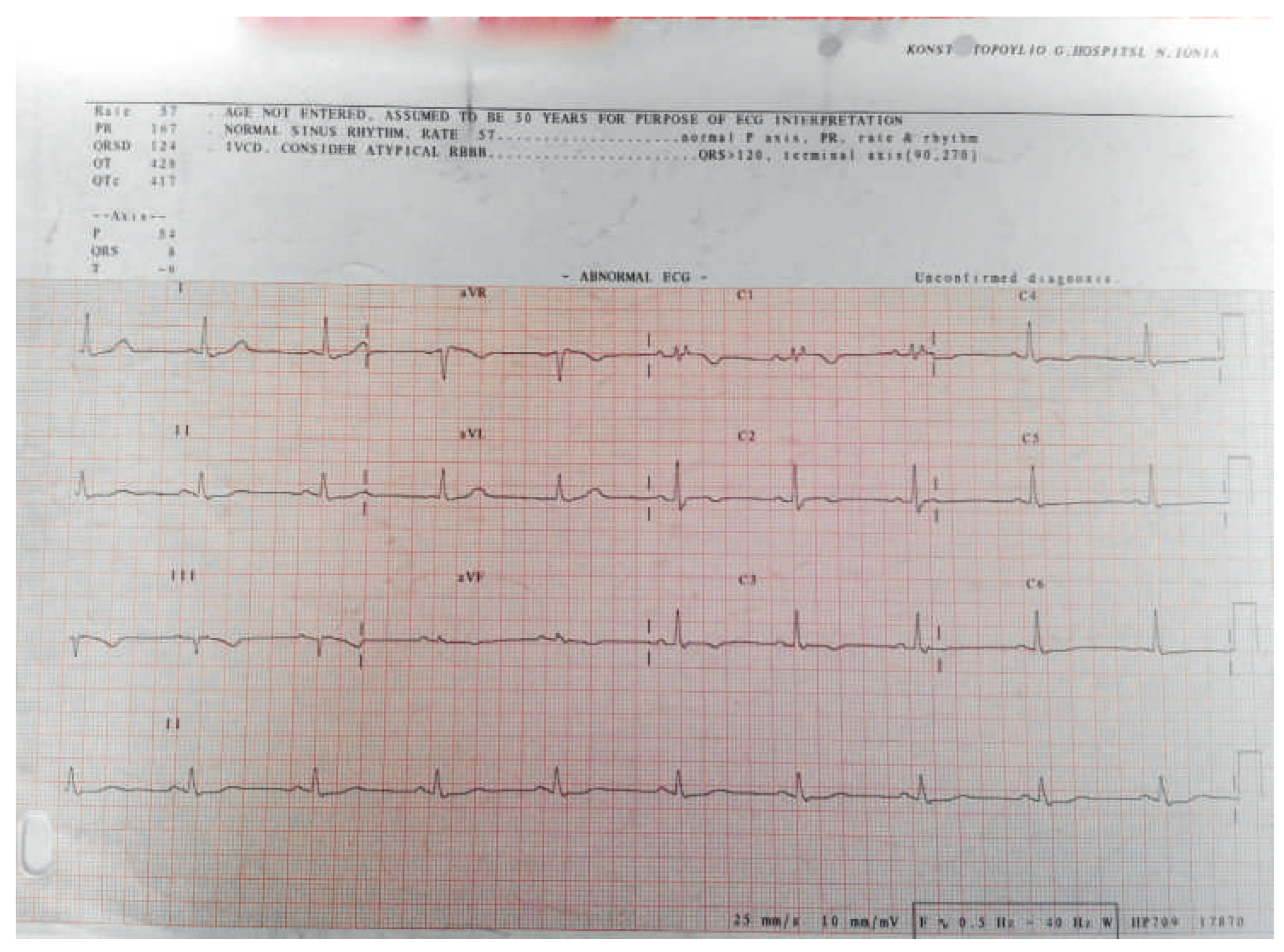
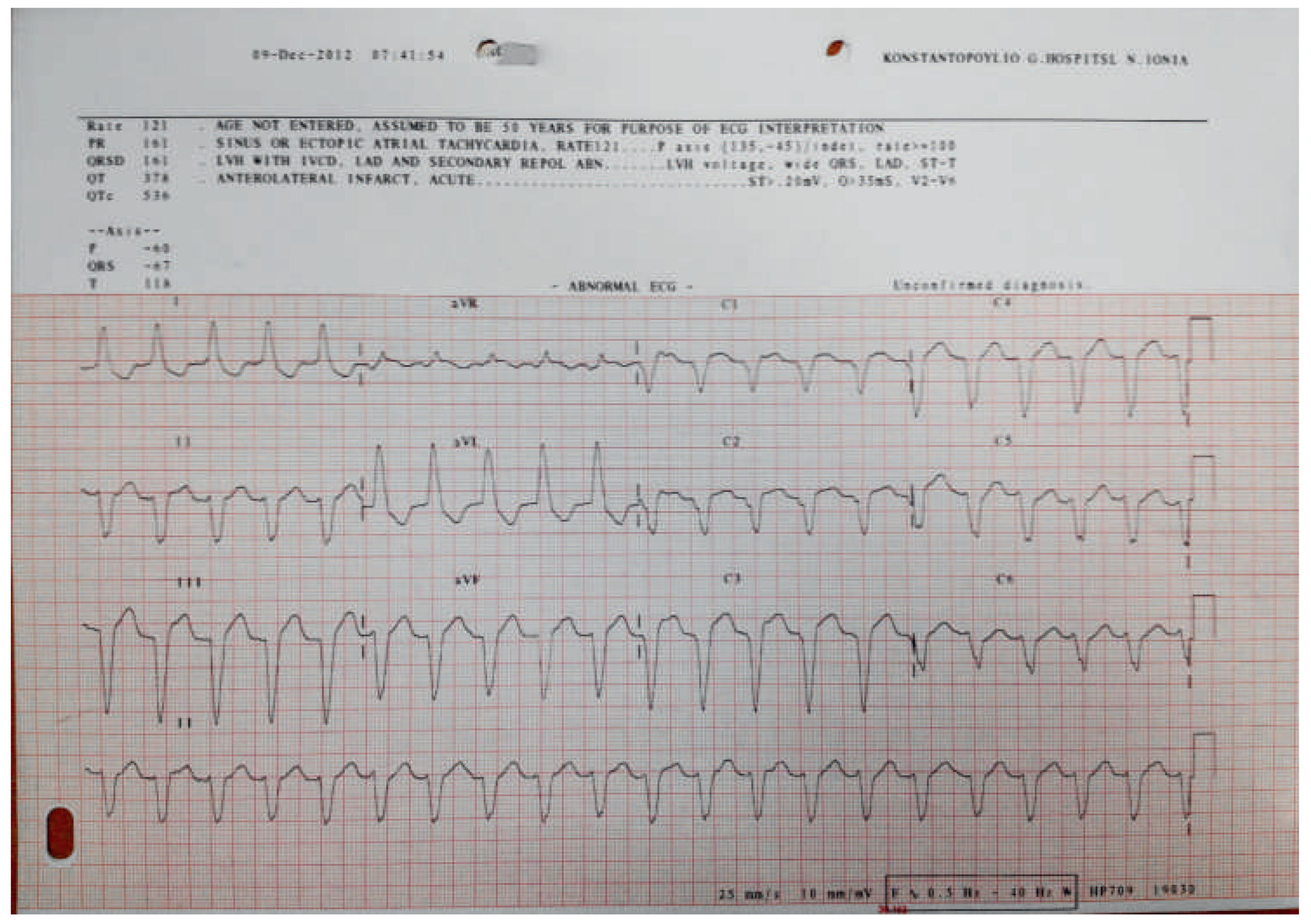
An 80-year-old female Caucasian patient presented at the emergency room (ER) with dizziness and presyncope. The 12-lead surface ECG (25 mm/s, 10 mm/mV) was as follows (Figure 1). A sustained regular monomorphic wide QRS complex tachycardia with left bundle branch block morphology and superior axis at a rate of 118 beats per minute was diagnosed. The patient was haemodynamically stable with arterial blood pressure 120/80 mm Hg, SatO2 99% and normal breathing rate. She was treated with an intravenous infusion of amiodarone and after 30 minutes the 12-lead surface ECG became as follows (Figure 2: 25 mm/s, 10 mm/mV).

Question

Which disease is most likely according to the two 12- lead ECGs shown?

Discussion

This patient was diagnosed in her early 60s with arrhythmogenic right ventricular cardiomyopathy (ARVC/D) fulfilling 5 major and 2 minor criteria according to the revised 2010 ARVC/D task force criteria (TFC), namely regional right ventricular dyskinesia (parasternal long-axis view RVOT-PLAX >32 mm) by 2D echocardiogram (major criterion), inverted T waves in right precordial leads V1–V3 in an adult >14 years old in the absence of right bundle branch block (with QRS wave >120 ms; major criterion), Epsilon waves (major criterion), non-sustained and sustained ventricular tachycardias with left bundle block morphology and superior axis (major criterion), and a terminal activation of duration ≥55 ms in V1–V3 measured from the nadir of the S wave to the end of the QRS complex, including R’ in V1,V2 or V3 in the absence of complete right bundle branch block (minor criterion). This patient also had regional right ventricular akinesia and dyssynchronous right ventricular contraction with right ventricular end-diastolic volume (RVEDV/BSA) ≥100 ml/m2 in cardiac MRI (major criterion) and right ventricular ejection fraction >40% and ≤45% in cardiac MRI (minor criterion). Due to previous ventricular tachycardia episodes, an ICD had been previously implanted. In this case, the ICD did not deliver any therapy, because the tachycardia rate shown in Figure 1, which corresponds to a ventricular tachycardia from the right ventricle and fulfils a major diagnostic criterion according to the TFC, was lower than the lower rate the device had been set to deliver anti-tachycardia pacing shocks.
Due to frequent ventricular tachycardia episodes, the patient was prescribed mexiletine per os and was discharged.
ARVC/D is a genetic form of cardiomyopathy, which is characterised by fibrofatty infiltration of the myocardium of the right ventricle. It is associated with up to 22% of sudden cardiac death cases in young athletes, and usually presents between adolescence and the age of 40. However, about 10% of the cases are diagnosed beyond this age range. Other diseases that may mimic ARVC/D, such as cardiac sarcoidosis and localised forms of myocarditis, have to be excluded, which can sometimes be very challenging. Genetic screening for desmosomal mutations and family screening is often necessary to confirm ARVC/D.
The characteristic 12-lead surface ECG findings in the absence of arrhythmias are:
-
A terminal activation of duration ≥55 ms in V1–V3 as shown in Figure 2 (measured from the nadir of the S wave to the end of the QRS complex, including R’, in V1, V2 or V3 in the absence of complete right bundle block; minor diagnostic criterion according to TFC).
-
Epsilon waves as shown in Figure 2 (arrow; reproducible low-amplitude signal between the end of the QRS complex to the onset of the T wave in V1–V3; major diagnostic criterion according to TFC).
-
T wave inversions in V1, V2 and V3 or beyond in individuals older than 14 years of age as shown in Figure 2.

Funding/potential competing interests
No financial support and no other potential conflict of interest relevant to this article were reported.
Figure 1. ECG recording when the patient was symptomatic.
Cardiovascmed 17 00189 g001
Figure 2. ECG recording when the patient recovered from the wide complex tachycardia.
Cardiovascmed 17 00189 g002