Next Article in Journal
Book Review: Eugene Braunwald und die Moderne Medizin
Previous Article in Journal
Extrakorporale Membranoxygenierung (ECMO) bei Patienten mit Akutem Koronarsyndrom als Unterstützung bei Primärer PCI
 
 
Cardiovascular Medicine is published by MDPI from Volume 28 Issue 1 (2025). Previous articles were published by another publisher in Open Access under a CC-BY (or CC-BY-NC-ND) licence, and they are hosted by MDPI on mdpi.com as a courtesy and upon agreement with Editores Medicorum Helveticorum (EMH).
Interesting Images

Which Arrhythmia Would You Prefer?

by
Peter Jäggi
and
Stephan Andreas Müller-Burri
*
Klinik für Kardiologie, Stadtspital Triemli, Birmensdorferstrasse, 497 CH-8063 Zürich, Switzerland
*
Author to whom correspondence should be addressed.
Cardiovasc. Med. 2014, 17(5), 156; https://doi.org/10.4414/cvm.2014.00243
Submission received: 28 February 2014 / Revised: 28 March 2014 / Accepted: 28 April 2014 / Published: 28 May 2014

Case report

A 92-year-old female was admitted to the hospital because of paroxysmal episodes of weakness and shortness of breath. She had a history of arterial hypertension and had been treated with sotalol and verapamil for many years for paroxysmal supraventricular tachycardia (SVT). In the emergency room, repeated shortlasting episodes of a narrow complex tachycardia (Figure 1) occurred reproducing the patient’s symptoms. Administration of adenosine resulted in conversion of the tachycardia to sinus rhythm. What is the diagnosis and how should this patient be treated?

Discussion

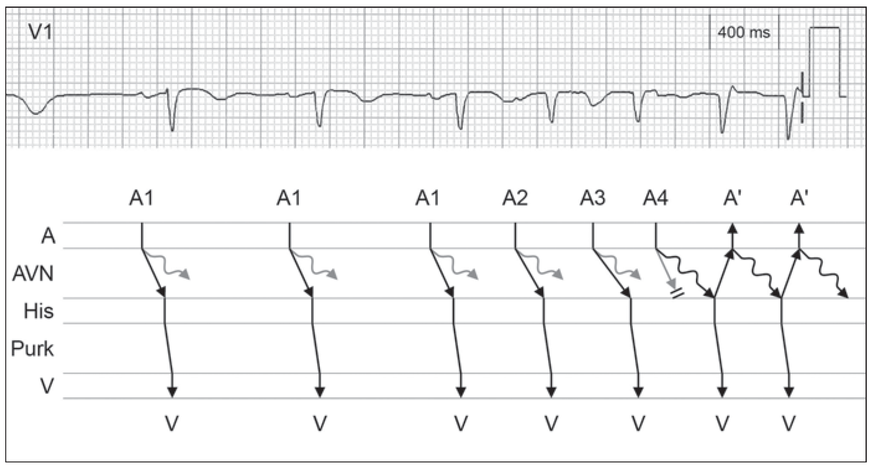
The ECG at the onset of the tachycardia (Figure 1A) shows three premature atrial complexes (PAC) with a significant prolongation of the PQ interval after the third PAC. The following QRS complex is the first beat of a regular narrow complex tachycardia (Figure 1B). During tachycardia the morphology of QRS complex slightly changes showing an Rr’ configuration in leads V1. The second r’ in V1 represents the retrograde activation of the atrium inscribed at the end of the QRS complex (Figure 2). These findings as well as the response to adenosine are compatible with the diagnosis of a typical AV nodal re-entry tachycardia (AVNRT).
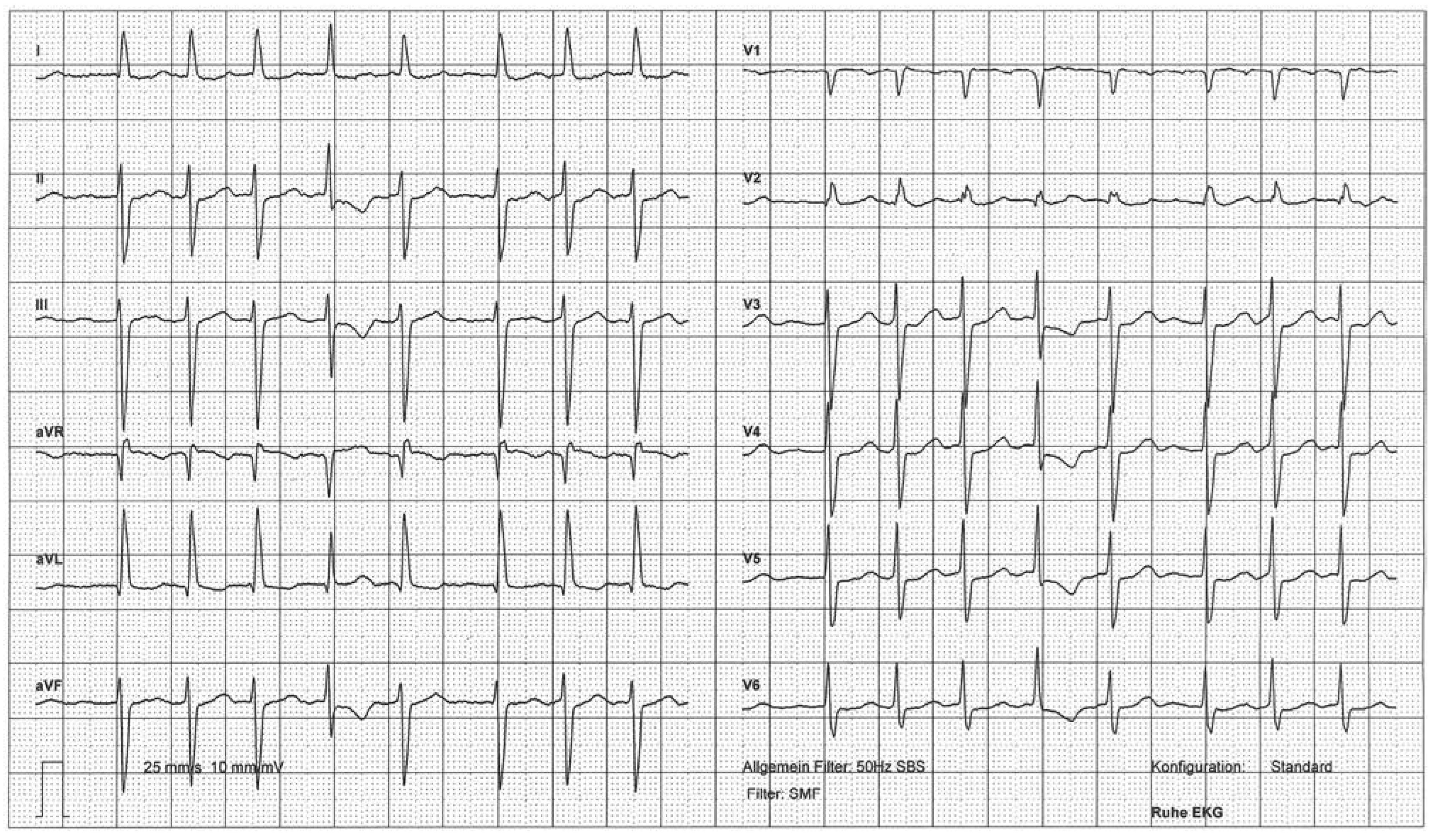
Considering the ineffectiveness of the dual anti-arrhythmic therapy with sotalol and verapamil, an electrophysiological study (EPS) and catheter ablation was discussed with the patient. In preparation the two anti-arrhythmics were replaced by a low dose of bisoprolol. The next day, the patient was asymptomatic, had a normal but irregular heart rate, and the ECG showed atrial fibrillation (AF) (Figure 3). Because of the high likelihood of recurrence and the absence of symptoms in AF, the patient was discharged on rate control therapy and oral anticoagulation.
The association between AF and circus movement tachycardia using an accessory pathway is well known [1]. However, every organised SVT (e.g., atrial flutter, focal atrial tachycardia, AVNRT, etc.) can degenerate to AF [2]. The very fast and chaotic atrial activation during AF prevents diagnosis and mapping during the EPS but also the recurrence of the regular SVT.
This 92-year-old female suffering from highly symptomatic AVNRT was effectively treated by the occurrence of an asymptomatic permanent AF. As in this case, sufficient rate control in AF can often be achieved easily while pharmacological rhythm control of SVT is difficult and less effective [3].

Funding/potential competing interests

No financial support and no other potential conflict of interest relevant to this article were reported.

References

  1. Centurión, O.A.; Shimizu, A.; Isomoto, S.; Konoe, A. Mechanisms for the genesis of paroxysmal atrial fibrillation in the Wolff Parkinson White syndrome: intrinsic atrial muscle vulnerability vs. electrophysiological properties of the accessory pathway. Europace 2008, 10, 294–302. [Google Scholar] [CrossRef] [PubMed]
  2. Lip, G.Y.; Tse, H.F.; Lane, D.A. Atrial fibrillation. Lancet 2012, 379, 648–661. [Google Scholar] [CrossRef] [PubMed]
  3. Camm, A.J.; Lip, G.Y.; De Caterina, R.; Savelieva, I.; Atar, D.; Hohnloser, S.H.; et al. 2012 focused update of the ESC Guidelines for the management of atrial fibrillation: an update of the 2010 ESC Guidelines for the management of atrial fibrillation. Developed with the special contribution of the European Heart Rhythm Association. Eur. Heart J. 2012, 33, 2719–2747. [Google Scholar] [CrossRef] [PubMed]
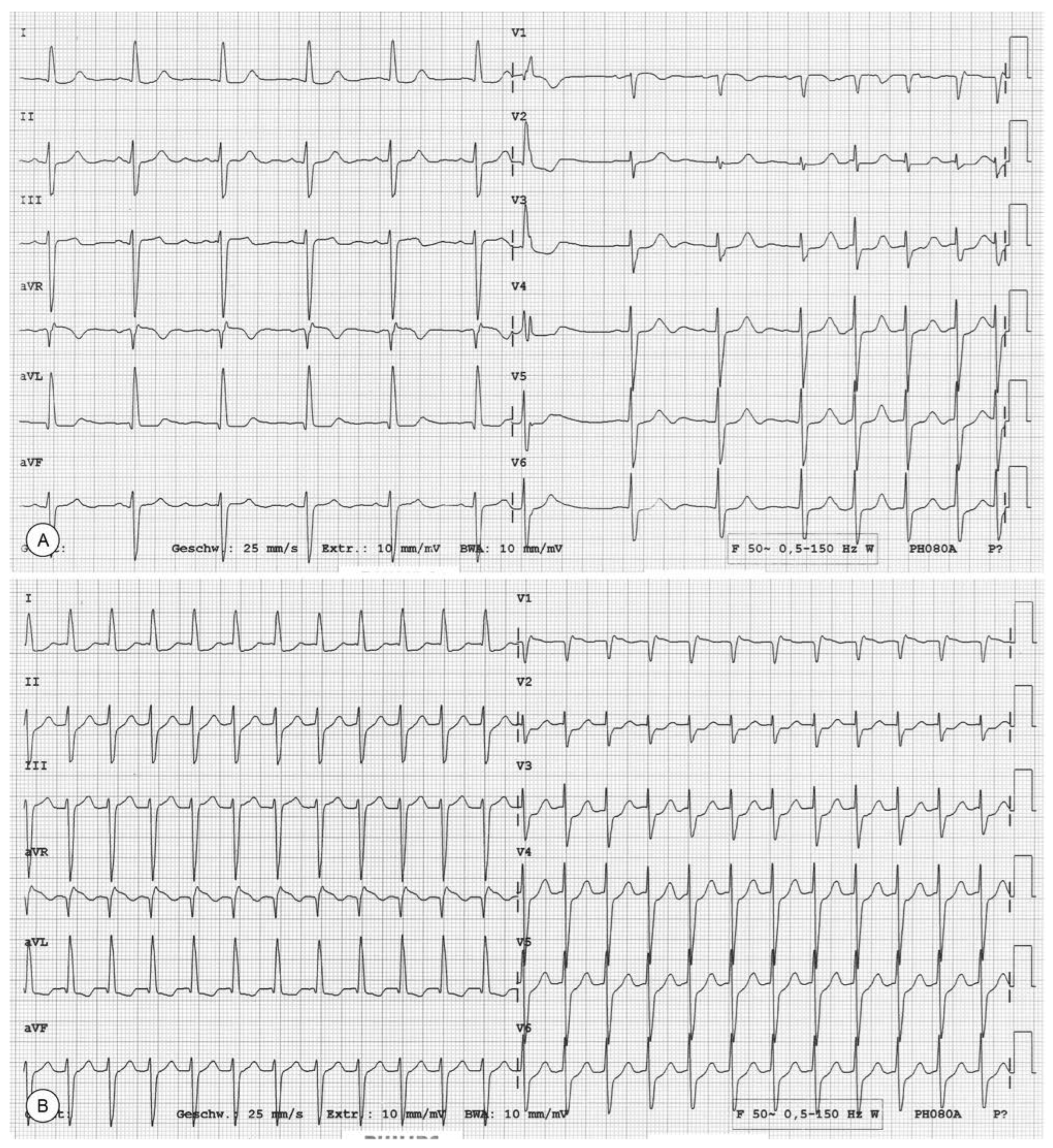
Figure 1. Onset of the clinical tachycardia. Initiation of the tachycardia by three consecutive premature atrial complexes (PAC) (A). Ongoing regular narrow QRS complex tachycardia (heart rate 143 bpm, cycle length 420ms) (B).
Cardiovascmed 17 00156 g001
Figure 2. Mechanism of AV nodal reentry tachycardia. The ECG tracing of lead V1 and the corresponding ladder diagram illustrate the jump of the AV-conduction from the fast pathway during sinus rhythm (A1) and the first two premature atrial complexes (A2, A3) to the slow pathway after the third PAC (A4). At the time this impulse reaches the AV node (AVN), the fast pathway has recovered its excitability and conducts the impulse back to the atria (A’). From here return conduction over the slow pathway to the AV node occurs and initiates the AV nodal reentry tachycardia. A denotes atrium, Purk Purkinje system, V ventricle.
Cardiovascmed 17 00156 g002
Figure 3. Atrial fibrillation. The diagnosis of atrial fibrillation is based on the absolute arrhythmia of the QRS complexes and the fast and changing P waves.
Cardiovascmed 17 00156 g003