Next Article in Journal
Coronary Spasm Provocation Testing: Still Useful?
Previous Article in Journal
Quantification of Differential ECMO Return Flow Through an Axillary Artery Anastomosis Graft with Spectral Doppler Echocardiographyphy
 
 
Cardiovascular Medicine is published by MDPI from Volume 28 Issue 1 (2025). Previous articles were published by another publisher in Open Access under a CC-BY (or CC-BY-NC-ND) licence, and they are hosted by MDPI on mdpi.com as a courtesy and upon agreement with Editores Medicorum Helveticorum (EMH).
Interesting Images

Unusual Complication of Transradial Coronary Angiography

by
Panneerselvam Arunkumar
Consultant Cardiologist, Department of Cardiology, Eugene Clinic, Mettupalayam 641301, India
Cardiovasc. Med. 2014, 17(2), 54; https://doi.org/10.4414/cvm.2014.00214
Submission received: 19 November 2013 / Revised: 19 December 2013 / Accepted: 19 January 2014 / Published: 19 February 2014

Case report

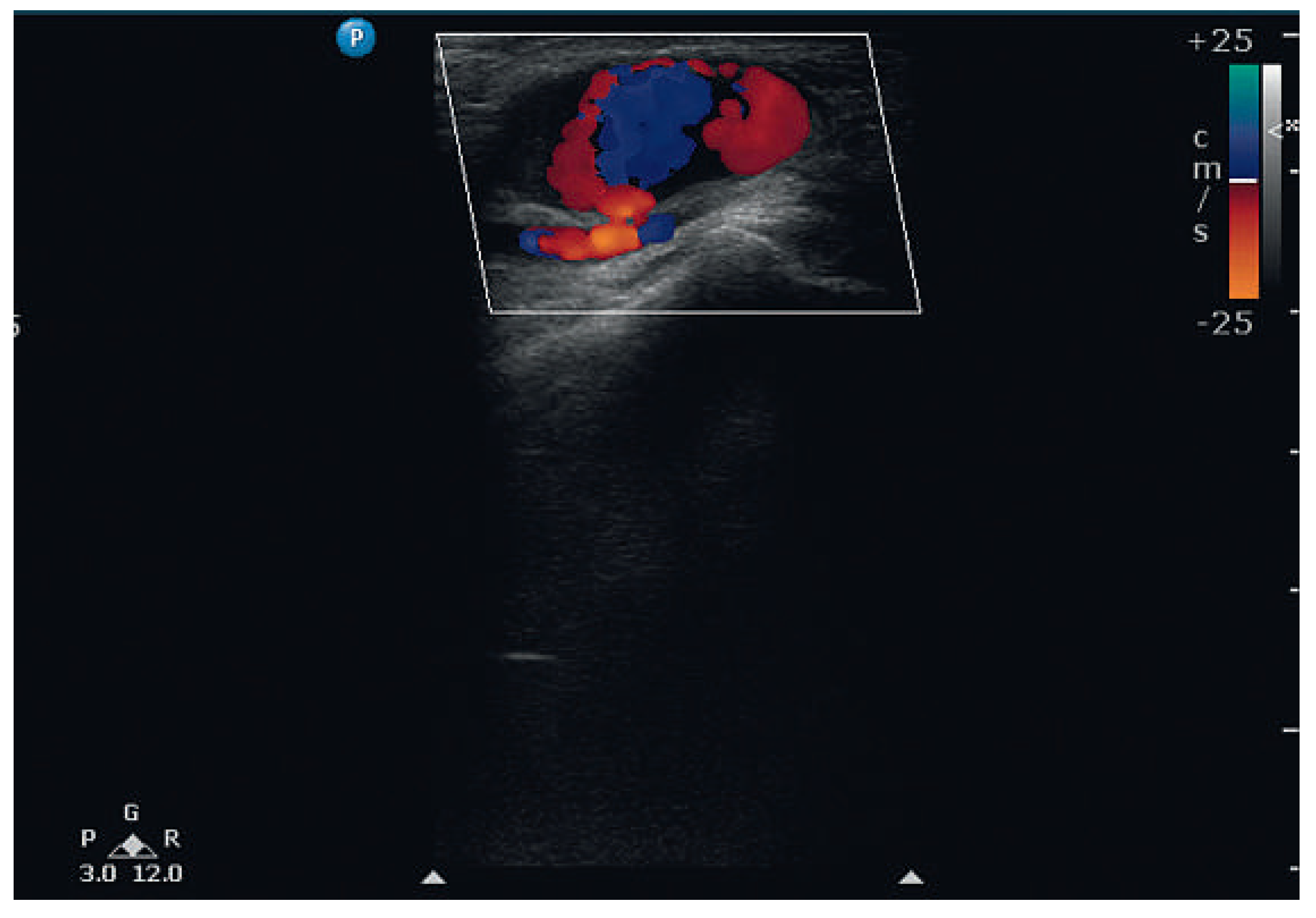
A 68-year-old male with diabetes and hypertension underwent a coronary angiogram, through the right radial artery approach, because of inducible ischaemia on stress testing. He had significant triple vessel disease for which revascularisation was advised. Two weeks later he presented with progressively increasing swelling at the radial puncture site. A 2.4 × 1.3 cm pulsatile swelling was seen (Figure 1, movie 1 (You can find the movies on: http://www.cardiovascmed.ch/for-readers/multimedia)). The skin over the swelling was stretched and shiny. Colour Doppler imaging revealed a pseudo-aneurysm originating from radial artery through a 3 mm neck (Figure 2, movie 2 (You can find the movies on: http://www.cardiovascmed.ch/for-readers/multimedia)). There was no evidence of infection or distal thromboembolism. On application of firm manual pressure for 15 minutes followed by a pressure bandage the pseudoaneurysm was completely obliterated.
Radial artery pseudo-aneurysm is an uncommon complication of transradial interventionw. Inadequate application of pressure following the coronary angiogram is the probable cause of this pseudoaneurysm. Early recognition and treatment is important to prevent infection, thromboembolism to the digits and rupture.

Funding / potential competing interests

No financial support and no other potential conflict of interest relevant to this article was reported.
Figure 1. The pulsatile swelling at the radial artery puncture site in the right forearm. The skin is stretched and shiny.
Cardiovascmed 17 00054 g001
Figure 2. Colour Doppler imaging showing a pseudo-aneurysm arising from radial artery. The prominent styloid process of the radius can also be seen.
Cardiovascmed 17 00054 g002