Next Article in Journal
Surviving Acute Traumatic Transection of the Ascending Aorta and Proximal Aortic Arch
Previous Article in Journal
Fishing for P-Waves During Wide-Complex Tachycardia
 
 
Cardiovascular Medicine is published by MDPI from Volume 28 Issue 1 (2025). Previous articles were published by another publisher in Open Access under a CC-BY (or CC-BY-NC-ND) licence, and they are hosted by MDPI on mdpi.com as a courtesy and upon agreement with Editores Medicorum Helveticorum (EMH).
Case Report

"Congenital" Chest Pain–Anomalous Origin of the Left Anterior Descending Coronary Artery from the Pulmonary Artery

by
Jens Robert
1,
Alexander Kadner
2,
Stephan Windecker
3,
Andrea Rosskopf
4,
Bernhard Meier
3 and
Markus Schwerzmann
2,*
1
Kardiologie, Solothurner Spitäler AG, 4500 Solothurn, Switzerland
2
Congenital Heart Disease Center, University Hospital, Inselspital, 3010 Bern, Switzerland
3
Department of Cardiology, University Hospital, Inselspital, 3010 Bern, Switzerland
4
Department of Radiology, University Hospital, Inselspital, 3010 Bern, Switzerland
*
Author to whom correspondence should be addressed.
Cardiovasc. Med. 2013, 16(9), 247; https://doi.org/10.4414/cvm.2013.00179
Submission received: 25 June 2013 / Revised: 25 July 2013 / Accepted: 25 August 2013 / Published: 25 September 2013

Abstract

Anomalous origin of the Left main Coronary Artery from the Pulmonary Artery (also called ALCAPA or Bland-White-Garland syndrome) is a rare finding with an often fatal natural history due to left ventricular ischaemia. This case report describes the late diagnosis and therapy of an anomalous origin of the left anterior descending coronary artery from the pulmonary artery. The natural history and therapeutic options of an anomalous left coronary artery origin from the pulmonary artery, be it a partial (as in our case) or a complete “ALCAPA”, are discussed.

1. Case Report

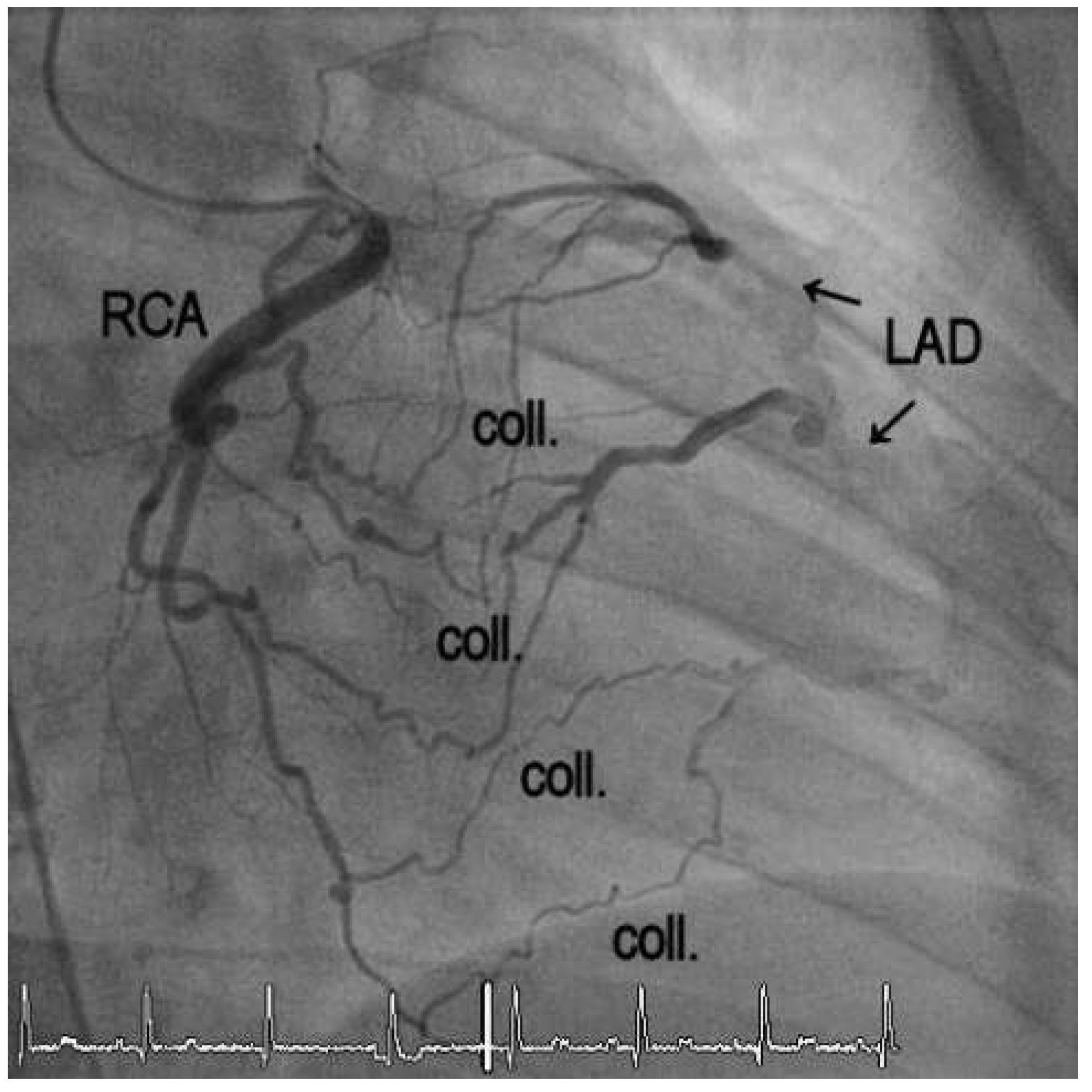
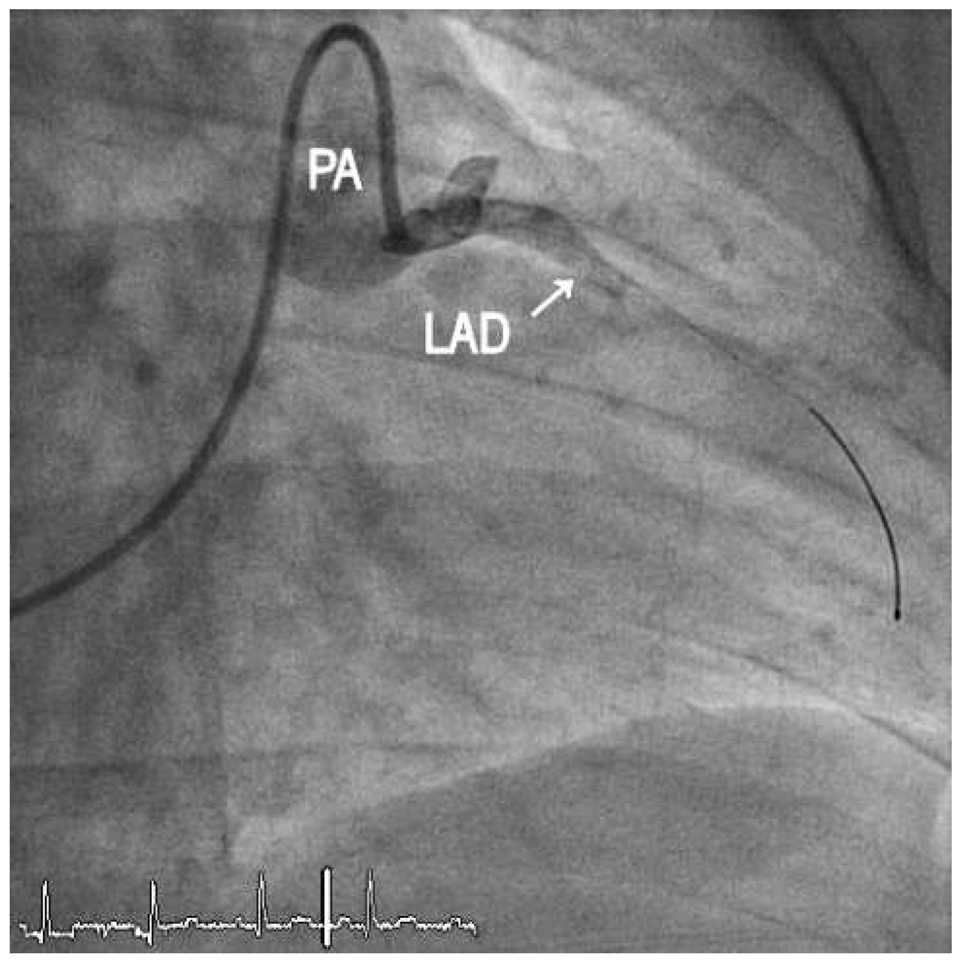
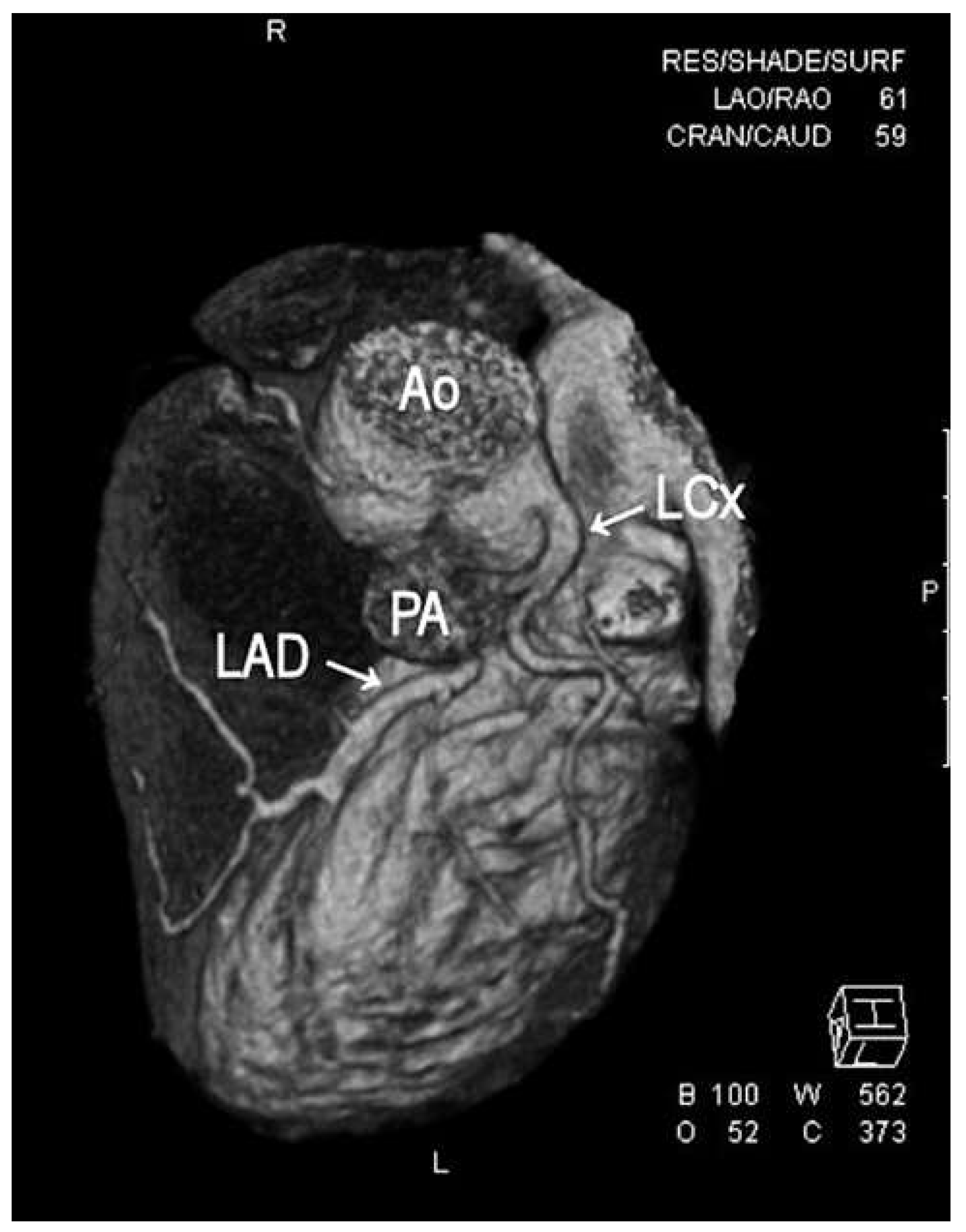
We report the case of a 27-year-old woman with a history of chest pain on exertion for several months. She had undergone surgical ligation of a patent ductus arteriosus (PDA) by the age of 12 in another country. Given her history of congenital heart disease, she was directly referred to our centre for congenital heart disease for further work-up. She had a history of two uneventful pregnancies, two and four years previously, with uncomplicated deliveries. On auscultation, there was a continuous murmur best heard on the back and at the left sternal border. During a standard exercise treadmill test, she reported chest pain at peak exercise, accompanied by >2 mm ST-segment depression in leads V4–6 (Figure 1). Transthoracic echocardiography showed normal biventricular size and function. Left ventricular diastolic and systolic function was preserved with no regional wall motion abnormalities. On colour Doppler, there was no sign of a residual PDA. The aortic valve was tricuspid with trace regurgitation. Further, Doppler flow signals were observed in the interventricular septum. A coronary fistula was presumed. Echo quality was insufficient to visualise the origin and size of the left and right coronary arteries in the parasternal short-axis view. Because of the positive exercise test and the suspicion of coronary fistulae, coronary angiography was performed. On angiography, the left circumflex (LCx) and the right coronary arteries (RCA) had their usual origins in the aortic root, but the left anterior descending coronary artery (LAD) was retrogradely perfused via contralateral collaterals from the RCA (Figure 2). The RCA was enlarged. The origin of the LAD could be selectively intubated with a 5 French left Amplatz catheter in the anterior aspect of the main pulmonary artery (Figure 3). Computed tomography confirmed the pathoanatomical situation (Figure 4).
Because of the extensive coronary collaterals between the RCA and LAD, our first approach was to improve retrograde LAD coronary artery flow by percutaneous occlusion of its pulmonary origin, thereby preventing coronary steal into the low pressure pulmonary circulation. This concept was based on pathophysiological considerations. There was no available literature that discussed treatment options for this rare clinical entity. Despite several manoeuvres (stabilisation of the guiding catheter at the pulmonary origin by a Magnum or Terumo wire placed in the LAD, inflation of a Maverick balloon in the LAD), it was not possible to create sufficient catheter back-up to plug the coronary ostium. A few weeks later, the patient underwent cardiac surgery with reinsertion of the LAD into the aorta by means of a venous interponate. The distance of the LAD origin from the aortic sinus did not allow direct reimplantation of the coronary vessel. The postoperative course was uncomplicated. Treadmill exercise testing was uneventful 3 and 24 months after the surgery and the patient remained free of symptoms.

2. Discussion

Anomalous origin of a coronary artery from the pulmonary artery is rare, and its incidence is not known. The most common of these anomalies is the origin of the left main coronary artery from the pulmonary artery (ALCAPA), followed by the origin of the single RCA or the LAD from the pulmonary artery, as shown by Yamanaka et al. in an analysis of 126,595 coronary angiographies between 1960 and 1988 [1]. Between 1971 and 1991, only 15 cases of a LAD from the pulmonary artery were reported [2], with an additional 20 more cases in the past 20 years.
In patients with ALCAPA, fatal left ventricular infarction, congestive heart failure and sudden cardiac death are common [3]. These adverse events are less severe or even absent in patients with only the LAD originating from the pulmonary artery, probably because less myocardium is in jeopardy from retrograde myocardial perfusion. As a result of decreased pressure in the LAD originating from the low pressure pulmonary artery system, reversed coronary flow develops with myocardial ischaemia promoting collateral vessel formation. Depending on the amount of collateral flow and pulmonal arterial resistance, some patients remain asymptomatic until adulthood. With this in mind, the presence of a PDA in the first years of our patient’s life may have contributed to a mild clinical course. Now, in adult life with new symptoms and objective myocardial ischaemia, further therapy became mandatory.
For patients with ALCAPA, the aim of surgery is to establish a two-coronary system with antegrade flow. The best results were shown for direct reimplantation of the anomalous coronary artery into the aorta. Other surgical options include ligation of the pulmonary origin of the anomalous coronary artery and, usually, grafting of the vessel with a saphenous vein graft or internal mammary artery graft. The Takeuchi procedure includes reinsertion of the anomalous coronary artery in the aorta by creating a transpulmonary tunnel [4]. So far, the optimal surgical option in patients with an isolated anomalous origin of the LAD from pulmonary artery is unclear.

Funding

No financial support.

Conflicts of Interest

No other potential conflict of interest relevant to this article was reported.

References

  1. Yamanaka, O.; Hobbs, R.E. Coronary artery anomalies in 126,595 patients undergoing coronary arteriography. Cathet Cardiovasc. Diagn. 1990, 21, 28–40. [Google Scholar] [CrossRef] [PubMed]
  2. Derrick, M.J.; Moreno-Cabral, R.J. Anomalous origin of the left anterior descending artery from the pulmonary artery. J. Card. Surg. 1991, 6, 24–28. [Google Scholar] [CrossRef] [PubMed]
  3. Wesselhoeft, H.; Fawcett, J.S.; Johnson, A.L. Anomalous origin of the left coronary artery from the pulmonary trunk. Its clinical spectrum, pathology, and pathophysiology, based on a review of 140 cases with seven further cases. Circulation 1968, 38, 403–425. [Google Scholar] [CrossRef] [PubMed]
  4. Azakie, A.; Russell, J.L.; McCrindle, B.W.; Van Arsdell, G.S.; Benson, L.N.; Coles, J.G.; et al. Anatomic repair of anomalous left coronary artery from the pulmonary artery by aortic reimplantation: Early survival, patterns of ventricular recovery and late outcome. Ann. Thorac. Surg. 2003, 75, 1535–1541. [Google Scholar] [CrossRef] [PubMed]
Figure 1. Exercise electrocardiogram showing the development of ST-segment-depressions in leads V4–6 in the early recovery time.
Cardiovascmed 16 00247 g001
Figure 2. Angiogram of the right coronary artery (RCA) with a 4 French left Amplatz 4 catheter showing the collaterals (coll.) from the RCA to the left anterior descending coronary artery (LAD).
Cardiovascmed 16 00247 g002
Figure 3. Angiogram of the left anterior descending coronay artery (LAD) via the pulmonary artery (PA) with a 0.014 inch Magnum wire placed in the LAD.
Cardiovascmed 16 00247 g003
Figure 4. Three-dimensional reconstruction of the coronary computed tomography angiography showing the origin of the left anterior descending coronary artery (LAD) from the pulmonary artery (PA) and the origin of the left circumflex coronary artery (LCx) from the aorta (Ao).
Cardiovascmed 16 00247 g004