Case Report
A 65-year-old male presented with acute anterior wall myocardial infarction for which he received fibrinolytic therapy. In addition he was given 1 mg/kg enoxaparin twice daily. On the 3rd day of hospitalisation he underwent coronary angiography through the right radial approach and received 70 U/kg heparin intra-arterially. The angiogram revealed residual 70% stenosis of the proximal left anterior descending artery and the patient was advised to undergo myocardial perfusion imaging during follow-up to determine revascularisation. He was discharged on the 5th day. Two days later he presented with pain and swelling of the right upper limb. Multiple blisters were noted at the right radial puncture site (Figure 1). Similar blisters were seen at the sites of subcutaneous enoxaparin injections. There was warmth, tenderness and induration of the right forearm but no compartmental syndrome. The platelet count fell by 30% from baseline. Heparininduced skin necrosis was provisionally diagnosed. As there was evidence of superficial venous thrombosis involving the entire right upper limb, the patient was started on oral anticoagulant to achieve therapeutic anticoagulation. In addition he received optimal doses of aspirin, atorvastatin, ACE inhibitor and beta blocker. The blisters subsequently ruptured and the necrosed skin areas healed gradually over a period of 4 weeks without the need for skin grafting. Heparininduced skin necrosis due to intraarterial heparin injection in a radial artery can lead to significant limb oedema with blisters and possible limb loss. Prompt recognition and treatment can be limband lifesaving.
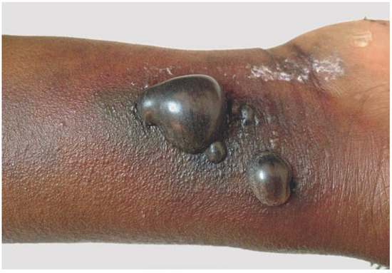
Figure 1.
Blisters seen at the site of radial puncture on the 7th day after initiation of heparin therapy.
Funding
No financial support.
Conflicts of Interest
No other potential conflict of interest relevant to this article was reported.
© 2013 by the author. Attribution - Non-Commercial - NoDerivatives 4.0.