Next Article in Journal
Changes in Ejection Fraction After an Ablate and Pace Procedure in Patients with Atrial Fibrillation
Previous Article in Journal
Assessment of Myocardial Viability After the STICH-Trial: Still Viable?
 
 
Cardiovascular Medicine is published by MDPI from Volume 28 Issue 1 (2025). Previous articles were published by another publisher in Open Access under a CC-BY (or CC-BY-NC-ND) licence, and they are hosted by MDPI on mdpi.com as a courtesy and upon agreement with Editores Medicorum Helveticorum (EMH).
Case Report

Endovascular Treatment of a Complex Type B Aortic Dissection

Department of Vascular Surgery, the Second Affiliated Hospital of Suzhou University, Suzhou, China
*
Author to whom correspondence should be addressed.
Cardiovasc. Med. 2013, 16(10), 267; https://doi.org/10.4414/cvm.2013.00187
Submission received: 23 July 2013 / Revised: 23 August 2013 / Accepted: 23 September 2013 / Published: 23 October 2013

Summary

Aortic dissection is a rare disease with high morbidity and mortality, especially when involving multiorgan malperfusion such as lower extremity and visceral ischaemia. Revascularisation should be performed to restore perfusion immediately. We report a case of acute Stanford type B aortic dissection complicated by lower extremity, renal and superior mesenteric ischaemia. The right iliac artery was occluded because of compression of the false lumen, resulting in thrombosis. The patient was successfully treated with endovascular treatments, including thrombus extraction and iliac artery, superior mesentic artery and aortic stenting combined with fenestration.

Introduction

Aortic dissection is a rare disease with an estimated incidence ranging from 2.6–3.5 cases per 100,000 person/year [1]. Approximately two-thirds of patients with aortic dissection are male, with an average age of 65 years. Almost 72% of patients have a history of hypertension. The morbidity and mortality of acute type B aortic dissection are often related to the patient's clinical condition and presentation, and are increased by limb, renal or visceral ischaemia, aortic rupture, shock and older age [2]. Aortic dissection inducing multiorgan ischemia is exceptionally rare in the literature. We present a case of an acute aortic dissection (AD) involving the lower extremity arteries, left renal artery and superior mesenteric artery (SMA).

Case report

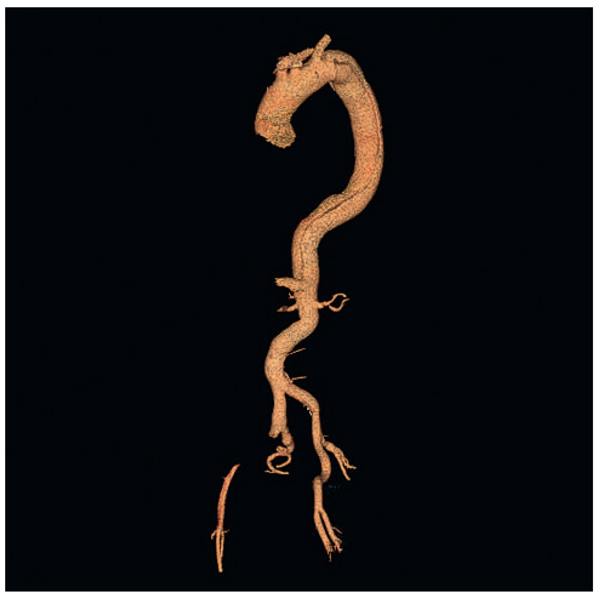
A 56-year-old man presented with an acute onset of lumbar back pain and dyspnoea, accompanied by pain, paleness and coldness of the right lower extremity. In addition, the femoral pulse was absent. The blood pressure was 189/94 mm Hg on admission. Urgent contrast-enhanced computed tomography angiography (CTA) confirmed the diagnosis of acute type B AD, extending from the origin of descending aorta to the bilateral external iliac arteries (Figure 1 and Figure 2). The true lumen of the descending aorta was narrowed. The right external iliac artery was completely occluded (Figure 2), and stenosis was detected in the left renal artery and the SMA. Tests of renal function and electrolytes showed a creatinine of 1.65 mg/dl, and potassium of 5.14 mmol/l.
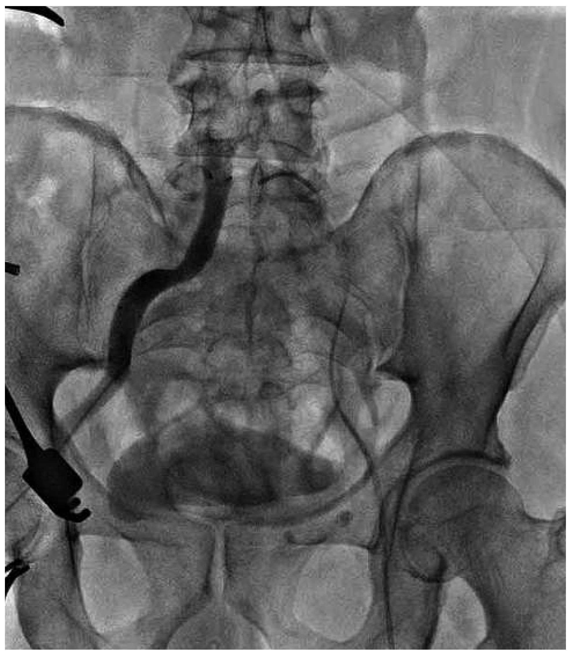
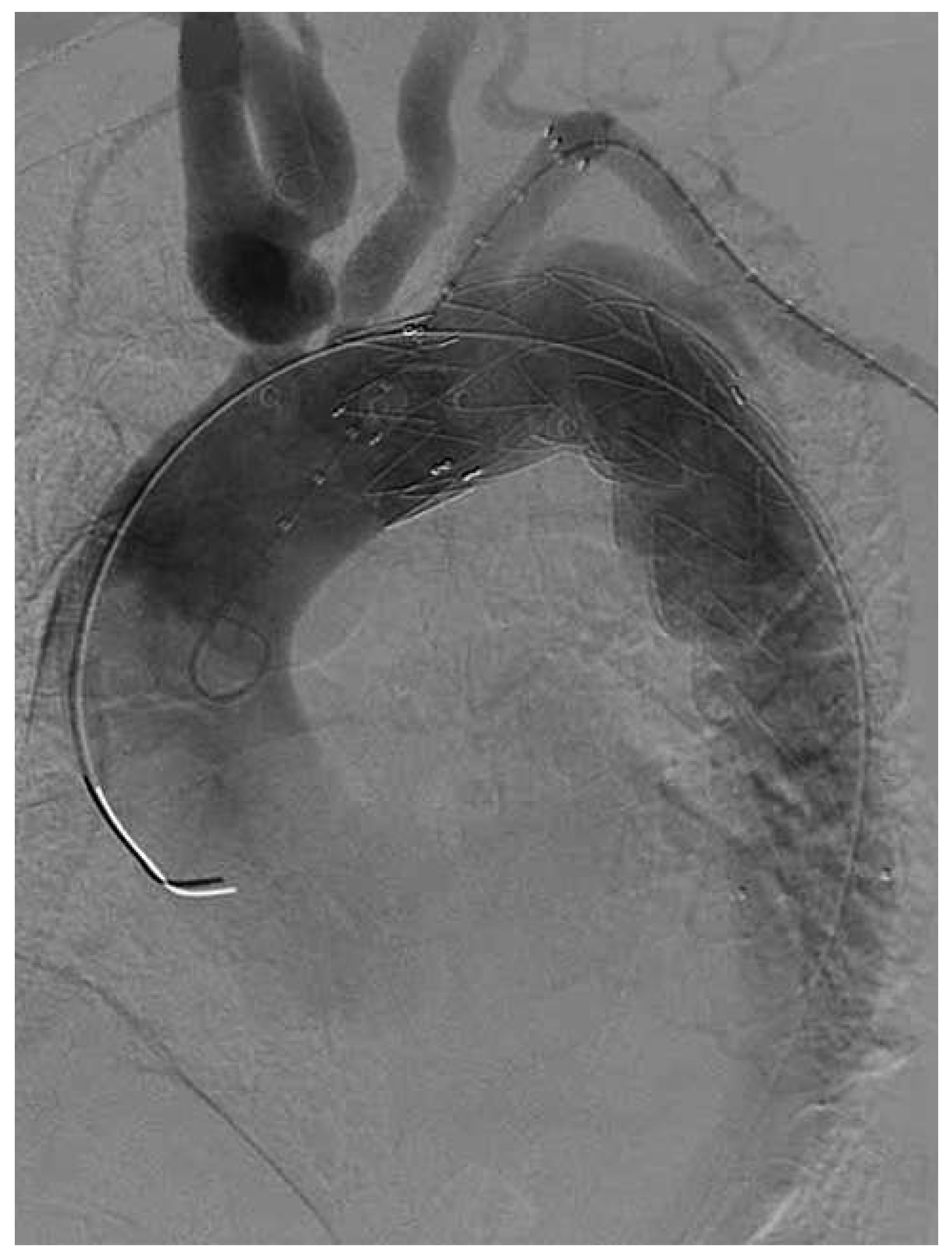
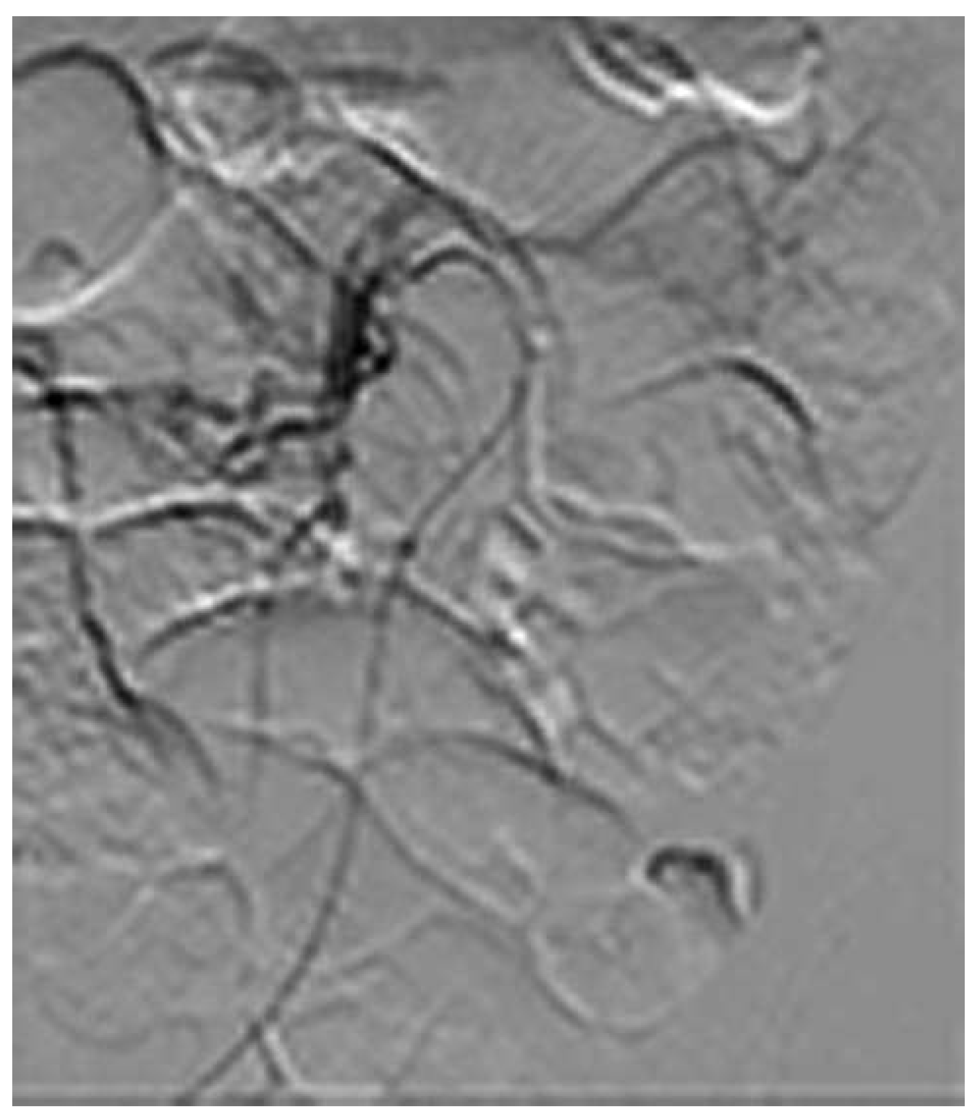
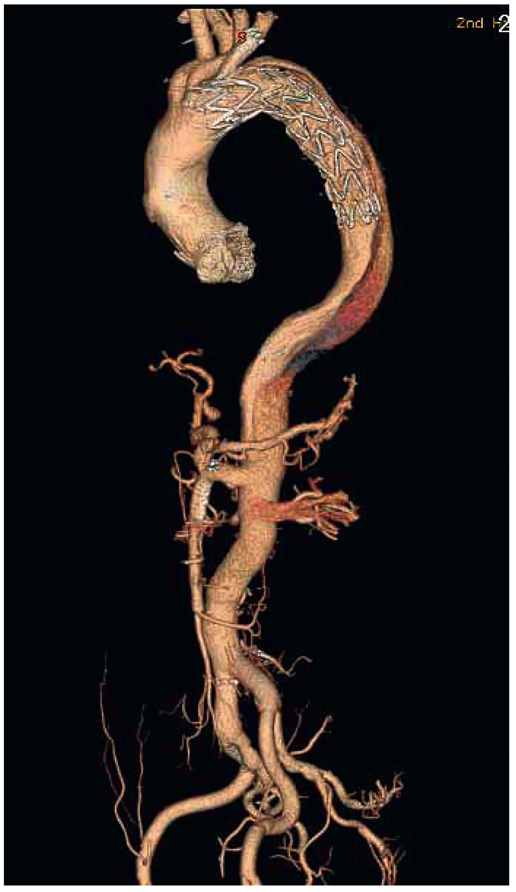
Medical management, including antihypertensive and analgesic drugs, was started immediately. An aortogram revealed that the blood flow in the SMA, bilateral renal artery and right iliac artery were provided by the true lumen, but the left iliac artery was supplied by the false lumen. Thrombosis was found in the right femoral artery. Because of the critical limb ischaemia in the right lower extremity, arteriotomy plus embolectomy with 4F and 5F Fogarty catheters (Edvards Lifesciences llc. Inc.) was performed in the right femoral artery. As we expected, a dark-red thrombus with a length of approximate 6cm was extracted. Subsequently a stent (10mm*80 mm, Bard graft, Inc.) was implanted in the right iliac artery to relieve persistent distal limb ischaemia (Figure 3). This restored blood flow peripheral artery as well as the pulses and the temperature of the skin recovered. However, two days later the patient became anuric, with abdominal colicky pain and distension. The repeated laboratory tests showed a further decline of renal function. The serum creatinine and potassium had increased to 4.55 mg/dl and 5.3 mmol/l, respectively. Endovascular repair (Valiant graft, TF3636C150X, Medtronic Inc.) to seal the intimal tear and fenestration (10mm*40mm, Bard graft, Inc.) in the left subclavian artery were performed (Figure 4). An aortogram after the procedure showed that the true lumen of SMA was severely stenotic because of compression by the false lumen (Figure 5). To correct malperfusion of the intestine, a stent (6mm*40mm, Bard, Inc.) was inserted into SMA. After the patient was sent back to the intensive care unit, the bowel ischaemia improved dramatically. Ten days later, the creatinine had decreased to 1.48 mg/dl. One week later, follow-up CTA showed that the stents were well expanded without restenosis or migration (Figure 6).

Discussion

The more complicated the dissection, the higher rate of mortality. Endovascular repairs including stent grafting and fenestration may be a superior method to achieve better short-term outcomes for the patients who present with life-threatening complications, including malperfusion syndrome, progression of dissection, aneurysmal dilatation, intractable pain, and impending rupture [3]. If there is sudden onset of pain or migrating pain, prompt invasive measures should be considered. Compared with conventional surgical approaches, endovascular procedures provide an effective and less invasive alternative for reperfusion of compromised organs, and have a higher success rate and lower mortality rate [2]. The false lumen can be obliterated by sealing the intimal tear with an aortic stent-graft and protected the from enlarging.
Acute limb ischaemia caused by type B aortic dissection is rare, with a reported incidence of 6%–7.5% [4]. In addition, the cases of lower limb ischaemia are less likely to present with chest and back pain, which makes diagnosis difficult. The blood flow to unilateral or bilateral lower extremities is obstructed, which may be caused by propagation of the dissection flap into the ilio-femoral vessels, and thrombosis of the true lumen or distal embolisation. The occlusion usually appears at the distal aortic or iliac level. Kristofe et al. proposed that lower limb ischaemia could be used as a surrogate marker for more extensive and severe dissection, because mortality was three-fold higher in patients with lower limb ischaemia than those without it [4]. Peripheral revascularisation should be carried out prior to the proximal aortic repair, because acute renal failure or myocardial infarction could occur if there were not a timely and effective intervention. Branch artery stenting and extra-anatomic bypass may be reliable techniques for achieving this goal. In this case, the right iliac artery was occluded, complicated by thrombosis in the distal. Arteriotomy of the femoral artery plus embolectomy was performed promptly. Furthemore, a stent was implanted in the right iliac artery. The distal limb ischaemia was thenrelieved.
Owing to obstruction or dissection of the aortic branches, about one-third of patients with AD develop visceral malperfusion [5]. Williams et al. classified the se obstructions as static, dynamic and mixed [6]. Static obstruction results from static extension of the dissection flap directly into a visceral branch; dynamic obstruction results from prolapse of the dissection flap across the origin of vessel; and the mixed type is consisted of the two above types. Serious visceral malperfusion caused by static obstruction may require visceral stenting to maintain lumen patency. Bare-metal stents may be the best choice for correcting residual distal malperfusion [7]. In our case, abdominal pain gave a hint of mesenteric ischaemia and the aortogram showed static obstruction in the SMA. Thus, a bare-metal stent was placed.
Mesenteric ischaemia or infarction is a rare complication of acute dissection, with an incidence of less than 5% reported previously [5]. Diagnosis of mesenteric ischaemia by means of CTA is significant and specific. Once small bowel or colon infarction occurs, the overall survival rate will be disappointingly low. Diagnosing and correcting suspected visceral malperfusion may prevent end-organ failure and ultimately death.
Although preoperative renal failure in patients with acute type B dissection is a significantly independent risk factor for mortality, the pathophysiology is unclear up to now. In fact, renal failure is not common unless bilateral renal arteries are involved. It is inferred that ischaemia-reperfusion injury and systemic effects of metabolites could be responsible for the renal function deterioration. In our case, the left renal dissection was observed in the preoperative CTA. We did not repair it. However, renal function restored ten days later. In conclusion, complicated type B AD remains a challenging problem. Percutaneous treatments represent a promising alternative to conventional surgical repair.

Funding

No financial support.

Conflicts of Interest

No other potential conflict of interest relevant to this article was reported.

References

  1. Akin, I.; Kische, S.; Ince, H.; Nienaber, C. Penetrating aortic ulcer, intramural hematoma, acute aortic syndrome: when to do what. J Cardiovasc Surg (Torino). 2012, 53 (Suppl. 1), 83–90. [Google Scholar] [PubMed]
  2. Trimarchi, S.; Tolenaar, J.L.; Tsai, T.T.; Froehlich, J.; Pegorer, M.; Upchurch, G.R.; et al. Influence of clinical presentation on the outcome of acute B aortic dissection: evidences from IRAD. J Cardiovasc Surg (Torino). 2012, 53, 161–168. [Google Scholar] [PubMed]
  3. Tsai, T.T.; Fattori, R.; Trimarchi, S.; Isselbacher, E.; Myrmel, T.; Evangelista, A.; et al. Long-term survival in patients presenting with type B acute aortic dissection: insights from the International Registry of Acute Aortic Dissection. Circulation. 2006, 114, 2226–2231. [Google Scholar] [CrossRef] [PubMed]
  4. Henke, P.K.; Williams, D.M.; Upchurch, G.R., Jr.; Proctor, M.; Cooper, J.V.; Fang, J.; et al. Acute limb ischemia associated with type B aortic dissection: clinical relevance and therapy. Surgery. 2006, 140, 532–539. [Google Scholar] [CrossRef] [PubMed]
  5. Di Eusanio, M.; Trimarchi, S.; Patel, H.J.; Hutchison, S.; Suzuki, T.; Peterson, M.D.; et al. Clinical presentation, management, and short-term outcome of patients with type A acute dissection complicated by mesenteric malperfusion: observations from the International Registry of Acute Aortic Dissection. J Thorac Cardiovasc Surg. 2013, 145, 385–390. [Google Scholar] [CrossRef] [PubMed]
  6. Williams, D.M.; Lee, D.Y.; Hamilton, B.H.; Marx, M.V.; Narasimham, D.L.; Kazanjian, S.N.; et al. The dissected aorta: part III. Anatomy and radiologic diagnosis of branch-vessel compromise. Radiology. 1997, 203, 37–44. [Google Scholar] [CrossRef] [PubMed]
  7. Nienaber, C.A.; Kische, S.; Zeller, T.; Rehders, T.C.; Schneider, H.; Lorenzen, B.; et al. Provisional extension to induce complete attachment after stentgraft placement in type B aortic dissection: the PETTICOAT concept. J Endovasc Ther. 2006, 13, 738–746. [Google Scholar] [CrossRef] [PubMed]
Figure 1. Computed tomography angiography showed that the site of the tear was the descending aorta, just past the origin of the subclavian artery.
Cardiovascmed 16 00267 g001
Figure 2. The dissection membrane extended to both external iliac arteries. The true lumen of thedescending aorta was narrowed. The right external iliac artery was completely occluded.
Cardiovascmed 16 00267 g002
Figure 3. Angiogram of the right iliac artery after stent placement showed the flow in the right iliac artery restored.
Cardiovascmed 16 00267 g003
Figure 4. After endovascular repair of the descending aorta and fenestration of the left subclavian artery, an aortogram showed the correct sites and shapes of the two stents.
Cardiovascmed 16 00267 g004
Figure 5. The true lumen of superior mesenteric artery was narrowed.
Cardiovascmed 16 00267 g005
Figure 6. Three-dimensional reformats image one week later showed that the stents were well expanded without restenosis or migration.
Cardiovascmed 16 00267 g006