Next Article in Journal
Prix de la Fondation Suisse de Cardiologie 2011 Preis der Schweizerischen Herzstiftung 2011
Previous Article in Journal
Zusammenarbeitsvertrag Zwischen dem Cardiocentro Ticino und der Universität Sowie dem UniversitätsSpital Zürich
 
 
Cardiovascular Medicine is published by MDPI from Volume 28 Issue 1 (2025). Previous articles were published by another publisher in Open Access under a CC-BY (or CC-BY-NC-ND) licence, and they are hosted by MDPI on mdpi.com as a courtesy and upon agreement with Editores Medicorum Helveticorum (EMH).
Case Report

Cardiac Memory Following Idiopathic Fascicular Left Ventricular Tachycardia

Division of Cardiology, Department of Internal Medicine, University Hospital of Geneva, Geneva, Switzerland
*
Author to whom correspondence should be addressed.
Cardiovasc. Med. 2012, 15(7), 224; https://doi.org/10.4414/cvm.2012.01684
Submission received: 29 May 2012 / Revised: 29 June 2012 / Accepted: 29 July 2012 / Published: 29 August 2012

Abstract

Cardiac memory (CM), also called Chatterjee phenomenon, is characterised by transient negative T-waves during sinus rhythm on the surface electrocardiogram (ECG). This phenomenon reflects a change in ventricular activation repolarisation induced by prolonged abnormal electrical activation (e.g., cardiac pacing). We report a case of a 28-year old patient with repolarisation abnormalities due to CM in response to idiopathic left fascicular ventricular tachycardia (IFLVT).

Case report

A 28-year-old healthy male without structural heart disease complained of five episodes of palpitations over a 2-year period, which were never documented.
Electrocardiography (ECG) in sinus rhythm soon after spontaneous cardioversion of a palpitation episode lasting for about 15 h, showed negative T-waves in inferior leads and V4–V6, which were absent on a comparative ECG two years prior (Figure 1A).
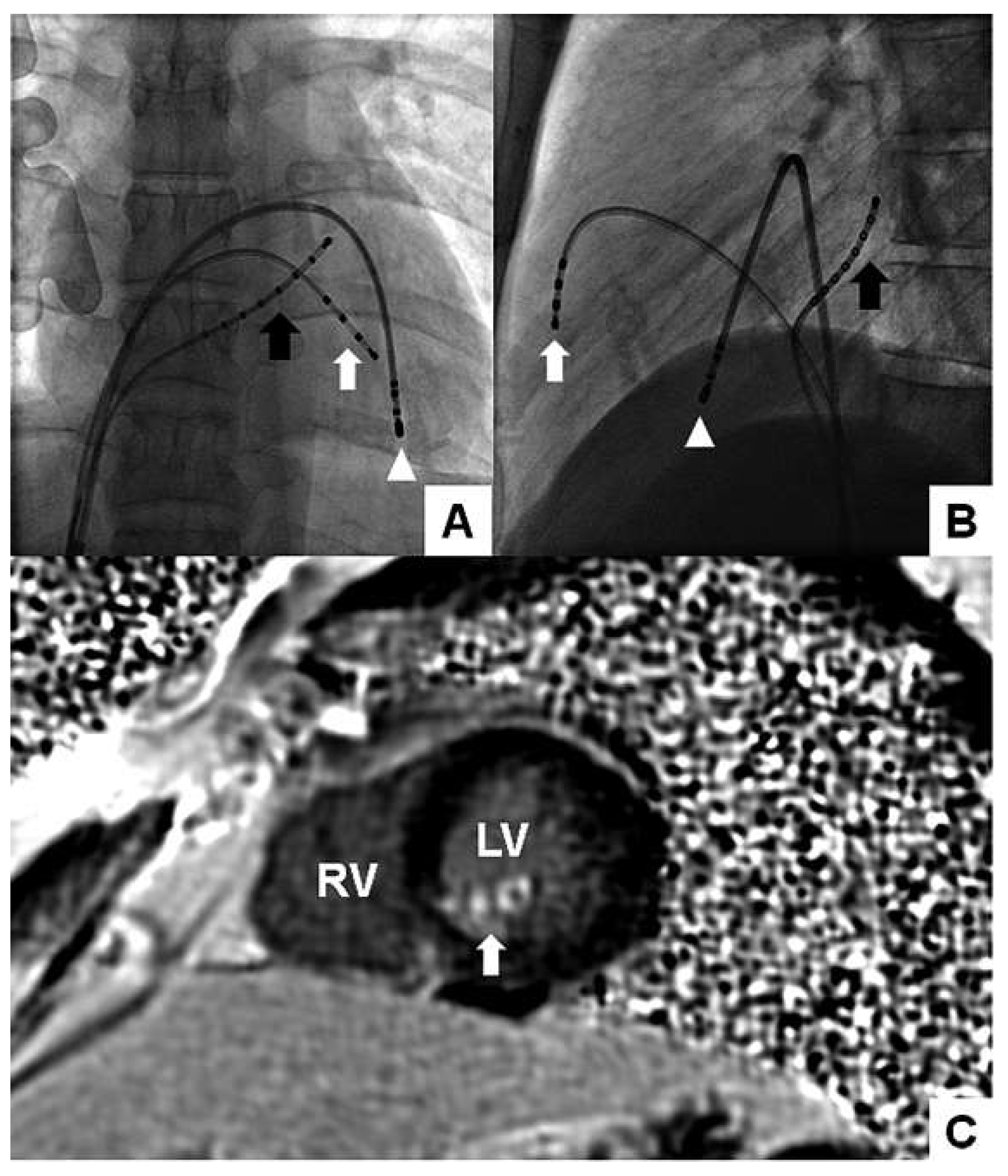
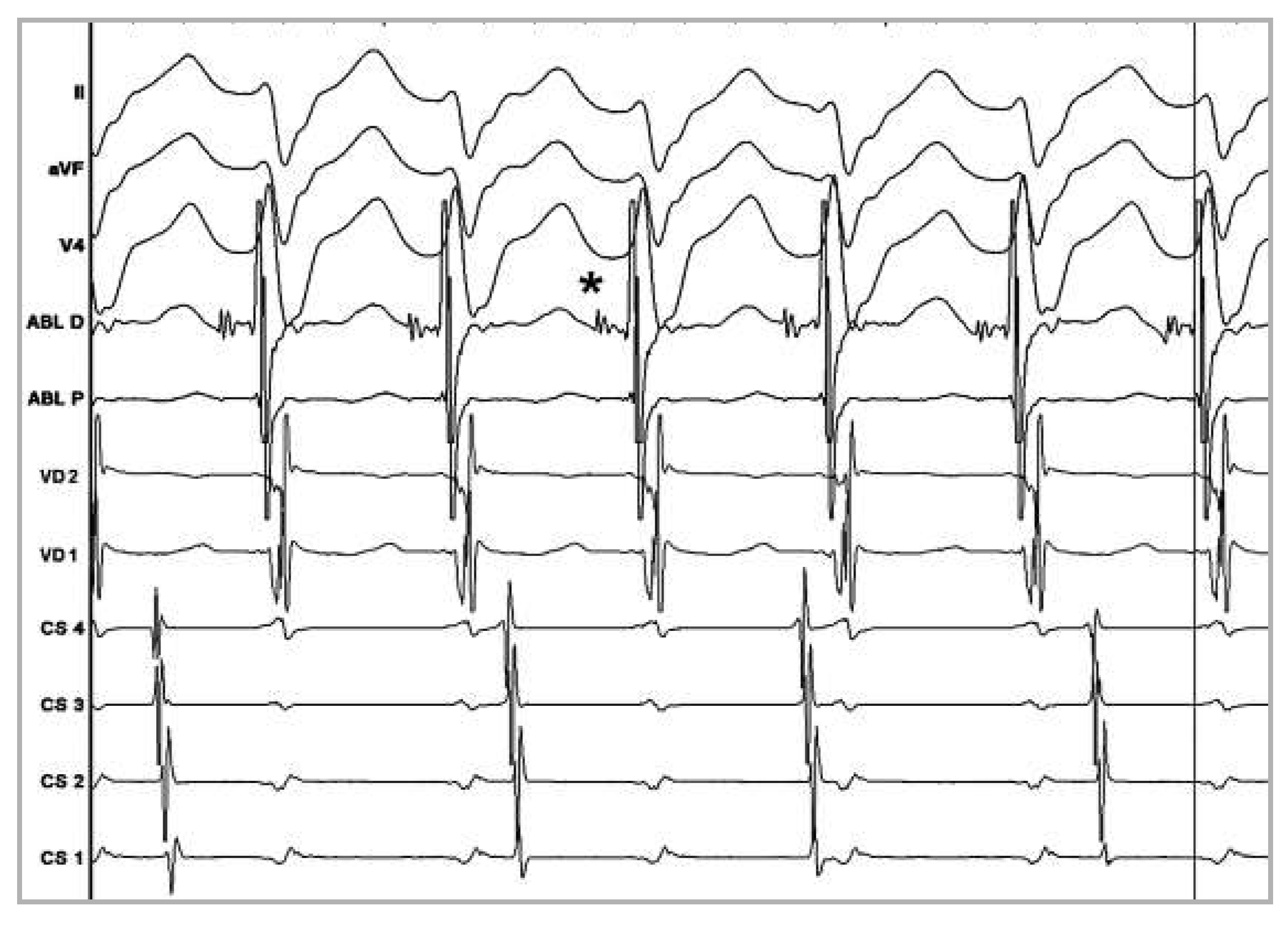
An electro-physiological study with programmed stimulation under isoproterenol infusion induced a sustained tachycardia with a right bundle branch block pattern and left-axis deviation compatible with an idiopathic fascicular left ventricular tachycardia (IFLVT) (Figure 1B). The tachycardia was successfully ablated with nine applications of radiofrequency (RF) current in the basal and mid inferoseptal LV segments (Figure 2A,B), where a late diastolic potential preceding ventricular activation was recorded (Figure 3). The arrhythmia was no longer inducible and fifteen months after the patient remains asymptomatic. A cardiac magnetic resonance (CMR) the day after the ablation showed subendocardial late gadolinium hyper-enhancement of the inferoseptal wall of LV (Figure 2C), reflecting inflammation following RF energy delivery. The underlying mechanism of IFLVT is thought to be re-entry with a slow conducting area, close to the Purkinje fibre network of the left posterior fascicle [1]. The locations visualised with CMR corresponded precisely to the sites where the tachyarrhythmia was successfully terminated.
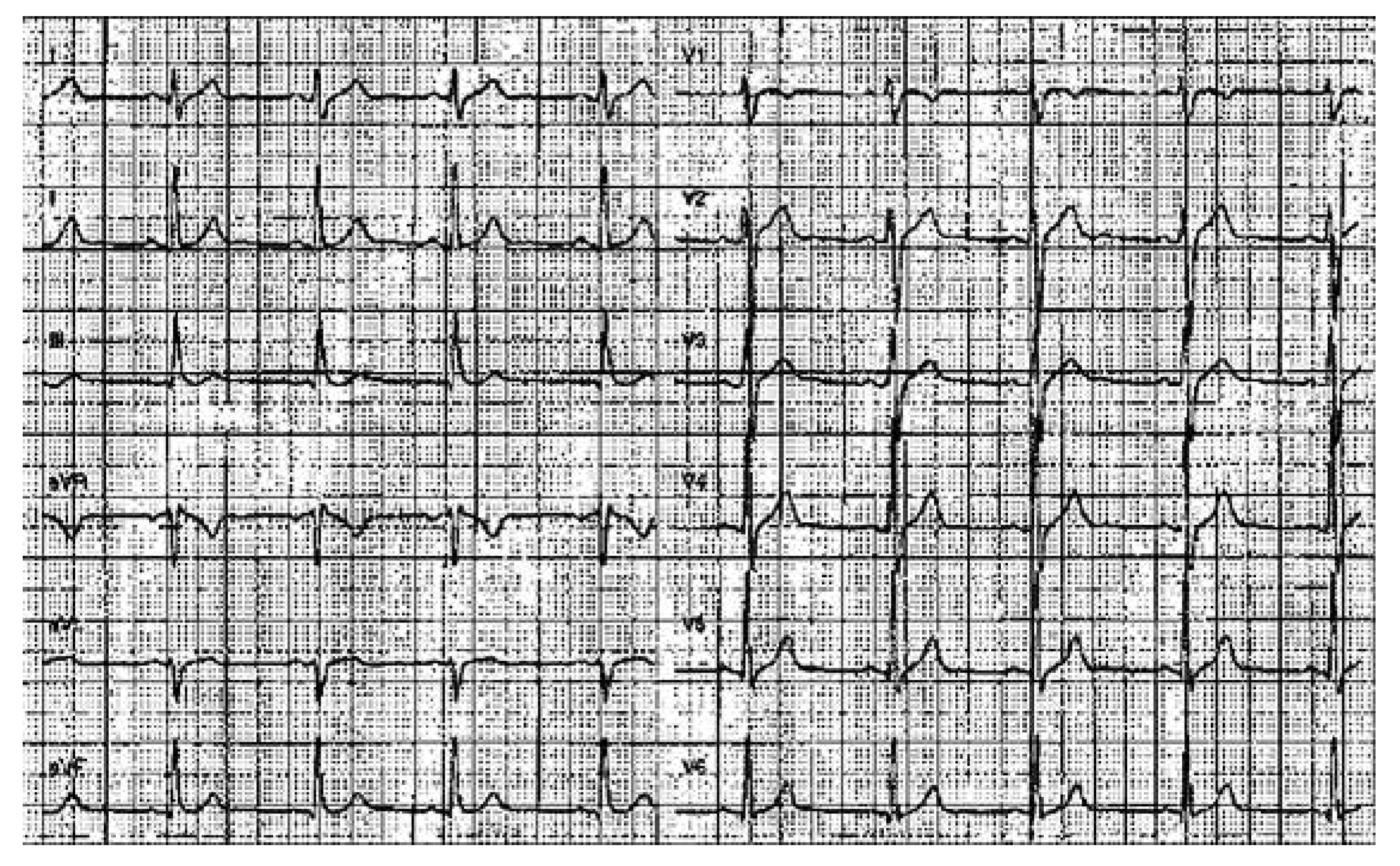
The T-waves abnormalities shown in the initial ECG are probably related to abnormal cardiac activation during IFLVT reflecting cardiac memory (CM) [2]; the T-wave vector shows the same direction as the QRS vector during the IFLVT (Figure 1A,B). These abnormalities were no longer visible three months after the intervention confirming this hypothesis (Figure 4).
In the differential diagnosis of T-wave inversion in young adults, CM due to tachyarrhythmia must be considered when hypertrophic cardiomyopathy, myopericarditis or cardiac ischaemia have been excluded.

Funding/potential competing interests

No financial support and no other potential conflict of interest relevant to this article were reported.

References

  1. Tsuchiya, T.; Okumura, K.; Honda, T.; Iwasa, A.; Yasue, H.; Tabuchi, T. Significance of late diastolic potential preceding Purkinje potential in verapamil-sensitive idiopathic left ventricular tachycardia. Circulation 1999, 99, 2408–2413. [Google Scholar] [CrossRef] [PubMed]
  2. Rosenbaum, M.B.; Blanco, H.H.; Elizari, M.V.; Lazzari, J.O.; Davidenko, J.M. Electrotonic modulation of the T wave and cardiac memory. Am J Cardiol. 1982, 50, 213–222. [Google Scholar] [CrossRef] [PubMed]
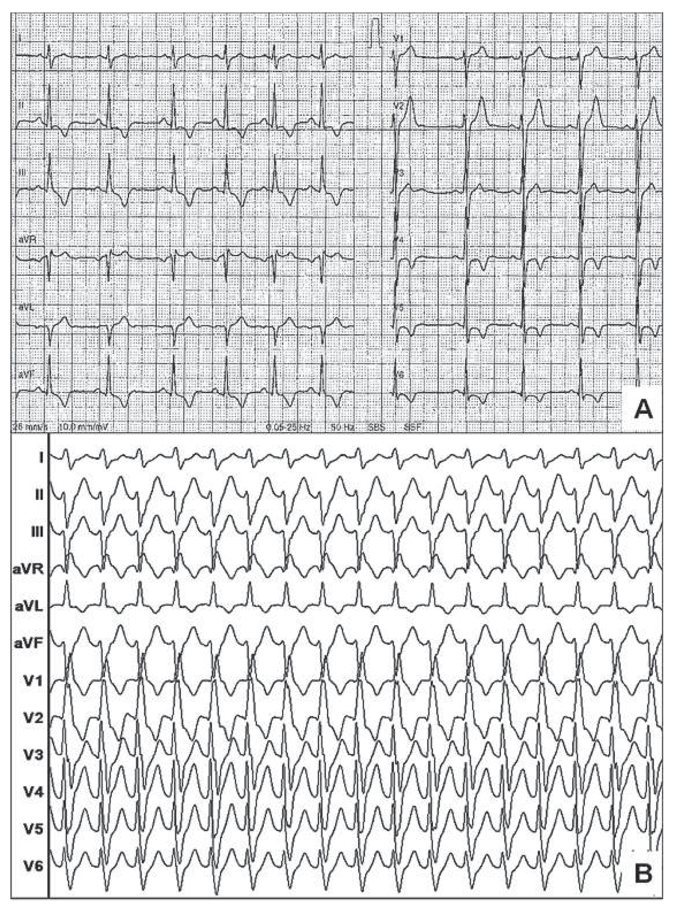
Figure 1. (A) Twelve-lead ECG obtained soon after spontaneous cardioversion demonstrating a sinus rhythm with negative T-waves in inferior and in V4 to V6 leads. (B) Induction by atrial pacing, under isoproterenol infusion of ventricular tachycardia with a right bundle branch block pattern and left-axis deviation compatible with an IFLVT.
Cardiovascmed 15 00224 g001
Figure 2. (A) Antero-posterior. (B) Lateral fluoroscopic views showing ablation catheter – antergradely inserted via trans-septal puncture – in the mid inferoseptal segment of the LV (white arrowhead). White arrow = quadripolar catheter placed at the anterior wall of the right ventricle; black arrow = octapolar catheter inserted into the coronary sinus; AP = anteroposterior; LAT = lateral. (C) CMR phase sensitive short axis slice of the mid ventricle showing subendocardial late gadolinium enhancement of the inferoseptal wall and posterior papillary muscle (arrow).
Cardiovascmed 15 00224 g002
Figure 3. Intracardiac recording during IFLVT showing a presystolic potential (*) 56 ms before the onset of the QRS at the mid inferoseptal segment of left ventricle. We can also observe atrioventricular dissociation in the coronary sinus electrogram, confirming the diagnosis of VT.
Cardiovascmed 15 00224 g003
Figure 4. Twelve-lead electrocardiogram performed three months after the procedure, showing the disappearance of the negative T-waves in inferior leads and V4–V6 confirming the hypothesis of cardiac memory.
Cardiovascmed 15 00224 g004