Next Article in Journal
Swiss Society of Cardiology and Swiss Society of Cardiac and Vascular Thoracic Surgery Award Ceremony 2012
Previous Article in Journal
A Dangerous Spike: Cause or Coincidence?
 
 
Cardiovascular Medicine is published by MDPI from Volume 28 Issue 1 (2025). Previous articles were published by another publisher in Open Access under a CC-BY (or CC-BY-NC-ND) licence, and they are hosted by MDPI on mdpi.com as a courtesy and upon agreement with Editores Medicorum Helveticorum (EMH).
Case Report

Left Ventricular Apical Ballooning Syndrome with Extensive Myocardial Late Gadolinium Enhancement: Tako-Tsubo Cardiomyopathy, Perhaps Not as Benign as Previously Thought?

by
Vincent Gabus
*,
Juerg Schwitter
,
Eric Eeckhout
and
Didier Locca
Service of Cardiology, Department of Internal Medicine, CHUV, Rue du Bugnon, CH-1011 Lausanne, Switzerland
*
Author to whom correspondence should be addressed.
Cardiovasc. Med. 2012, 15(11), 325; https://doi.org/10.4414/cvm.2012.00120
Submission received: 21 August 2012 / Revised: 21 September 2012 / Accepted: 21 October 2012 / Published: 21 November 2012

Summary

Aim: In patients meeting the criteria for LV apical ballooning syndrome, CMR typically identifies myocardial oedema without Late Gadolinium Enhancement (LGE). However, LGE does not exclude LV apical ballooning syndrome and small necrotic areas can be seen in the acute setting resolving at follow-up. Results: We describe the case of a 61 year-old woman admitted with a 2 hour history of retrosternal chest pain. ECG demonstrated Q waves and dynamic ST segment elevation. Cardiac enzymes on admission were elevated. Coronary angiography showed unobstructed coronary arteries. Left ventriculography showed apical ballooning. Cardiac Magnetic Resonance (CMR) revealed antero-septal and anterior akinesia. Myocardial oedema was seen in the septal wall. Late gadolinium enhancement (LGE) was seen in the same territory indicating the presence of necrosis. A follow up CMR scan was performed at 3 months showing a complete resolution of the septal oedema. However, on LGE sequences there was evidence of persistent extensive fibrotic scar located in the septal wall. The size of this fibrotic scar was 20% bigger than on the previous scan. Conclusion: Detection and quantification of a scar is important, as scar tissue on LGE images has been described as a predictor of major adverse cardiac events. Therefore, this case suggests that the spectrum of Tako-tsubo cardiomyopathy may include a more severe variant than previously described. CMR examinations could serve as a useful means to identify severe cases and predict potential complications of this pathology as well as to drive therapeutic decisions.

Case Report

A 61-year-old woman with known essential hypertension was admitted to the emergency room with a 2 hour history of retrosternal chest pain. She described severe fatigue and emotional stress during the previous week and recalled a similar episode of pain 10 years ago. Current medical therapy at the time of admission included progesterone, enalapril and hydrochlorothiazide. On physical examination she was distressed with a heart rate of 91 beats per minute and blood pressure of 174/100 mm Hg. Clinical examination was otherwise unremarkable. Serial 12 lead ECGs at admission (Figure 1A) demonstrated sinus rhythm with Q waves and dynamic ST segment elevation in V1–V3 leads. 24 hours later ECG demonstrated T waves inversion (Figure 1B). An urgent transthoracic echocardiogram was performed showing apical, antero-septal akinesia and antero-lateral hypokinesia with a left ventricular ejection fraction (LVEF) estimated at 50% compatible with apical ballooning. Cardiac enzymes on admission were elevated (Troponin I: 3.55 µg/l [<0.04]; CK: 339 U/l [25–140]) with a peak five hours later (Troponin I: 26.37 µg/l; CK: 825 U/l). Urgent coronary angiography showed unobstructed coronary arteries without any features of spasm (Figure 2A). Left ventriculography (Figure 2B) confirmed the echocardiogram findings showing apical ballooning. Cardiac Magnetic Resonance (CMR) scan (Siemens 1.5T, Erlangen, Germany) was performed 5 days after admission. LVEF calculated from the cine CMR was 50% with antero-septal and anterior akinesia. On the STIR images (T2 weighted sequences, short tau inversion recovery) myocardial oedema was seen in the antero-septal and inferior-septal wall at the mid ventricular level. (Figure 3A, arrows). Late gadolinium enhancement (LGE) was seen in the same territory indicating the presence of necrosis (Figure 4A). Clinical evolution was uneventful and the patient was discharged 9 days post admission. A follow up CMR scan was performed at 3 months showing a preserved LVEF without akinesia and complete resolution of the septal oedema (Figure 3B). However, on LGE sequences there was evidence of extensive persistent, patchy, diffuse subendocardial, mid wall and epicardial fibrotic scar located in the antero-septal and inferior-septal segments (Figure 4B, arrows). The amount of fibrotic tissue was 20% increased in comparison to the previous scan.
Patients meeting the criteria for LV apical ballooning syndrome usually have a modest rise in cardiac enzymes [1]. CMR in these patients typically identifies myocardial oedema without LGE [2]. However, LGE does not exclude LV apical ballooning syndrome and small necrotic areas can be seen in the acute setting resolving at follow-up [3]. Detection and quantification of a scar is important as scar tissue on LGE images has been described as a predictor of major adverse cardiac events [4]. It is unlikely that in our case the territories with LGE in the acute phase represent an incidental finding due to a previous cardiac event, since oedema was detected in the same territories identifying these lesions as acute damage. Therefore, this case suggests that the spectrum of Tako-tsubo cardiomyopathy may include a more severe variant than previously described. CMR examinations of clinical cases presenting with symptoms as described here, could serve as a useful means to identify severe cases and predict potential complications of this pathology as well as to drive therapeutic decisions.

Funding/Potential Competing Interests

No financial support and no other potential conflict of interest relevant to this article were reported.

References

  1. Prasad, A.; Lerman, A.; Rihal, C.S. Apical ballooning syndrome (Tako-Tsubo or stress cardiomyopathy): a mimic of acute myocardial infarction. Am Heart J. 2008, 155, 408–417. [Google Scholar] [CrossRef] [PubMed]
  2. Eitel, I.; Behrendt, F.; Schindler, K.; Kivelitz, D.; Gutberlet, M.; Schuler, G.; et al. Differential diagnosis of suspected apical ballooning syndrome using contrast-enhanced magnetic resonance imaging. Eur Heart J. 2008, 29, 2651–2659. [Google Scholar] [CrossRef] [PubMed]
  3. Rolf, A.; Nef, H.M.; Möllmann, H.; Troidl, C.; Voss, S.; Conradi, G.; et al. Immunohistological basis of the late gadolinium enhancement phenomenon in tako-tsubo cardiomyopathy. Eur Heart J. 2009, 30, 1635–1642. [Google Scholar] [CrossRef] [PubMed]
  4. Flett, A.; Westwood, M.; Davies, L.; Mathur, A.; Moon, J. The prognostic implications of Cardiovascular Magnetic Resonance. Circ Cardiovasc Imaging 2009, 2, 243–250. [Google Scholar] [CrossRef] [PubMed]
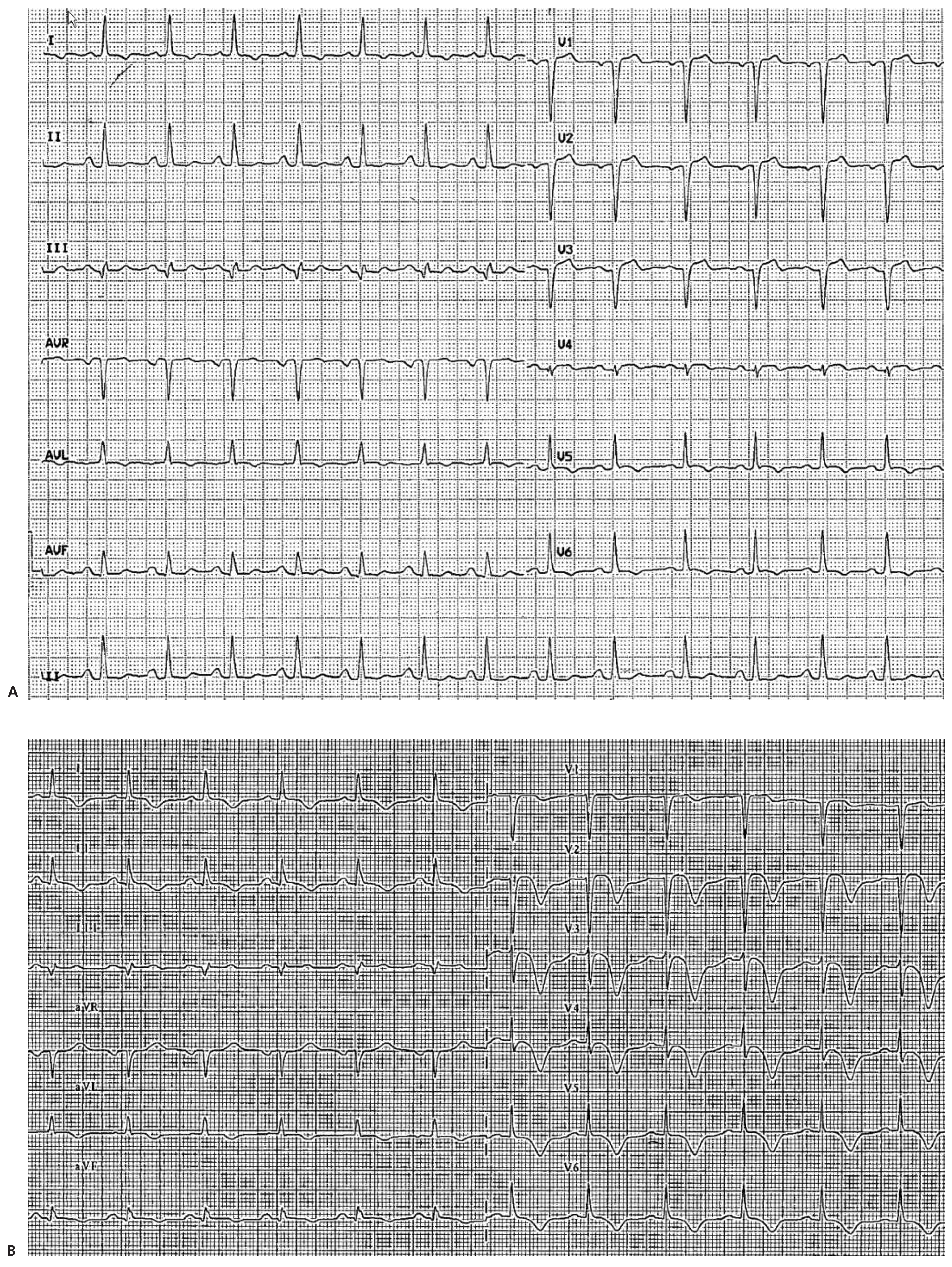
Figure 1. A, B Serial 12 lead ECGs demonstrating sinus rhythm with Q waves and dynamic ST segment elevation in V1–V3 lead.
Cardiovascmed 15 00325 g001
Figure 2. A. Urgent coronary angiography showed unobstructed coronary arteries without any features of spasm. B. Left ventriculography showing apical ballooning.
Cardiovascmed 15 00325 g002
Figure 3. A. Cardiac Magnetic Resonance (CMR) scan (Siemens 1.5T, Erlangen, Germany) 5 days after admission. On the STIR images (T2 weighted sequences, short tau inversion recovery) myocardial oedema was seen in the antero-septal and inferior-septal wall at the mid ventricular level (arrows). B. Follow up CMR scan performed at 3 months showing complete resolution of the septal oedema.
Cardiovascmed 15 00325 g003
Figure 4. A. Cardiac Magnetic Resonance (CMR) scan (Siemens 1.5T, Erlangen, Germany) 5 days after admission showing Late gadolinium enhancement (LGE) in the antero-septal and inferiorseptal wall indicating the presence of necrosis. B. Follow up CMR scan performed at 3 months. On LGE sequences there is evidence of extensive persistent, patchy, diffuse subendocardial, mid wall and epicardial fibrotic scar located in the antero-septal and inferior-septal segments.
Cardiovascmed 15 00325 g004