Next Article in Journal
Richtlinien 2011 zur Herzschrittmacher—und Defibrillatortherapie und Perkutanen Katheterablation
Previous Article in Journal
Typical Signs of Secundum Atrial Septal Defect in a Young Man
 
 
Cardiovascular Medicine is published by MDPI from Volume 28 Issue 1 (2025). Previous articles were published by another publisher in Open Access under a CC-BY (or CC-BY-NC-ND) licence, and they are hosted by MDPI on mdpi.com as a courtesy and upon agreement with Editores Medicorum Helveticorum (EMH).
Case Report

Acute Myocardial Infarction in a 19 Year-Old Woman: Sometimes Hoofbeats are Zebras

by
Diego Arroyo
1,
Tobias Rutz
2,* and
Jean-Christophe Stauffer
3
1
Department of Internal Medicine, Hopital Cantonal de Fribourg, 1708 Fribourg, Switzerland
2
Department of Cardiology, Inselspital Bern, 3010 Bern, Switzerland
3
Department of Cardiology, Hopital Cantonal de Fribourg, 1708 Fribourg, Switzerland
*
Author to whom correspondence should be addressed.
Cardiovasc. Med. 2011, 14(3), 95; https://doi.org/10.4414/cvm.2011.01576
Submission received: 23 December 2010 / Revised: 23 January 2011 / Accepted: 23 February 2011 / Published: 23 March 2011

Abstract

We report the case of a 19 year-old woman who presented with chest pain at the emergency department. Cardiac risk factors were obesity, dyslipidemia, cigarette smoking and a sedentary lifestyle. Investigations revealed an acute myocardial infarction with ST-segment elevation. A thrombotic occlusion of the mid left anterior descending artery was visualised on coronary angiography, the cause of which remains unknown.

Case report

A 19 year-old woman arrived at the emergency department complaining of left parasternal thoracic pain for the past 24 h. Respiration enhanced pain, while leaning forward alleviated it and she described left arm radiation. She also complained of nausea, vomiting and diarrhoea for the past week. She had no past medical or surgical history. Cardiac risk factors were active cigarette smoking (2 pack-years), obesity (BMI 33.3 kg/m2) and a sedentary life-style. Usual medication was oral estroprogestative contraception introduced 2 weeks prior to admission and paracetamol. History was inconsistent with past or present drug abuse.
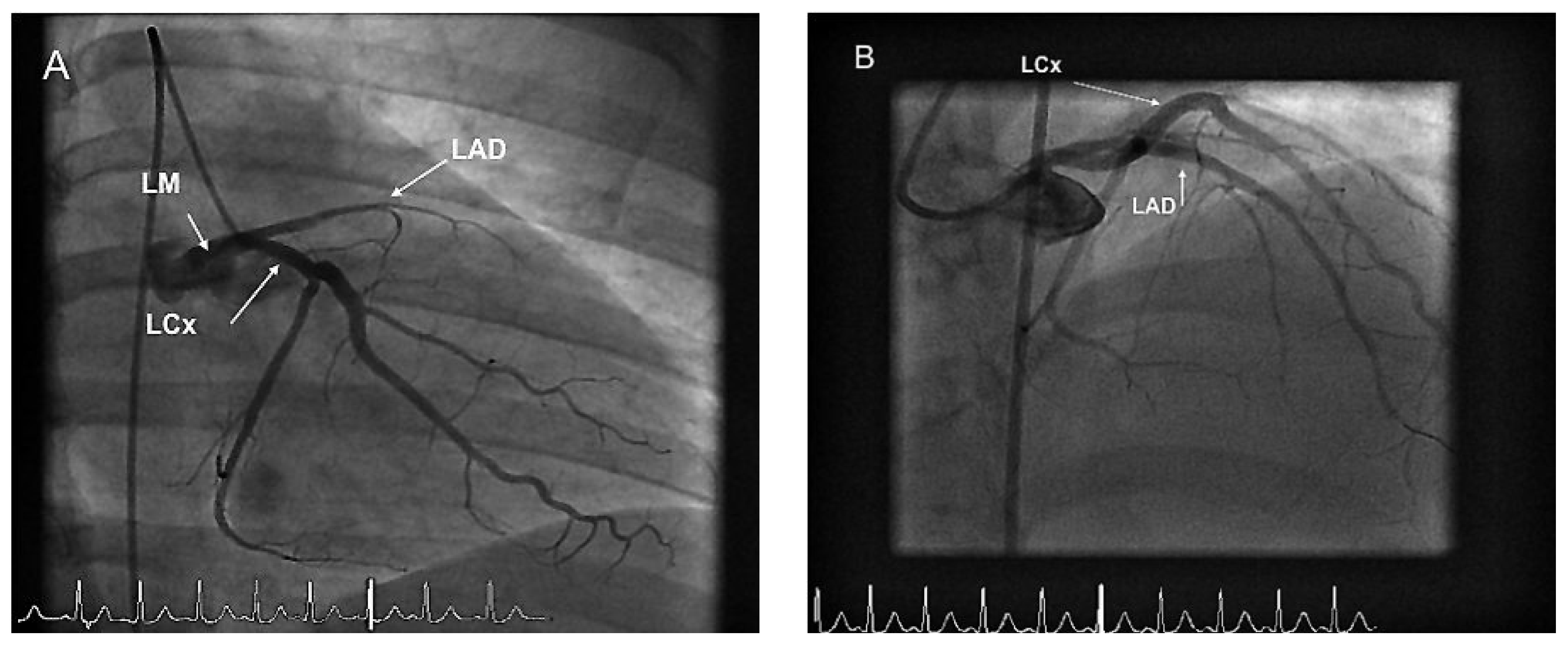
A physical examination revealed elevated blood pressure at 160/80 mm Hg and regular tachycardia at 110 beats per minute. Respiratory frequency, temperature and oxygen saturation were however normal. Pulmonary and cardiac auscultation were normal, as well an abdominal examination. A chest X ray was unremarkable. Initial ECG showed sinus tachycardia with anterolateral ST elevation as well as inferior ST depression (Figure 1). Laboratory reports revealed an elevated troponine Ic at 0.69 µg/l, CK was within normal limits and CRP was slightly elevated at 19 mg/l; the rest of the workup, including a complete toxicological-screen, was normal and a pregnancy test was negative. Although presentation suggested pericarditis, an urgent transthoracic echocardiography showed mid-sep-tum and anterior wall hypokinesia. Ejection fraction was estimated by Simpson’s method to be 40%. Further investigation by coronary angiogram revealed an occlusion of the proximal left anterior descending artery as well as a minor lesion of the circumflex artery (Figure 2A). The lesion was treated by thromboaspiration, balloon pre-dilatation and implantation of two zotarolimuseluting stents (Endeavour Resolute 2.5/18 mm and 3.0/15 mm) efficiently covering the proximal anterior inter-ventricular segment (Figure 2B). There were no periprocedural complications. Post-procedure ECG demonstrated partial ST-segment resolution. A treatment of lifelong aspirin, one-year clopidogrel, atorvastatine, metoprolol and lisinopril were initiated. The patient remained symptom free and was discharged on day five for cardiac rehabilitation. A 3-month control echocardiography was consistent with a slightly diminished ejection fraction at 50% and anteroseptal, as well as anterolateral, akinesia.
A complementary laboratory workup showed that cholesterol and triglyceride levels were within limits but HDL-cholesterol was low at 0.75 mmol/l (normal 1.0–2.2 mmol/l). Complete immunological and thrombophilia workup were unremarkable. An additional transoesophageal echocardiography showed no patent foramen ovale.

Discussion

Acute myocardial infarction (AMI) in the young adult, usually defined as younger than 45 years old, is a rare event. It represents 0.7 to 4% of all AMIs and affects mostly men [1,2]. There is however no reliable data as to the prevalence and incidence of this event. Pathogenesis and risk factors differ slightly from those in older patients. Atherosclerotic disease remains the principal cause, others being non-atherosclerotic coronary disease, hypercoagulability states and drug abuse.
Conventional risk factors remain identical. Positive family history, obesity, cigarette smoking are all more frequent in young adults. Cigarette smoking remains the main risk factor and is present in 65 to 92% of patients, which is higher than in older ones [3]. Diabetes and hypertension are preponderant in older patients. Less than 10% of young adults with AMI have diabetes [4]. Oral contraception only becomes a risk factor if associated with tobacco smoking [5].
Congenital anomalies are rare and account for less than 4% of AMIs in young adults [6]. Spontaneous coronary dissection occurs primarily in women during the peripartum period. Other rare causes are large-vessel vasculitis such as Kawasaki’s disease [7] and systemic lupus erythematosus. Pathogenic mechanisms include coronary arteritis and early coronary atherosclerosis due to hypertension and hyperlipidaemia [8].
Coagulation anomalies such as antiphospholipid antibody syndrome or hyperhomocysteinaemia induce early atherosclerosis, platelet hyper-aggregation and consequently thrombotic occlusions [9,10].
Cocaine is the main cause of AMI related to drug abuse. The underlying mechanisms being tachycardia, hypertension, vasospasm and platelet hyperaggregation [11,12]. Amphetamines, cannabis and alcohol are associated with an increased risk for AMI for reasons presently unknown.
Clinical presentation of AMI in the young is often atypical. Angina pectoris is usually absent but up to 90% present with chest pain during the week before the infarct [2,13]. The underlying pathophysiology is thought to be due to a highly unstable lipid-rich lesion with little or no calcification. Initial ECG shows preferential ST-elevation [14].
Angiography reveals, in a majority of cases, a complex one-vessel disease. Three-vessel disease is rare; 14% in the young as opposed to 39% in the older patients [15]. The amount of diseased vessels does not change overall mortality [16].
Prognosis is however better in the younger patient with in-hospital mortality ranging from 1 to 6%. Complications such as cardiogenic shock, stroke or lifethreatening arrhythmias are infrequent. The 7-year survival rate is 84% in men and 90% in woman [16]. Ejection fraction of less than 30% and diabetes are both negative prognostic factors [17].
The two most important differential diagnoses with similar clinical presentation are myopericarditis and pulmonary embolism. While these are more frequent in young people, myocardial ischemia remains a possibility.
This case was exceptional considering the very young age of the patient and the apparently inexplicable cause for the coronary occlusion. The patient presented multiple cardiac risk factors including tobacco smoking associated with oral contraception, obesity, a sedentary lifestyle and low HDL-cholesterol favouring the hypothesis of atherosclerotic disease. The latter, in the absence of intravascular coronary ultrasound (IVUS), cannot be proven. According to literature, the clinical presentation was typical of a myocardial infarction in the young adult. One should stress the importance of conventional cardiac risk factors, all of which should alert physicians to a possible myocardial ischemia and efforts should be made on primary prevention. Substance abuse and other rare non-atherosclerotic coronary diseases (e.g., spontaneous coronary dissection, congenital anomalies, spasms, coagulopathies) should be considered in the differential diagnosis.

Conflicts of Interest

The authors certify that there is no actual or potential conflict of interest in relation to this article.

References

  1. Doughty, M.; Mehta, R.; Bruckman, D.; et al. Acute myocardial infarction in the young—The University of Michigan experience. Am Heart J. 2002, 143, 56. [Google Scholar] [CrossRef] [PubMed]
  2. Schoenenberger, A.W.; Radovanovic, D.; Stauffer, J.C.; Windecker, S.; Urban, P.; Niedermaier, G.; et al. Acute coronary syndromes in young patiente: Presentation, treatment and outcome. Int J Cardiol. 2009. [CrossRef]
  3. Mukherjee, D.; Hsu, A.; Moliterno, D.J.; Lincoff, A.M.; Goormastic, M.; Topol, E.J. Risk factors for premature coronary artery disease and determinants of adverse outcomes after revascularisation in patients less than 40 years old. Am J Cardiol. 2003, 92, 1465–1467. [Google Scholar] [CrossRef] [PubMed]
  4. Jalowiec, D.A.; Hill, J.A. Myocardial infarction in the young and in women. Cardiovascular Clinics. 1989, 20, 197–206. [Google Scholar] [PubMed]
  5. Rosenberg, L.; Palmer, J.R.; Rao, R.S.; Shapiro, S. Low-dose oral contraceptive use and the risk of myocardial infarction. Arch Intern Med. 2001, 161, 1065. [Google Scholar] [CrossRef] [PubMed]
  6. Warren, S.E.; Thompson, S.I.; Vieweg, W.V. Historic and angiographic features of young adults surviving myocardial infarction. Chest. 1979, 75, 667–670. [Google Scholar] [CrossRef] [PubMed]
  7. Smith, B.A.; Grider, D.J. Sudden death in a young adult: sequelae of childhood Kawasaki disease. Am J Emer Med. 1993, 11, 381–383. [Google Scholar] [CrossRef] [PubMed]
  8. Wilson, V.E.; Eck, S.L.; Bates, E.R. Evaluation and treatment of acute myocardial infarction complicating systemic lupus erythematosis. Chest. 1992, 101, 420–424. [Google Scholar] [CrossRef] [PubMed]
  9. Vaarala, O. Antiphospholipid antibodies and athrtrosclerosis. Lupus. 1996, 5, 442–447. [Google Scholar] [CrossRef] [PubMed]
  10. Nygard, O.; Nordrehaug, J.E.; Refsum, H.; Ueland, P.M.; Farstad, M.; Vollset, S.E. Plasma homocysteine levels and mortality in patients with coronary artery disease. N Engl J Med. 1997, 337, 230–236. [Google Scholar] [CrossRef] [PubMed]
  11. Hollander, J.E.; Todd, K.H.; Green, G.; et al. Chest pain associated with cocaine: an assessment of prevalence in suburban and urban emergency departments. Ann Emerg Med. 1995, 26, 671–676. [Google Scholar] [CrossRef] [PubMed]
  12. Lange, R.A.; Hills, C.D. Cardiovascular complications of cocaine use. N Engl J Med. 2001, 345, 351–358. [Google Scholar] [CrossRef] [PubMed]
  13. Fournier, J.A.; Sanchez, A.; Quero, J.; et al. Myocardial infarction in men aged 40 years or less: a prospective clinical-angiographic study. Clin Cardiol. 1996, 19, 631. [Google Scholar] [CrossRef] [PubMed]
  14. Morillas, P.; Bartomeu, V.; Pahon, P.; et al. Characteristics and outcome of acute myocardial infarction in young patients. The PRIAMHO II study, Cardiology. 2006, 107, 217–225. [Google Scholar] [CrossRef] [PubMed]
  15. Zimmerman, F.H.; Cameron, A.; Fisher, L.D. Myocardial infarction in young adults: angiographic characterization, risk factors and prognosis (Coronary Artery Surgery Study Registry). J Am Coll Cardiol. 1995, 26, 654. [Google Scholar] [CrossRef] [PubMed]
  16. Porter, A.; Wurzel, M.; Ben-Gal, T.; et al. Long-term prognosis of 210 patients who underwent coronary angiography before 40 years of age. Am J Cardiol. 1998, 81, 1168. [Google Scholar] [CrossRef] [PubMed]
  17. Cole, J.H.; Miller, J.I.; Sperling, L.S.; Weintraub, W.S. Longe-term follow up of coronary artery disease presenting in young adults. J Am Coll Cardiol. 2003, 41, 521. [Google Scholar] [CrossRef] [PubMed]
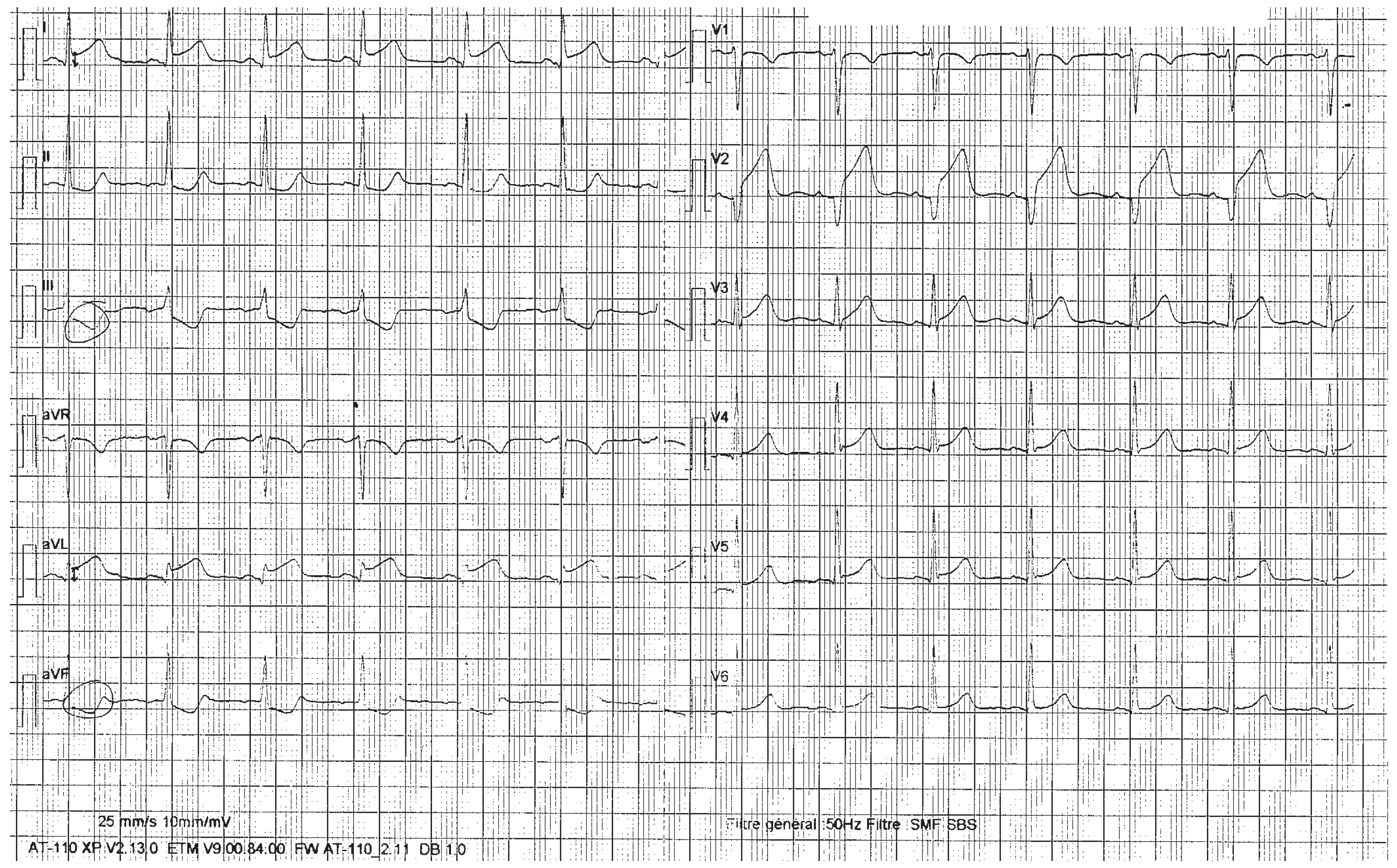
Figure 1. A Initial ECG showing ST segment elevation in I, avL, V2–V3 and mirror image ST segment depression in II, III, avF.
Cardiovascmed 14 00095 g001
Figure 2. A RAO view. Image showing occlusion of the mid-LAD. LM = left main coronary artery; LAD = left anterior descending artery; LCx = Left circumflex artery with minor irregularities. B Anteroposterior view. Image showing successful reperfusion and stenting of the LAD.
Cardiovascmed 14 00095 g002