Next Article in Journal
Typical Signs of Secundum Atrial Septal Defect in a Young Man
Previous Article in Journal
Collateral Function Changes During Primary Percutaneous Coronary Intervention in Acute Myocardial Infarction: Effect on Infarct Size
 
 
Cardiovascular Medicine is published by MDPI from Volume 28 Issue 1 (2025). Previous articles were published by another publisher in Open Access under a CC-BY (or CC-BY-NC-ND) licence, and they are hosted by MDPI on mdpi.com as a courtesy and upon agreement with Editores Medicorum Helveticorum (EMH).
Case Report

Iatrogenic Left Main Stem Stenosis After Surgical Aortic Valve Replacement

by
Jens Robert
1,*,
David Tüller
2 and
Stephan Windecker
1
1
Swiss Cardiovascular Centre Bern, University Hospital, CH-3010 Bern, Switzerland
2
Stadtspital Triemli, 8063 Zurich, Switzerland
*
Author to whom correspondence should be addressed.
Cardiovasc. Med. 2011, 14(3), 101; https://doi.org/10.4414/cvm.2011.01578
Submission received: 23 December 2010 / Revised: 23 January 2011 / Accepted: 23 February 2011 / Published: 23 March 2011

Abstract

Surgical aortic valve replacement (SAVR) represents the gold standard in the treatment of symptomatic severe aortic valve stenosis as reflected by the class I indication assigned in the ACC/AHA and ESC guidelines. SAVR effectively relieves symptoms, improves quality of life as well as prognosis of affected patients. As with any therapeutic intervention, SAVR is associated with some short and long term adverse events including death, stroke, myocardial infarction, renal failure, bleeding, as well as structural and non-structural valve deterioration. We present two cases of iatrogenic left main stem stenosis within less than one year after SAVR, describe the management and discuss the literature.

Case presentations

Case 1

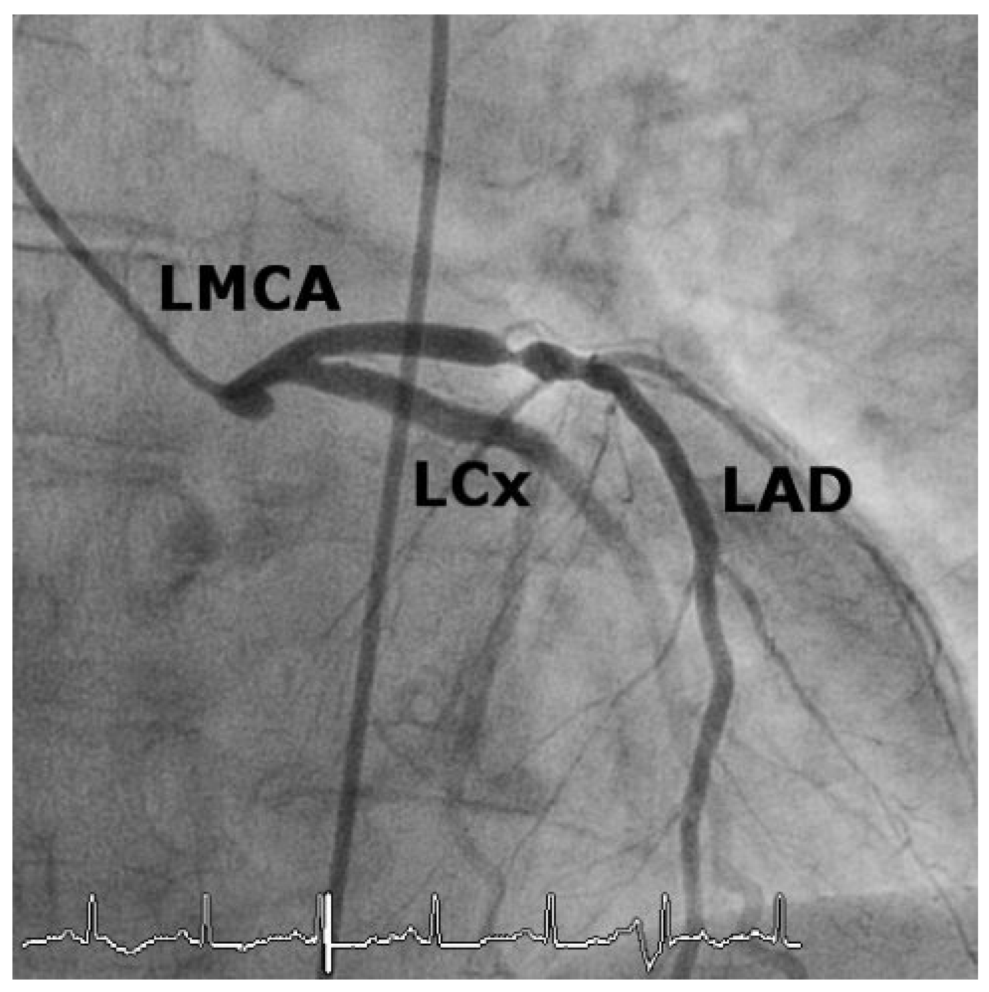
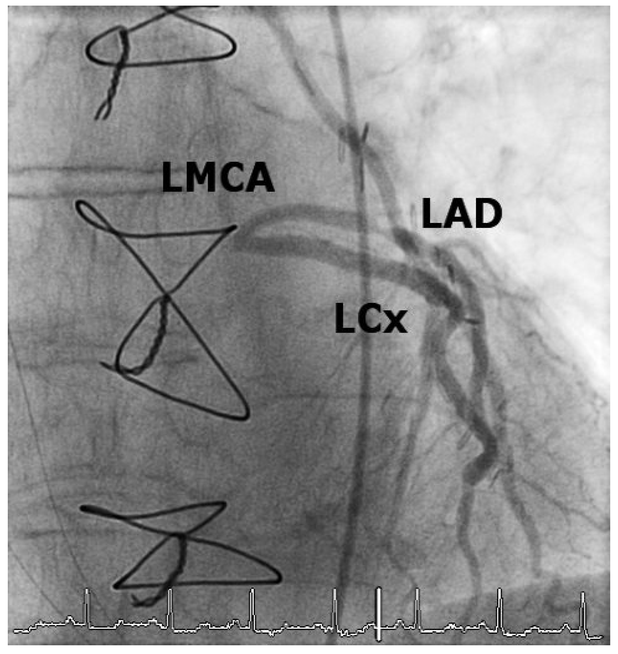
A 74-year-old woman with symptomatic, severe aortic valve stenosis (mean-gradient 38 mm Hg, effective valve orifice area = 0.4 cm2) and single vessel coronary artery disease with a significant stenosis of the proximal left anterior descend-ing artery (LAD) underwent surgical aortic valve replacement (SAVR) with a bioprosthesis (porcine stentless prosthesis 23 mm Shelhigh NR2000, Shelhigh, Inc., NJ, USA) and coronary artery bypass grafting with a single graft of the left internal mammary artery to the distal LAD. Eight months after the procedure the patient presented to a local emergency room with unstable angina and dyspnoea. The electrocardiogram showed ST-segment depression in leads I, aVL, V4–V6 (Figure 1), and cardiac biomarkers including CK (416 U/l, ULN = 140 U/l) and troponin (0.48 ng/mL, ULN ≤ 0.10 ng/mL) were elevated. The diagnosis of non-ST-elevation myocardial infarction was established and the patient was referred to our institution for further evaluation. Coronary angiography revealed occlusion of the previously patent left main stem (Figure 2 and Figure 3). The arterial graft to the distal LAD was patent, but retrograde flow to the proximal LAD and the circumflex artery was diminished owing to the high grade stenosis of the proximal LAD (Figure 4).
The left main lesion was crossed with a hydrophilic guide wire (ChoICEPT®, 0,014”, Boston Scientific, MA, USA), then dilated with a balloon dilatation catheter (Maverick 3,0 × 15 mm, Boston Scientific, MA, USA). A drug-eluting stent (Biomatrix 3.5 × 11 mm, Biosensors Interventional Technologies Pte Ltd., Singapore) was placed with a satisfactory final result (Figure 5). The patient was discharged on dual antiplatelet therapy with acetylsalicylic acid and clopidogrel. A control coronary angiography 11 months later revealed a patent stent with no signs of restenosis.

Case2

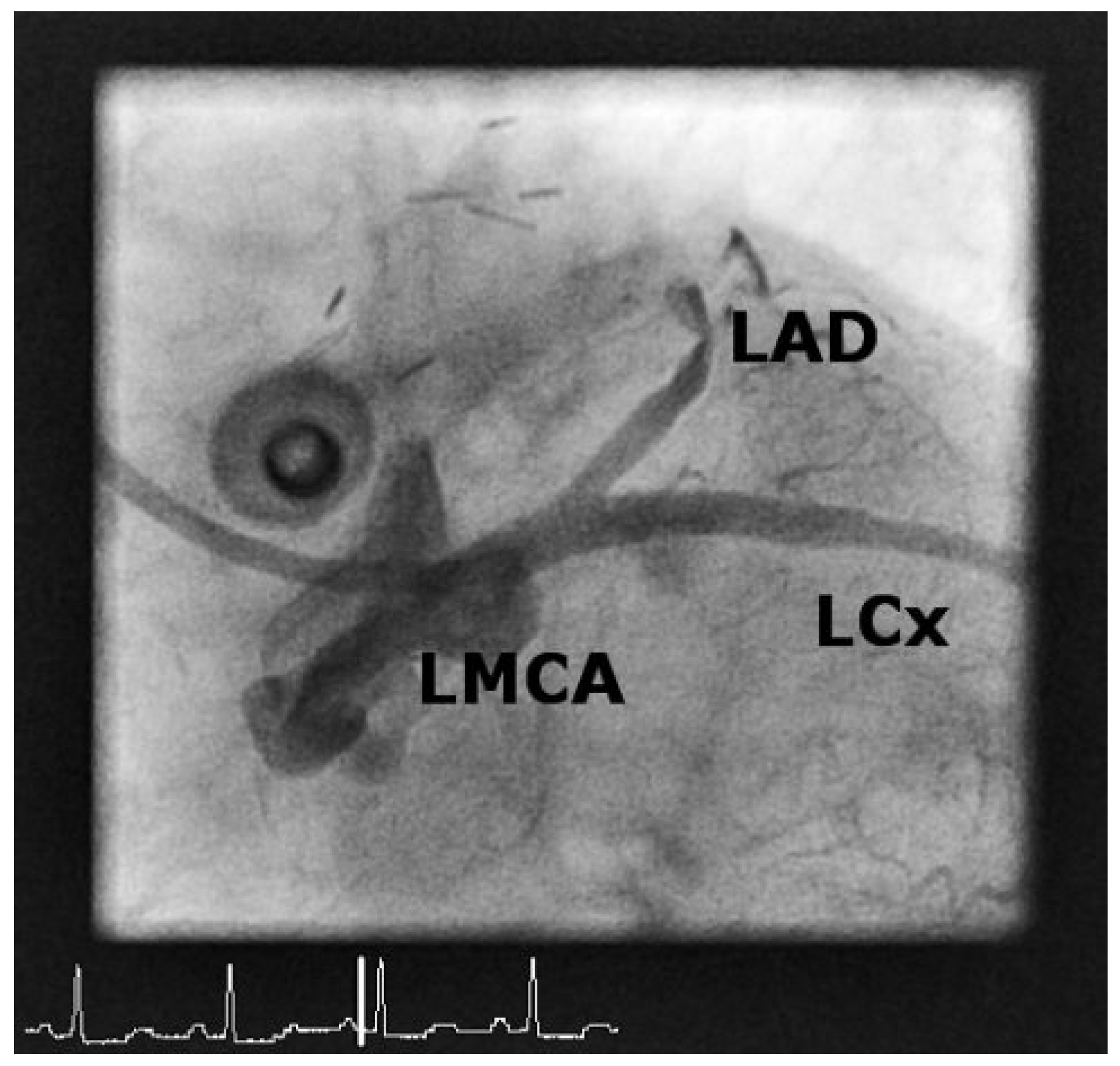
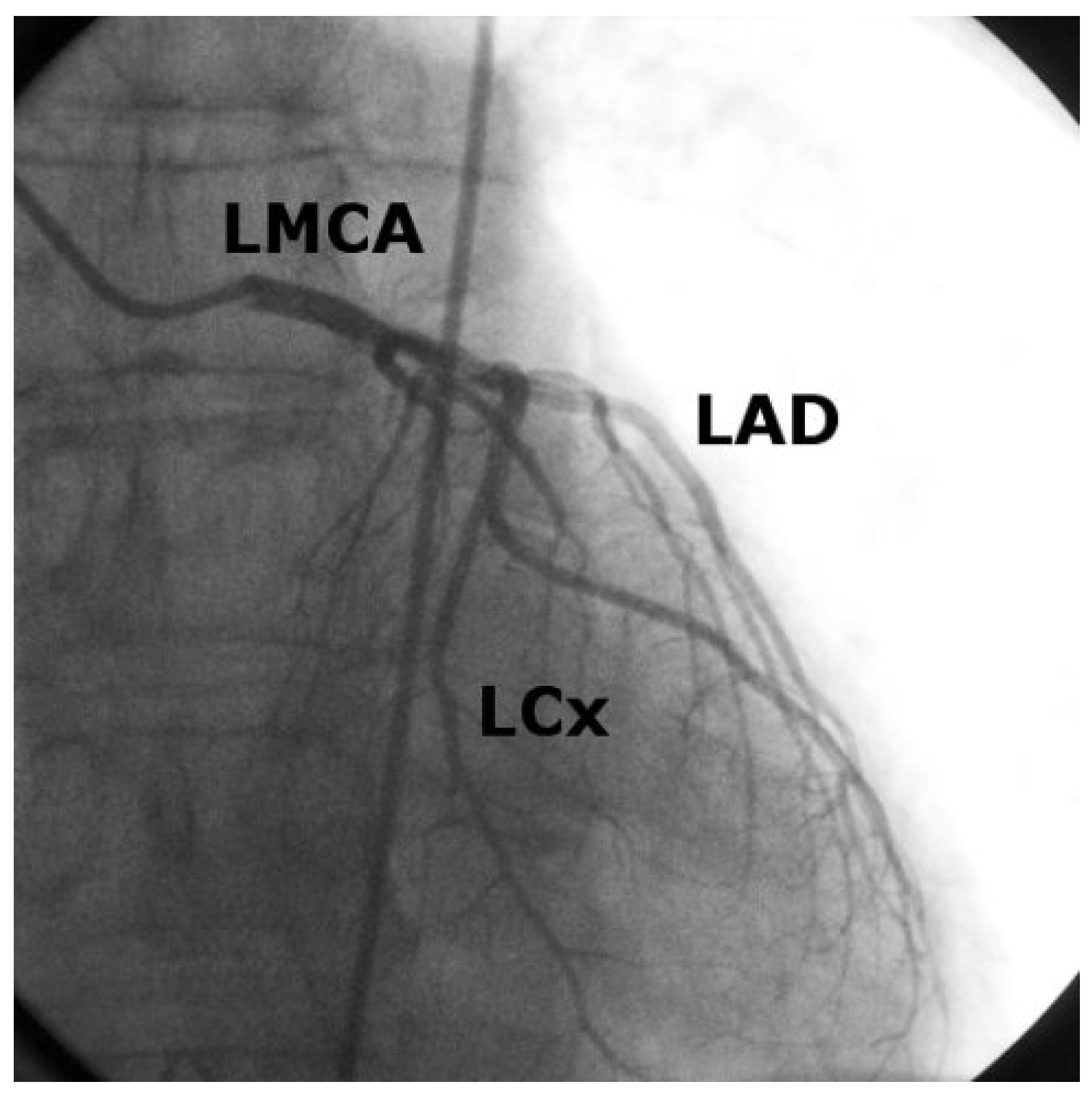
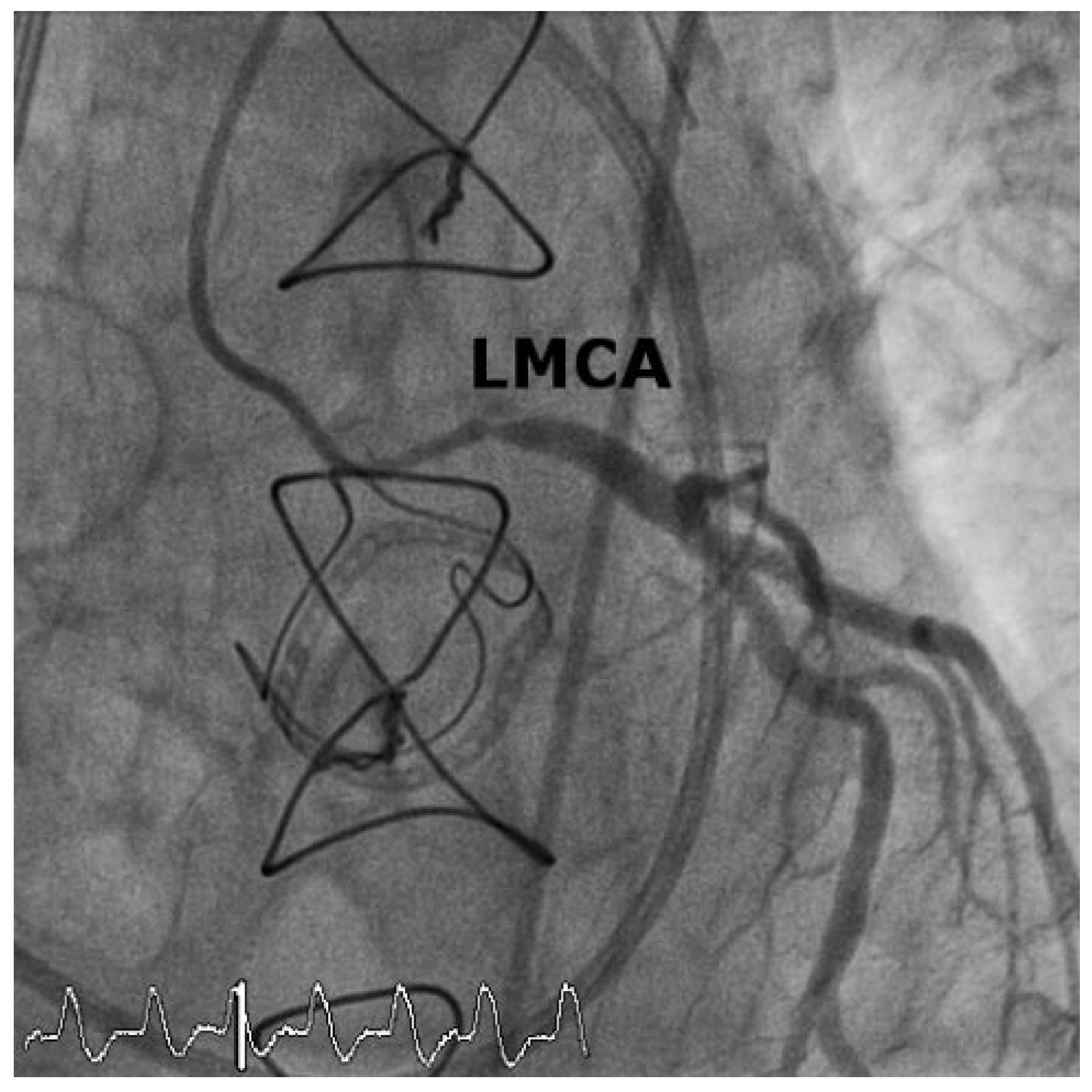
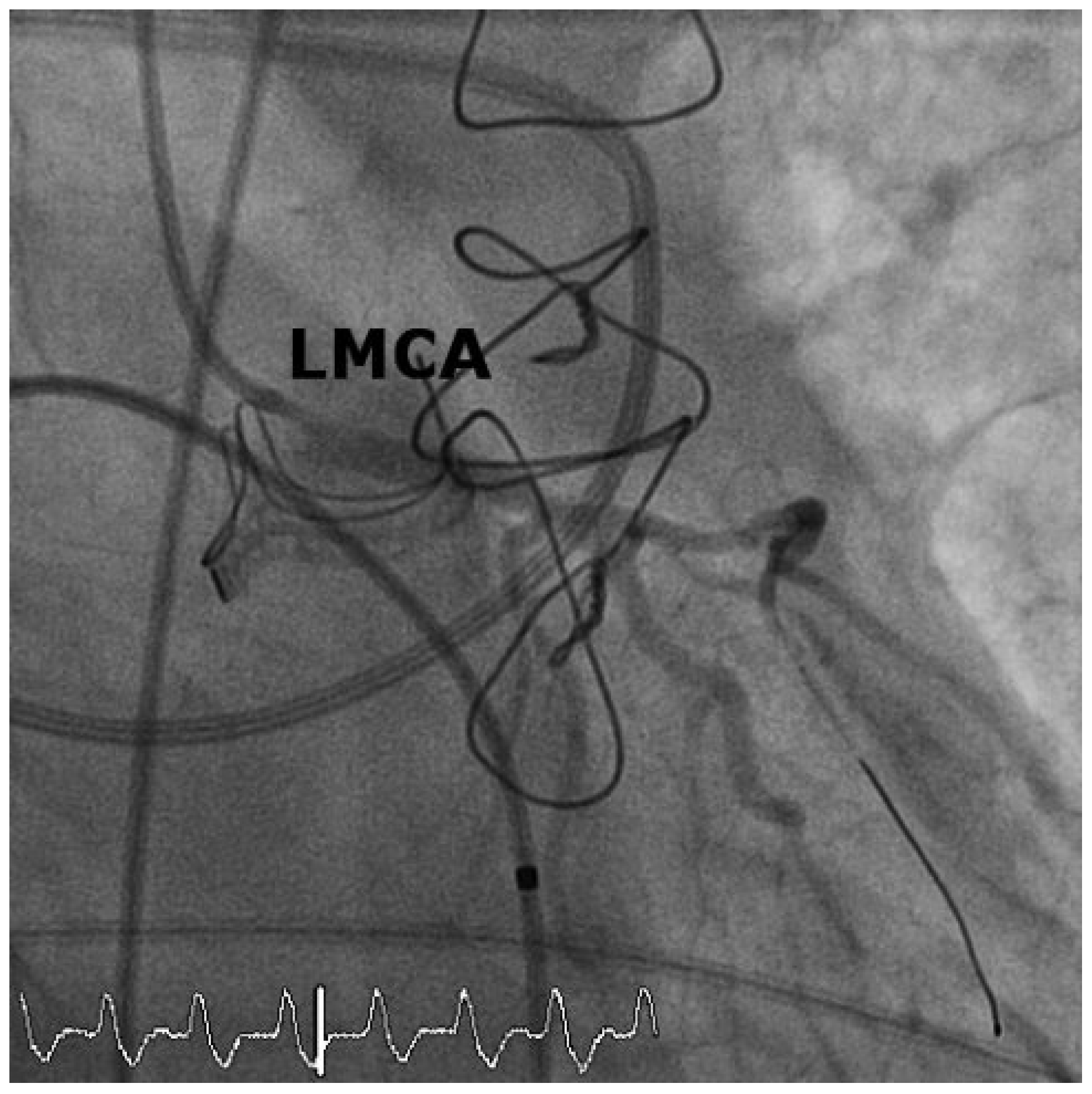
A 53-year-old male with symptomatic, severe paradoxical low-flow, low-gradient aortic valve stenosis due to restrictive physiology after thoracic radiotherapy (meangradient 23 mm Hg, effective valve orifice area 0.8 cm2) underwent SAVR with a bioprosthesis (23 mm Edwards Lifesciences Perimount, model 2900, Edwards Lifesciences, CA, USA). Preoperative coronary angiography showed no significant coronary stenosis (Figure 6). Eleven months later the patient presented with angina pectoris, dyspnoea and signs of pulmonary oedema. The electrocardiogram was non-conclusive because of a complete left bundle branch block, but cardiac biomarkers were elevated (CK 193 U/l, ULN = 190 U/l, and troponin T 0.685 µg/l, ULN < 0.010 µg/l). Transoesophageal echocardiography revealed severe central mitral valve regurgitation and normal left ventricular function. Coronary angiography showed a high grade ostial stenosis of the left main stem (Figure 7). The stenosis was crossed with a guide wire (Magnum 0,014”, Schneider, Switzerland) and dilated with a balloon dilatation catheter (Pantera 3.0 × 15 mm, Biotronik AG, Switzerland). This was followed by drug-eluting stent implantation with a Biomatrix stent (Biosensors Interventional Technologies Pte Ltd., Singapore) (Figure 8). Transoesophageal echocardiography after percutaneous coronary intervention showed a reduction in the severity of mitral valve regurgitation from severe to moderate. The patient successfully completed a rehabilitation program and had no signs of cardiac ischaemia or heart failure symptoms three months after the procedure. Follow-up echocardiography revealed only mild mitral regurgitation.

Discussion

Since the first description of iatrogenic coronary left main stem stenosis following SAVR in 1969 by Trimble et al. [1] several cases have been reported in the literature [2,3,4,5,6,7,8]. The incidence of this complication is rare and ranges from 0.3 to 3% [5,6]. Most of the affected patients develop sudden onset of angina pectoris 3–6 months after SAVR, but cases with onset of symptoms as late as 30 months after SAVR have been reported. Until the 1990s coronary artery bypass grafting (CABG) was performed to alleviate ischaemia with the attendant complications of redo surgery. In 1984, treatment of iatrogenic left main stem stenosis after SAVR by balloon angioplasty was described for the first time by Simarro et al. [4]. Since then several cases of successful percutaneous coronary intervention have been described. Hadjimiltiades et al. published the first case of ostial left main stent implantation after SAVR, which was protected by a left internal mammary artery graft to the distal LAO in 2005 [8].
Several registry studies have shown that percutaneous coronary intervention (PCI) of the ostial left main coronary artery is associated with a low procedural risk and a low risk of mortality and repeat revascularisation with the use of DES during long term follow-up [9,10]. More recently, data from the SYNTAX trial comparing PCI with CABG among patients with multivessel coronary artery disease showed comparable outcomes in terms of mortality and repeat revascularisation in the small subgroup of patients with isolated left main disease [11].
The pathological mechanism leading to iatrogenic left main stem stenosis is poorly understood.
Aortic root fibrosis with involvement of the left and right coronary main stem ostium secondary to turbulent flow around ball prostheses has been proposed by Roberts and Morrow in 1967 [12]. The development of heart valve prostheses with more laminar flow patterns should have attenuated the risk of aortic root fibrosis.
Another hypothesis relates to local pressure necrosis with subsequent intimal proliferation due to the insertion of perfusion catheters and turbulences related to cardioplegia [1,5]. In 1968 the problems of coronary injury due to cannulation for antegrade perfusion during CABG were recognized by Fishman et al. [13] and cannulas and coronary perfusion techniques improved. Silver et al. described three cases of iatrogenic ostial coronary stenosis in patients, who underwent SAVR with high pressure coronary artery perfusion. It was felt that the high antegrade pressure perfusion caused injury of the media and consecutive repair mechanisms led to coronary artery stenosis [2]. Cases of iatrogenic left main stem stenosis in the absence of coronary cannulation have also been reported [3].
Winkelmann et al. found evidence for a genetic predisposition to an increased proliferative response after arterial injury in seven cases of iatrogenic left main stem stenosis after SAVR. The investigator observed a signif-icantly higher incidence of the ε4 allele of the apolipoprotein E phenotype [5].
Finally an immunological reaction after SAVR with heterografts has been postulated by Tsukiji et al., who reported one case of bilateral coronary ostial narrowing. Their examination with intravascular ultrasonography demonstrated localised, membranous, homogeneous, and severe stenoses in the ostium of the right and left coronary artery. Histological examination showed intimal hypertrophy, mucinous degeneration, and hyaline degeneration without reactive changes, and signs of atherosclerosis were not found, though the authors suggested an immunological reaction to the heterograft as the mechanism [7].

Conclusion

Iatrogenic left main stenosis after SAVR is a rare but potentially life threatening complication. Clinical signs are angina pectoris, dyspnoea and potentially lethal arrhythmias. Whenever symptoms recur after SAVR, left or right ostial main stem stenosis should be suspected, even in cases with previously normal coronary arteries. Percutaneous coronary intervention is feasible and the therapy of choice in order to circumvent redo surgery.

Conflicts of Interest

The authors certify that there is no actual or potential conflict of interest in relation to this article.

References

  1. Trimble, A.S.; Bigelow, W.G.; Wigle, E.D.; Silver, M.D. Coronary ostial stenosis: a late complication of coronary perfusion in open-heart surgery. J Thorac Cardiovasc Surg. 1969, 57, 792–795. [Google Scholar] [CrossRef] [PubMed]
  2. Silver, M.D.; Wigle, E.D.; Trimble, A.S.; Bigelow, W.G. Iatrogenic coronary ostial stenosis. Arch Pathol. 1969, 88, 73–77. [Google Scholar] [PubMed]
  3. Force, T.L.; Raabe, D.S.; Jr Coffin, L.H.; DeMeules, J.D. Coronary ostial stenosis following aortic valve replacement without continuous coronary perfusion. J Thorac Cardiovasc Surg. 1980, 80, 637–641. [Google Scholar] [PubMed]
  4. Simarro Martin, E.; Moris de la Tassa, C.; Rodriguez Lambert, J.L.; Sieres Felgueres, M.; Gutierrez Rodriguez, J.A.; Cortina Llosa, A. Percutaneous transluminal coronary angioplasty in a case of iatrogenic stenosis of the left coronary ostium. Rev Esp Cardiol. 1984, 37, 366–367. [Google Scholar] [PubMed]
  5. Winkelmann, B.R.; Ihnken, K.; Beyersdorf, F.; Eckel, L.; Skupin, M.; Marz, W.; et al. Left main coronary artery stenosis after aortic valve replacement: genetic disposition for accelerated arteriosclerosis after injury of the intact human coronary artery? Coron Artery Dis. 1993, 4, 659–667. [Google Scholar] [CrossRef] [PubMed]
  6. Chavanon, O.; Carrier, M.; Cartier, R.; Hebert, Y.; Pellerin, M.; Perrault, L.P. Early reoperation for iatrogenic left main stenosis after aortic valve replacement: a perilous situation. Cardiovasc Surg. 2002, 10, 256–263. [Google Scholar] [CrossRef] [PubMed]
  7. Tsukiji, M.; Akasaka, T.; Wada, N.; Okahashi, N.; Kume, T.; Yoshitani, H.; et al. Bilateral coronary ostial stenosis after aortic valve replacement with freestyle stentless bioprosthesis: a case report. J Cardiol. 2004, 44, 207–213. [Google Scholar] [PubMed]
  8. Hadjimiltiades, S.; Harokopos, N.; Papadopoulos, C.; Gourassas, I.; Spanos, P.; Louridas, G. Left main coronary artery stenosis after aortic valve replacement. Hellenic J Cardiol. 2005, 46, 306–309. [Google Scholar] [PubMed]
  9. Chieffo, A.; Park, S.J.; Valgimigli, M.; Kim, Y.H.; Daemen, J.; Sheiban, I.; et al. Favorable long-term outcome after drug-eluting stent implantation in nonbifurcation lesions that involve unprotected left main coronary artery: a multicenter registry. Circulation. 2007, 116, 158–162. [Google Scholar] [CrossRef] [PubMed]
  10. Kim, Y.H.; Dangas, G.D.; Solinas, E.; Aoki, J.; Parise, H.; Kimura, M.; et al. Effectiveness of drug-eluting stent implantation for patients with unprotected left main coronary artery stenosis. Am J Cardiol. 2008, 101, 801–806. [Google Scholar] [CrossRef] [PubMed]
  11. Serruys, P.W.; Morice, M.C.; Kappetein, A.P.; Colombo, A.; Holmes, D.R.; Mack, M.J.; et al. Percutaneous coronary intervention versus coronary-artery bypass grafting for severe coronary artery disease. N Engl J Med. 2009, 360, 961–972. [Google Scholar] [CrossRef] [PubMed]
  12. Roberts, W.C.; Morrow, A.G. Late postoperative pathological findings after cardiac valve replacement. Circulation. 1967, 35 (Suppl. 4), I48–62. [Google Scholar] [CrossRef] [PubMed]
  13. Fishman, N.H.; Youker, J.E.; Roe, B.B. Mechanical injury to the coronary arteries during operative cannulation. Am Heart J. 1968, 75, 26–33. [Google Scholar] [CrossRef] [PubMed]
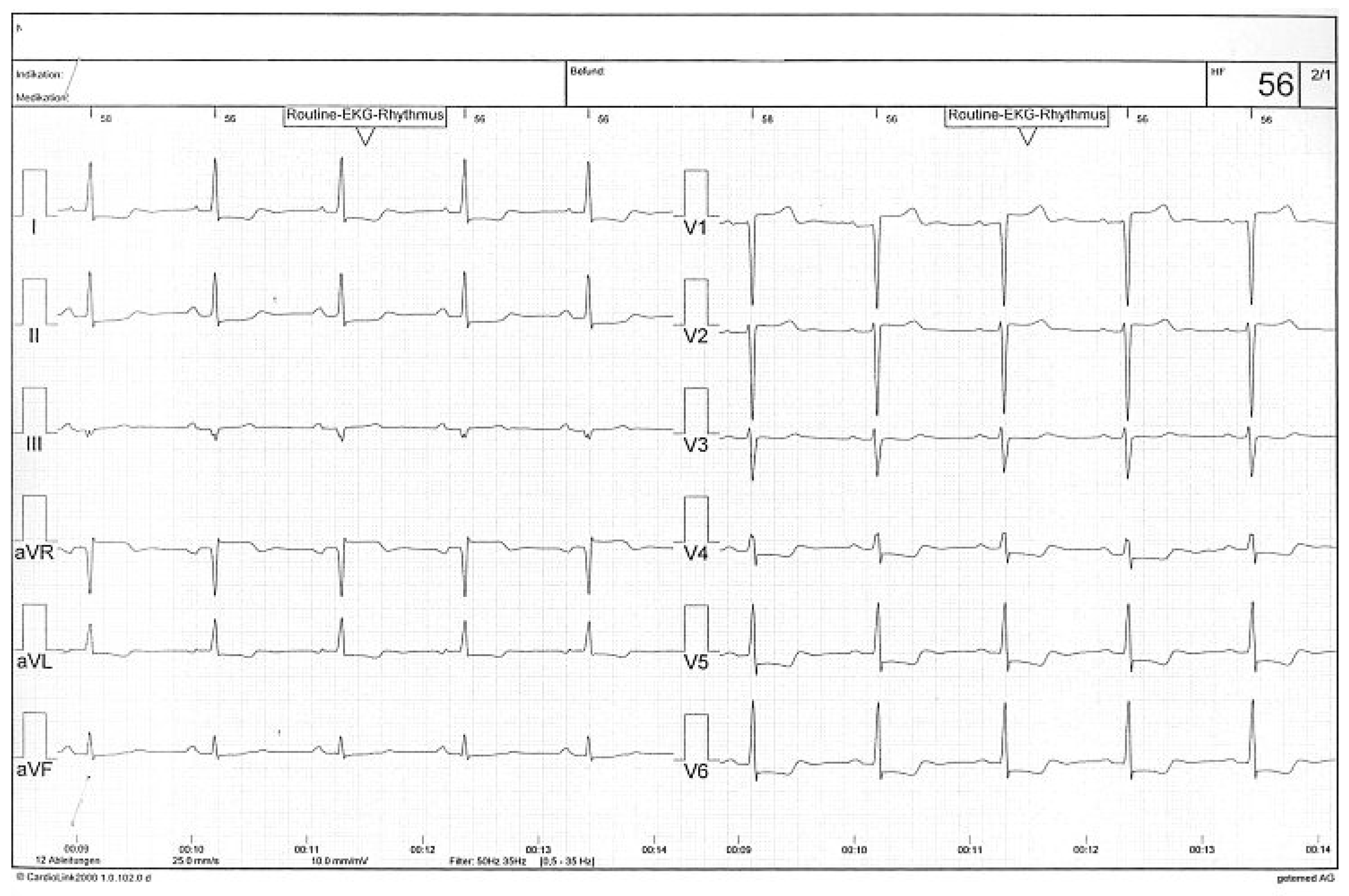
Figure 1. Patient 1: Electrocardiogram revealing severe ST-segment depression in I, aVL, V4–V6.
Cardiovascmed 14 00101 g001
Figure 2. Patient 1: Coronary angiogram of the left coronary artery before surgical aortic valve replacement. The left main coronary artery (LMCA) is patent. The proximal left anterior descending artery (LAD) shows a significant stenosis. LMCA = left main coronary artery; LAD = left anterior descending artery; LCx = left circumflex artery.
Cardiovascmed 14 00101 g002
Figure 3. Patient 1: Aortography eight months after aortic valve replacement shows an occlusion of the left main stem. LMCA = left main coronary artery.
Cardiovascmed 14 00101 g003
Figure 4. Patient 1: Coronary angiogram of the left internal mammary artery with retrograde opacification of the proximal LAD and LCx. Occlusion of the left main stem at the ostium is apparent. LMCA = left main coronary artery; LAD = left anterior descending artery; LCx = left circumflex artery.
Cardiovascmed 14 00101 g004
Figure 5. Patient 1: Coronary angiogram of the left main stem after recanalisation, balloon angioplasty and drug-eluting stent implantation. LMCA = left main coronary artery; LAD = left anterior descending artery; LCx = left circumflex artery.
Cardiovascmed 14 00101 g005
Figure 6. Patient 2: Coronary angiogram before surgical aortic valve replacement showing a pristine left main stem. LMCA = left main coronary artery; LAD = left anterior descending artery; LCx = left circumflex artery.
Cardiovascmed 14 00101 g006
Figure 7. Patient 2: Coronary angiogram 11 months after surgical aortic valve replacement shows a severe ostial main stem stenosis. LMCA = left main coronary artery.
Cardiovascmed 14 00101 g007
Figure 8. Patient 2: Coronary angiogram after percutaneous coronary intervention and drug-eluting stent implantation of the main stem lesion. LMCA = left main coronary artery.
Cardiovascmed 14 00101 g008