Next Article in Journal
Left Main Stem Thrombosis in a 37-Year-Old Man
Previous Article in Journal
A Case for Incidental Coronary Angiography During Percutaneous Closure of the Patent Foramen Ovale?
 
 
Cardiovascular Medicine is published by MDPI from Volume 28 Issue 1 (2025). Previous articles were published by another publisher in Open Access under a CC-BY (or CC-BY-NC-ND) licence, and they are hosted by MDPI on mdpi.com as a courtesy and upon agreement with Editores Medicorum Helveticorum (EMH).
Case Report

Transcoronary Alcohol Ablation—On Behalf of Three Cases with Hypertrophic Obstructive Cardiomyopathy

by
Alexander Breitenstein
*,
Jasmina Alibegovic
,
David Hürlimann
,
Patric Biaggi
,
Roberto Corti
and
Thomas F. Lüscher
Cardiology, Cardiovascular Center, University Hospital Zurich, Rämistrasse 100, CH-8091 Zurich, Switzerland
*
Author to whom correspondence should be addressed.
Cardiovasc. Med. 2011, 14(10), 290; https://doi.org/10.4414/cvm.2011.01615
Submission received: 26 July 2011 / Revised: 26 August 2011 / Accepted: 26 September 2011 / Published: 26 October 2011

Abstract

Hypertrophic cardiomyopathy (HCM) is a common genetic disorder with an estimated prevalence of 1:500. About a quarter of the patients with HCM have left ventricular outflow tract obstruction referred to as hypertrophic obstructive cardiomyopathy (HOCM). Even with optimal medical treatment using negative inotropic drugs such as β-blockers and first generation calcium-antagonists, about 5–10% of patients with HOCM remain refractory to therapy. In such cases, reduction of the outflow gradient by either surgical myectomy or catheter-based alcohol septal ablation represent further treatment options. Even though myectomy historically is considered the gold standard, interventional alcohol ablation of the septum has become the treatment of choice in many centres. We present the history of three patients suffering from HOCM, who were successfully treated by catheter-based alcohol septal ablation.

Case 1

A 42-year-old man was diagnosed with HOCM 10 years ago. Despite medical treatment with metoprolol 100 mg twice daily, he complained of shortness of breath during exercise (NYHA class III). During physical examination, a 3/6 holosystolic murmur was audible. The echocardiography showed left ventricular hypertrophy with an elevated subvalvular pressure gradient across the left ventricular outflow tract.
At left and right heart catheterisation, the resting intraventricular peak pressure gradient was 38 mm Hg, with an increase to 110 mm Hg after application of isoproterenol (Figure 1A). Additionally, a 90% occlusion of the right coronary artery was identified during coronary angiography and treated first by PTCA and bare metal stent implantation (Skylor 3.5 × 16 mm). Next, the first major septal branch of the left anterior descending coronary artery was identified (Figure 1B) and catheterised with a guide wire (Whisper ES J) and an over-the-wire 2 mm angioplasty balloon catheter (Sprinter OTW NC). After inflation of the balloon and withdrawal of the guide wire, echo contrast agent (SonoVue®) was injected through the balloon catheter to visualise echocardiographically the myocardium supplied by this vessel (Figure 1C) followed by administration of 2 mL of absolute ethanol (96%). After five minutes, the balloon was deflated.
The invasive pressure measurements revealed a decrease in the pressure gradient from 110 mm Hg to 55 mm Hg (Figure 1D). Immediately after start of the alcohol injection, the patient reported moderate chest pain, which subsided shortly after administration.
After intervention, the patient was transferred to the cardiac care unit for 24 h. Total creatine kinase rose to a maximum of 1180 U/l. The patient could be discharged three days after intervention on low-dose aspirin and metoprolol 100 mg once daily. Echocardiographic control after five months showed a persistent decrease of the left ventricular outflow gradient, especially during exercise.

Case 2

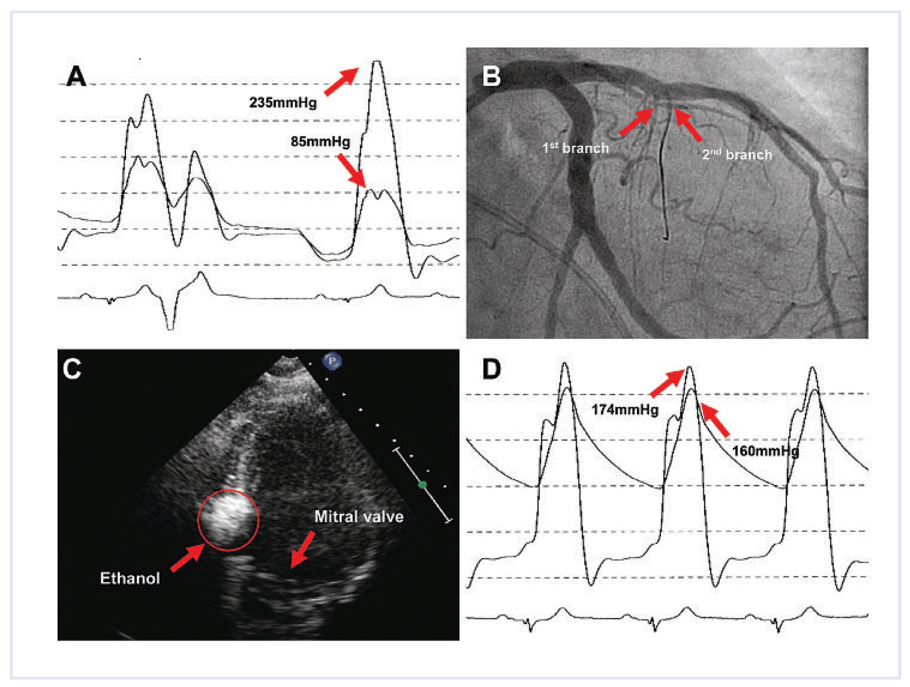
This 80-year-old woman presented initially with HOCM about seven years ago. Despite medical treatment with metoprolol 50 mg once daily, she complained of dyspnoea on exertion (NYHA class III). Echocardiography confirmed the diagnosis. The intraventricular pressure gradient at rest averaged 11 mm Hg and rose up to 50 mm Hg during exercise. Interestingly, the pressure gradient measured during catheterisation did not change after administration of isoproterenol as compared to the resting condition, while after induction of ventricular extrasystoles, the intraventricular pressure gradient rose to 150 mm Hg (Figure 2A). The second septal branch of the left main descending coronary artery was identified (Figure 2B) and inflated with a 2 mm balloon (Sprinter OTW NC). Application of an echo contrast agent (SonoVue®) provided visualisation of the myocardium supplied by this vessel (Figure 2C). After injection of 3 mL of absolute ethanol (96%) for five minutes, the postextrasystolic intraventricular pressure gradient had already decreased slightly. Nevertheless, it could be shown that application of the echo contrast agent (SonoVue®) into the very first septal branch revealed that this coronary vessel additionally supplied a clinically relevant part of the septal myocardium. Therefore, alcohol ablation of the septum was subsequently also performed through the first septal branch. 2 mL of absolute ethanol (96%) were injected and the balloon was kept inflated for an additional five minutes. The initially elevated pressure gradient was significantly reduced after the two procedures (Figure 2D). After uneventful catheterisation, the patient was transferred to the cardiac care unit and discharged two days after intervention. Maximal creatine kinase was 1241 U/l and no heart block was noticed. Two months after the procedure, the echocardiographic control revealed an intraventricular gradient of 8 mm Hg without an increase during exercise.

Case 3

HOCM was diagnosed ten years ago in this 71-year-old woman. The subvalvular pressure gradient across the left ventricular outflow tract remained stable at 30–40 mm Hg for several years, but had now increased to 74 mm Hg resulting in dyspnoea on exertion (NYHA III) despite medical treatment.
During catheterisation, the resting intraventricular mean pressure gradient was 78 mm Hg (Figure 3A). The first major septal branch of the left anterior descending coronary artery was identified (Figure 3B) and catheterised with a guide wire (Whisper ES J) followed by an over-the-wire balloon catheter (Sprinter OTW NC). By injection of echo contrast agent (SonoVue®), the myocardium supplied by this vessel was identified (Figure 3C). The artery was occluded by the balloon catheter and 1.5 mL of absolute ethanol (96%) was applied. After five minutes, the balloon was deflated. Immediately after the procedure, the intraventricular pressure gradient decreased from 78 to 17 mm Hg (Figure 3D). The patient was transferred to the cardiac care unit and was discharged home three days after the intervention. No new heart block was present and maximal creatine kinase rose to 1108 U/l. Clinical and echocardiographic control after three months revealed a persistent decrease of the outflow gradient.

Discussion

HCM is a well known primary myocardial disorder with a prevalence within the general population of 1:500. HCM is characterised by symmetric or asymmetric hypertrophy of the myocardium with or without obstruction of the left ventricular outflow tract, the later being referred to as HOCM. The annual mortality of HCM is 1% and 2% for patients suffering from HOCM [1]. Treatment strategies in patients suffering from HOCM include medical therapy with negative inotropic agents most commonly the use of β-blockers. Due to the reduction in myocardial contractility, including that of the septal wall, β-blockers decrease the septal bulge and therefore intraventricular pressure gradients [2]. In addition, in selected patients, implantation of a cardioverter-defîbrillator (ICD) [2,3,4] has to be considered to prevent sudden cardiac death.
In patients who remain symptomatic despite optimal medical treatment, reduction of the intraventricular pressure gradient has clinical and prognostic implications. For many years, surgical myectomy represented the gold standard. However, the surgical approach carries a significant morbidity and is sometimes associated with complications such as the occurrence of aortic regurgitation and dilation of the left ventricle [5]. Furthermore, the intervention requires general anaesthesia, extracorporeal circulation and opening of the chest. Therefore, in 1994 Sigwart introduced a new catheterbased therapy of HOCM [6]. By infusion of small amounts of absolute ethanol into the septal branch of the left anterior descending artery which supplies the offending myocardial bulge, a small myocardial infarction within the supplied area is induced and in most cases a reduction of the thickness of the intraventricular septum is achieved. This percutaneous intervention has gained substantial popularity and represents the treatment of choice in patients with refractory HOCM in many centres. Nevertheless, due to the rarity of this disease, no randomized clinical trial has been, nor probably will be, performed directly comparing septal ablation to surgical myectomy [7]. Data from non-randomized studies indicate that both procedures are equally effective with respect to haemodynamic and functional improvement, including NYHA class and exercise capacity [8,9]. Furthermore, two meta-analyses have documented no difference in total mortality between septal ablation and surgical myectomy [10,11]. Since septal ablation is a much more recently introduced procedure compared to surgical myectomy, data on long term benefit and outcome still remain unknown. Complications in general are rare and within the range of surgical myectomy. The most common side effect is a transient or permanent atrioventricular block [8,9]. About half of the patients experience a self-terminating atrioventricular block during the procedure, which recovers in the majority of cases within hours [12]. With increasing experience and the use of a smaller amounts of injected alcohol, the level of permanent pacemaker implantations has decreased significantly, but is still necessary in about 10% of the patients [13]. To further reduce the onset of atrioventricular block, some operators have started to use microspheres, polyvinyl alcohol or absorbable gelatine sponges instead of absolute alcohol [14]. Controversially discussed is the potential pro-arrhythmic effects of septal ablation [15]. Even though life-threatening ventricular arrhythmias are described after septal ablation, a recent meta-analysis comparing septal ablation to surgical myectomy revealed a similar incidence of ventricular arrhythmias with the two procedures [10,16].
Since its introduction by Sigwart at the end of the last century, the number of septal ablations performed world wide has now reached nearly 5000, which is numerically more than all septal myectomies in the last 50 years and thus underscoring the importance of this procedure. If performed by skilled cardiac interventionalists, the success rate (marked decrease of the leftventricular outflow tract gradient; LVOT) is usually more than 80% [12,17], with a persistence of the LVOT gradient reduction even after 12 months [18]. Furthermore, the reduction of the LVOT gradient is paralleled by an improvement in exercise capacity and NYHA class. Predictor for a poor outcome is an immediate residual LVOT gradient of ≥25 mm Hg [19]; data from the literature indicate that a second procedure is necessary in 9% of the patients initially treated by septal ablation [18]. In general, alcohol septal ablation represents a safe procedure with a cardiac mortality of 0.7% per year [20].
In the three patients reported above, who were treated earlier this year at the Zurich University Hospital, the intervention was both successful and safe. Despite its favourable outcome in most instances, the role of this procedure for the management of patients with HOCM and symptoms despite optimal medical therapy is still under intense scrutiny.

Funding/Potential Competing Interests

No financial support and no other potential conflict of interest relevant to this article was reported.

References

  1. Maron, M.S.; Olivotto, I.; Betocchi, S.; Casey, S.A.; Lesser, J.R.; Losi, M.A.; et al. Effect of left ventricular outflow tract obstruction on clinical outcome in hypertrophic cardiomyopathy. N Engl J Med. 2003, 348, 295–303. [Google Scholar] [CrossRef] [PubMed]
  2. Roberts, R.; Sigwart, U. Current concepts of the pathogenesis and treatment of hypertrophic cardiomyopathy. Circulation. 2005, 112, 293–296. [Google Scholar] [CrossRef] [PubMed]
  3. Fifer, M.A.; Sigwart, U. Hypertrophic obstructive cardiomyopathy: Alcohol septal ablation. Eur Heart J. 2011, 32, 1059–1064. [Google Scholar] [CrossRef] [PubMed]
  4. Maron, B.J.; Shen, W.K.; Link, M.S.; Epstein, A.E.; Almquist, A.K.; Daubert, J.P.; et al. Efficacy of implantable cardioverter-defibrillators for the prevention of sudden death in patients with hypertrophic cardiomyopathy. N Engl J Med. 2000, 342, 365–373. [Google Scholar] [CrossRef] [PubMed]
  5. Seiler, C.; Hess, O.M.; Schoenbeck, M.; Turina, J.; Jenni, R.; Turina, M.; et al. Long-term follow-up of medical versus surgical therapy for hypertrophic cardiomyopathy: A retrospective study. J Am Coll Cardiol. 1991, 17, 634–42. [Google Scholar] [CrossRef] [PubMed][Green Version]
  6. Sigwart, U. Non-surgical myocardial reduction for hypertrophic obstructive cardiomyopathy. Lancet. 1995, 346, 211–214. [Google Scholar] [PubMed][Green Version]
  7. Olivotto, I.; Ommen, S.R.; Maron, M.S.; Cecchi, F.; Maron, B.J. Surgical myectomy versus alcohol septal ablation for obstructive hypertrophic cardiomyopathy. Will there ever be a randomized trial? J Am Coll Cardiol. 2007, 50, 831–834. [Google Scholar] [CrossRef] [PubMed]
  8. Qin, J.X.; Shiota, T.; Lever, H.M.; Kapadia, S.R.; Sitges, M.; Rubin, D.N.; et al. Outcome of patients with hypertrophic obstructive cardiomyopathy after percutaneous transluminal septal myocardial ablation and septal myectomy surgery. J Am Coll Cardiol. 2001, 38, 1994–2000. [Google Scholar] [CrossRef] [PubMed]
  9. Nagueh, S.F.; Ommen, S.R.; Lakkis, N.M.; Killip, D.; Zoghbi, W.A.; Schaff, H.V.; et al. Comparison of ethanol septal reduction therapy with surgical myectomy for the treatment of hypertrophic obstructive cardiomyopathy. J Am Coll Cardiol. 2001, 38, 1701–1706. [Google Scholar] [CrossRef] [PubMed]
  10. Agarwal, S.; Tuzcu, E.M.; Desai, M.Y.; Smedira, N.; Lever, H.M.; Lytle, B.W.; et al. Updated meta-analysis of septal alcohol ablation versus myectomy for hypertrophic cardiomyopathy. J Am Coll Cardiol. 2010, 55, 823–834. [Google Scholar] [CrossRef] [PubMed]
  11. Alam, M.; Dokainish, H.; Lakkis, N.M. Hypertrophic obstructive cardiomyopathy-alcohol septal ablation vs. myectomy: A meta-analysis. Eur Heart J. 2009, 30, 1080–1087. [Google Scholar] [CrossRef] [PubMed]
  12. Faber, L.; Seggewiss, H.; Gleichmann, U. Percutaneous transluminal septal myocardial ablation in hypertrophic obstructive cardiomyopathy: Results with respect to intraprocedural myocardial contrast echocardiography. Circulation 1998, 98, 2415–2421. [Google Scholar] [CrossRef] [PubMed]
  13. Hess, O.M.; Sigwart, U. New treatment strategies for hypertrophic obstructive cardiomyopathy: Alcohol ablation of the septum: The new gold standard? J Am Coll Cardiol. 2004, 44, 2054–2055. [Google Scholar] [CrossRef] [PubMed]
  14. Fifer, M.A.; Sigwart, U. Controversies in cardiovascular medicine. Hypertrophic obstructive cardiomyopathy: Alcohol septal ablation. Eur Heart. J. 2011, 32, 1059–1064. [Google Scholar] [CrossRef] [PubMed]
  15. Maron, B.J. Controversies in cardiovascular medicine. Surgical myectomy remains the primary treatment option for severely symptomatic patients with obstructive hypertrophic cardiomyopathy. Circulation 2007, 116, 196–206; discussion 206. [Google Scholar] [CrossRef] [PubMed]
  16. Lawrenz, T.; Obergassel, L.; Lieder, F.; Leuner, C.; Strunk-Mueller, C.; Meyer, D.; Vilsendorf, Z.; et al. Transcoronary ablation of septal hypertrophy does not alter ICD intervention rates in high risk patients with hypertrophic obstructive cardiomyopathy. Pacing Clin Electrophysiol. 2005, 28, 295–300. [Google Scholar] [CrossRef] [PubMed]
  17. Gietzen, F.H.; Leuner, C.J.; Raute-Kreinsen, U.; Dellmann, A.; Hegselmann, J.; Strunk-Mueller, C.; et al. Acute and long-term results after transcoronary ablation of septal hypertrophy (TASH). Catheter interventional treatment for hypertrophic obstructive cardiomyopathy. Eur Heart J. 1999, 20, 1342–1354. [Google Scholar] [PubMed]
  18. Alam, M.; Dokainish, H.; Lakkis, N. Alcohol septal ablation for hypertrophic obstructive cardiomyopathy: A systematic review of published studies. J Interv Cardiol. 2006, 19, 319–327. [Google Scholar] [CrossRef] [PubMed]
  19. Chang, S.M.; Lakkis, N.M.; Franklin, J.; Spencer, W.H., 3rd; Nagueh, S.F. Predictors of outcome after alcohol septal ablation therapy in patients with hypertrophic obstructive cardiomyopathy. Circulation 2004, 109, 824–827. [Google Scholar] [CrossRef] [PubMed]
  20. Kuhn, H.; Lawrenz, T.; Lieder, F.; Leuner, C.; Strunk-Mueller, C.; Obergassel, L.; et al. Survival after transcoronary ablation of septal hypertrophy in hypertrophic obstructive cardiomyopathy (TASH): A 10 year experience. Clin Res Cardiol. 2008, 97, 234–243. [Google Scholar] [PubMed]
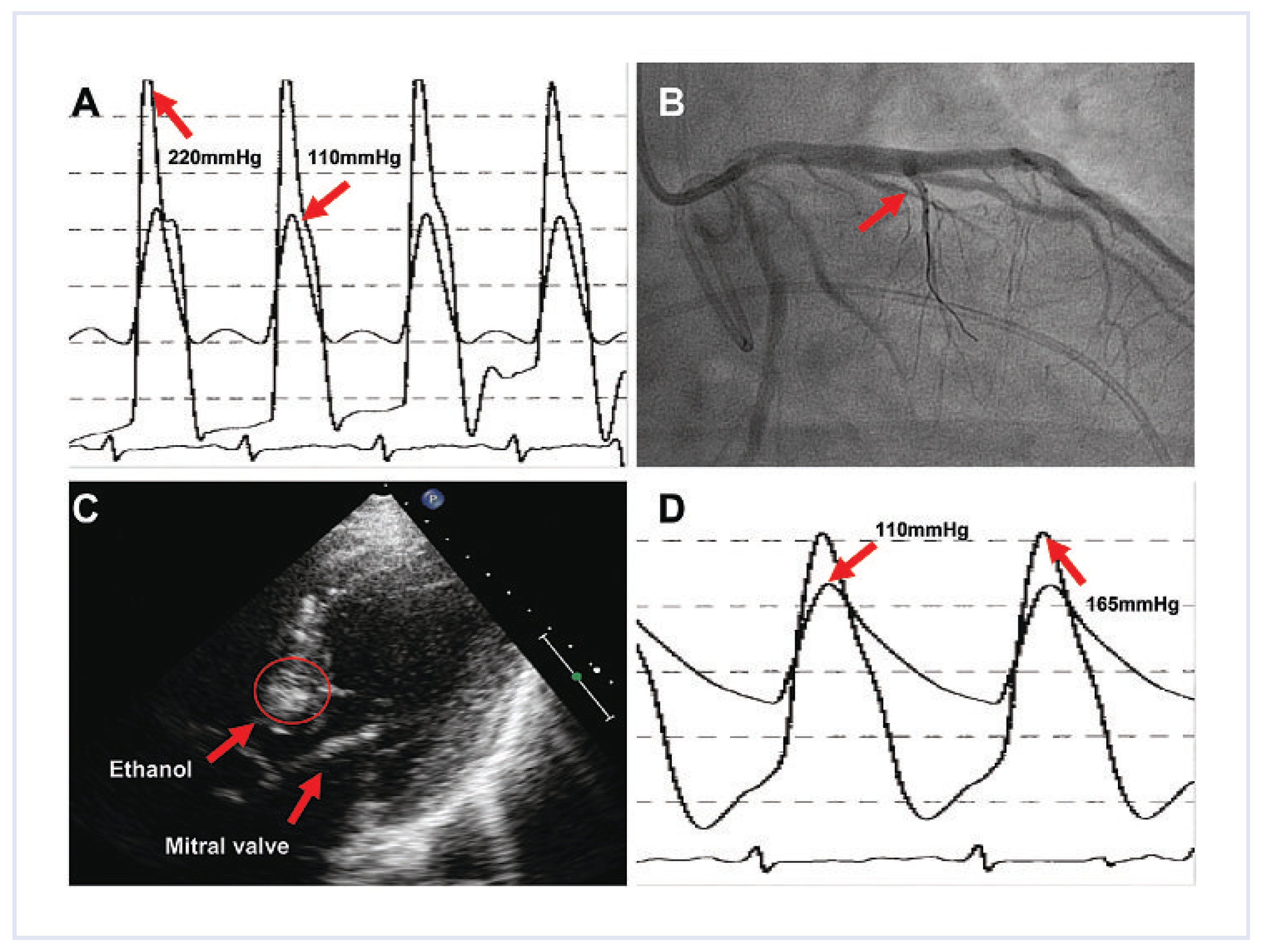
Figure 1. (A) Haemodynamic recording showing the peak pressure gradient of 110 mm Hg after application of isoprotenerol. (B) Catheterisation of the first septal branch of the LAD. (C) Application of an echo contrast media demonstrating the myocardium perfused by the vessel. (D) Haemodynamic recordings after ethanol application showing the reduced pressure gradient.
Cardiovascmed 14 00290 g001
Figure 2. (A) Postextrasystolic increase of the pressure gradient. (B) Catheterisation of the second septal branch of the. LAD. (C) Myocardium perfused by the vessel as demonstrated after application of an echo contrast media. (D) Reduced pressure gradient after the two ethanol applications.
Cardiovascmed 14 00290 g002
Figure 3. (A) Subvalvular peak pressure gradient of 78 mm Hg during intervention. (B) Catheterisation of the first septal branch of the LAD. (C) Echo contrast agent demonstrating the myocardium perfused by the vessel. (D) Reduced pressure gradient after ethanol application.
Cardiovascmed 14 00290 g003